| Size | Price | Stock | Qty |
|---|---|---|---|
| 5mg |
|
||
| 10mg |
|
||
| 25mg |
|
||
| 50mg |
|
||
| 100mg | |||
| 250mg | |||
| Other Sizes |
ZZW-115 (ZZW115) is a novel and potent NUPR1 inhibitor (Kd=2.1 uM) with antitumor activity. It is the TFP-derived compound in the family that is the most active. The protein NUPR1 has a completely disordered conformation and is an intrinsically disordered protein (IDP).
| Targets |
ZZW 115 targets nuclear protein 1 (NUPR1) (Ki = 0.7 μM; binding affinity Kd = 0.5 μM) [1]
ZZW 115 targets NUPR1 in pancreatic adenocarcinoma cells (IC50 for cell proliferation inhibition = 3.2 μM in PANC-1 cells) [2] |
|---|---|
| ln Vitro |
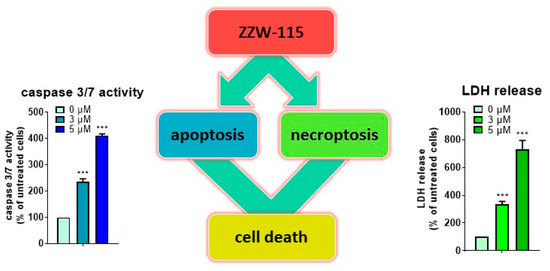
ZZW-115 (0.1-33 μM; 72 hours) effectively kills cancer cells, with an IC50 ranging from 0.84 μM (ANOR) to 4.93 μM (HN14)[1].
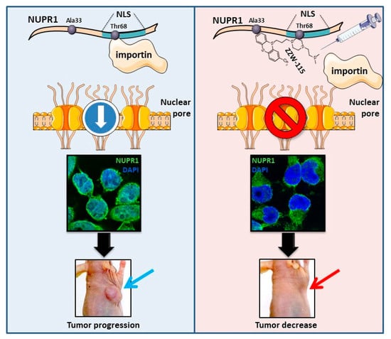
ZZW-115 (0-100 μM; 24-72 hours) effectively kills these cancerous cells, with an IC50 ranging from 0.42 μM (Hep2G cells) to 7.75 μM (SaOS-2 cells)[1]. ZZW-115 causes necrosis and apoptosis, which lead to pancreatic cell death. A decrease in ATP production and an increase in ROS production are brought on by ZZW-115 treatment[1]. ZZW-115-treated cells (MiaPaCa-2, 02-063, LIPC, Foie8b, and HN14 cells) release LDH at a concentration-dependently higher rate than untreated cells (LDH). Similar to this, cells treated with ZZW-115 exhibit increased caspase 3/7 activity. Through these tests, it was shown that ZZW-115 had pronecrotic and proapoptotic effects[1]. In human pancreatic adenocarcinoma cell lines (PANC-1, MIA PaCa-2, AsPC-1), ZZW 115 (1–20 μM) inhibits cell proliferation in a concentration-dependent manner, with IC50 values ranging from 3.2 to 7.5 μM. It induces necroptosis (not apoptosis) by upregulating the expression of RIP1, RIP3, and MLKL (necroptosis markers) and increasing their phosphorylation, as detected by Western blot. Pretreatment with necrostatin-1 (RIP1 inhibitor) reverses the anti-proliferative effect of ZZW 115 [1] - ZZW 115 binds directly to the N-terminal domain of NUPR1, suppressing its transcriptional regulatory activity. In PANC-1 cells, it downregulates the expression of NUPR1 target genes (Bcl-2, Survivin, XIAP) at both mRNA and protein levels (RT-PCR and Western blot). It also reduces colony formation ability: treatment with 5 μM ZZW 115 decreases colony number by ~60% compared to control [1] - In gemcitabine-resistant pancreatic adenocarcinoma cells (PANC-1/GemR), ZZW 115 (5–15 μM) restores sensitivity to gemcitabine, reducing the IC50 of gemcitabine from 40 μM to 8 μM. This synergistic effect is associated with inhibition of NUPR1-mediated DNA damage repair (downregulation of BRCA1 and RAD51) [2] - In normal human pancreatic ductal epithelial cells (HPDE), ZZW 115 at concentrations up to 20 μM shows no significant anti-proliferative effect, indicating selective toxicity to cancer cells [1,2] |
| ln Vivo |
ZZW-115 (0.5-5 mg/kg; injection; daily for 30 days) prevents the growth of pancreatic xenografted tumors[1].
Treatment with ZZW-115 (5 mg/kg for 30 days; immunocompetent C57BL/6 mice were orthotopically implanted with Panc02 cells) reveals that the tumor size is occasionally almost immeasurable[1].
In a subcutaneous xenograft model of pancreatic adenocarcinoma (PANC-1 cells implanted in nude mice), intraperitoneal administration of ZZW 115 (10 mg/kg/day) for 21 days inhibits tumor growth by ~70% compared to vehicle control. Tumor tissues show increased necrosis (H&E staining), upregulated phosphorylated RIP3/MLKL, and downregulated NUPR1 and Bcl-2 expression (Western blot and immunohistochemistry) [1] - In an orthotopic pancreatic adenocarcinoma model (MIA PaCa-2 cells implanted into mouse pancreas), oral administration of ZZW 115 (20 mg/kg/day) for 28 days reduces primary tumor weight by ~65% and inhibits liver metastasis (number of metastatic nodules reduced by ~80%). The treatment also prolongs mouse overall survival (median survival increased from 35 days to 58 days) [2] - In the gemcitabine-resistant xenograft model (PANC-1/GemR cells implanted in nude mice), combined treatment with ZZW 115 (10 mg/kg/day, intraperitoneal) and gemcitabine (20 mg/kg/week, intravenous) for 24 days inhibits tumor growth by ~85%, which is significantly higher than either agent alone (~30% for gemcitabine alone, ~60% for ZZW 115 alone) [2] |
| Enzyme Assay |
Fluorescence polarization (FP) assay for NUPR1 binding: Recombinant NUPR1 protein was incubated with a fluorescently labeled peptide probe (derived from NUPR1's target DNA sequence) in assay buffer. ZZW 115 was added at concentrations ranging from 0.01 to 50 μM, and the mixture was incubated at room temperature for 1 hour. FP signal (mP) was measured, and the binding affinity (Ki) was calculated by fitting the competition curve to the Hill equation [1]
- Isothermal titration calorimetry (ITC) assay: Purified NUPR1 protein was dialyzed into buffer, and ZZW 115 was dissolved in the same buffer. The drug solution was titrated into the protein solution at 25°C, and heat changes during binding were recorded. The dissociation constant (Kd) was determined by analyzing the ITC thermogram using Origin software [1] - NUPR1 transcriptional activity assay: HEK293T cells were cotransfected with NUPR1 expression plasmid, reporter plasmid (luciferase under the control of NUPR1-responsive promoter), and internal control plasmid (Renilla luciferase). After 24 hours, cells were treated with ZZW 115 (0.5–20 μM) for 12 hours. Luciferase activity was measured using a dual-luciferase assay kit, and the inhibition rate of NUPR1 transcriptional activity was calculated [2] |
| Cell Assay |
Cell proliferation assay: Pancreatic adenocarcinoma cells (5×10³ per well) were seeded in 96-well plates, incubated overnight, and treated with ZZW 115 (0.1–40 μM) for 48–72 hours. Cell viability was detected by CCK-8 assay (absorbance at 450 nm), and IC50 values were calculated by nonlinear regression [1,2]
- Necroptosis/apoptosis detection: PANC-1 cells were treated with ZZW 115 (10 μM) for 24 hours, stained with Annexin V-FITC/PI (for apoptosis) or PI/MLKL antibody (for necroptosis) and analyzed by flow cytometry. For morphological observation, cells were stained with Hoechst 33342 and propidium iodide, and necrotic cells (PI-positive, fragmented nuclei) were counted under fluorescence microscopy [1] - Western blot analysis: Cells treated with ZZW 115 (2.5–20 μM) for 12–24 hours were lysed to extract total protein. Equal amounts of protein were separated by SDS-PAGE, transferred to PVDF membranes, and probed with antibodies against NUPR1, RIP1, p-RIP1, RIP3, p-RIP3, MLKL, p-MLKL, Bcl-2, Survivin, or GAPDH (loading control). Protein bands were visualized by chemiluminescence and quantified by ImageJ [1,2] - Colony formation assay: Pancreatic adenocarcinoma cells (1×10³ per well) were seeded in 6-well plates, treated with ZZW 115 (2.5–10 μM) for 2 weeks, with medium changed every 3 days. Colonies were stained with crystal violet, and those with >50 cells were counted. The colony formation rate was calculated relative to control [1,2] - RT-PCR analysis: Total RNA was extracted from treated cells using TRIzol reagent, reverse-transcribed into cDNA. Quantitative real-time PCR was performed with specific primers for NUPR1 target genes (Bcl-2, Survivin, XIAP, BRCA1, RAD51) and GAPDH (reference gene). Relative mRNA expression was calculated using the 2-ΔΔCt method [2] |
| Animal Protocol |
Subcutaneous xenograft model: Nude mice (4-week-old, male) were subcutaneously injected with PANC-1 cells (5×10⁶ cells/mouse) into the right flank. When tumors reached ~100 mm3, mice were randomly divided into control (n = 6) and ZZW 115 treatment (n = 6) groups. ZZW 115 was dissolved in DMSO (5%) + saline (95%), administered via intraperitoneal injection at 10 mg/kg once daily for 21 days. Tumor volume was measured every 3 days (volume = length × width2 / 2), and mouse body weight was recorded weekly. At the end of treatment, mice were euthanized, tumors were excised, weighed, and stored for molecular and histological analysis [1]
- Orthotopic xenograft model: Nude mice (4-week-old, male) were anesthetized, and MIA PaCa-2 cells (1×10⁶ cells/mouse) were injected into the pancreatic tail. Seven days after implantation, mice were assigned to control (n = 8) and ZZW 115 treatment (n = 8) groups. ZZW 115 was dissolved in 0.5% carboxymethylcellulose sodium (CMC) solution, administered via oral gavage at 20 mg/kg once daily for 28 days. Mouse survival was recorded daily. At euthanasia, primary tumors and livers were collected for metastasis detection and histological examination [2] - Gemcitabine-resistant xenograft model: Nude mice (4-week-old, male) were subcutaneously implanted with PANC-1/GemR cells (5×10⁶ cells/mouse). When tumors reached ~120 mm3, mice were divided into four groups (n = 6 per group): control, gemcitabine alone (20 mg/kg, intravenous injection once weekly), ZZW 115 alone (10 mg/kg, intraperitoneal injection once daily), and combination group. ZZW 115 was dissolved in DMSO (3%) + saline (97%), and treatment lasted for 24 days. Tumor volume and body weight were measured every 3 days [2] |
| ADME/Pharmacokinetics |
In mice, the oral bioavailability of ZZW 115 (20 mg/kg) was 42%. The peak plasma concentration (Cmax) was 1.8 μg/mL and the time to peak concentration (Tmax) was 1.5 h. The elimination half-life (t1/2) was 6.8 h and the area under the plasma concentration-time curve (AUC0–24h) was 12.3 μg·h/mL [2] - In mice, after intravenous injection of ZZW 115 (10 mg/kg), the Cmax was 4.5 μg/mL, the t1/2 was 5.2 h, and the AUC0–24h was 18.7 μg·h/mL. The drug is widely distributed in various tissues, with the highest concentrations in the liver and pancreas (tumor-bearing mice) [2] - In vitro metabolic stability test: ZZW 115 was incubated with human liver microsomes for 0–60 minutes. The residual drug concentration was determined by HPLC-MS/MS, and the in vitro half-life (t1/2) was 85 minutes, indicating that it has good metabolic stability [2].
|
| Toxicity/Toxicokinetics |
In vitro toxicity: At concentrations up to 20 μM, ZZW 115 showed no significant cytotoxicity to normal human cells (HPDE, HUVEC) (CCK-8 assay), with cell viability >85% compared to the control group [1,2]. - Acute in vivo toxicity: Mice were given a single oral dose of ZZW 115 (up to 200 mg/kg) or an intraperitoneal injection (up to 100 mg/kg). No deaths or significant toxic symptoms (drowsiness, weight loss, diarrhea) were observed within 14 days [2]. - Long-term in vivo toxicity: Compared to the control group, mice treated with ZZW 115 (20 mg/kg/day orally for 28 days; or 10 mg/kg/day intraperitoneally for 21 days) showed no significant changes in body weight, liver function (ALT, AST), or kidney function (BUN, creatinine). Histological examination of the liver, kidneys, heart, and spleen revealed no abnormal lesions or inflammation [1,2]
- Plasma protein binding rate: determined by balanced dialysis, the plasma protein binding rate of ZZW 115 in mouse plasma was 89%, and in human plasma it was 91% [2] |
| References | |
| Additional Infomation |
ZZW 115 is a small molecule inhibitor of NUPR1, designed by ligand-based drug design based on the structure of the NUPR1 DNA binding domain [1]. NUPR1 is a stress-induced nucleoprotein that is overexpressed in pancreatic adenocarcinoma, promoting cancer cell survival, proliferation, and chemotherapy resistance. ZZW 115 exerts its anticancer effect by binding to NUPR1, inhibiting its transcriptional activity, and inducing necroptosis through the RIP1/RIP3/MLKL pathway [1]. ZZW 115 and gemcitabine (a first-line chemotherapy drug for pancreatic cancer) showed synergistic anticancer activity in gemcitabine-resistant cells and xenograft models, providing a potential treatment strategy for chemotherapy-resistant pancreatic adenocarcinoma [2]. The drug has good water solubility and oral bioavailability, making it suitable for clinical oral administration [2].
|
| Molecular Formula |
C24H31F3N4S
|
|---|---|
| Molecular Weight |
464.589954614639
|
| Exact Mass |
464.222
|
| Elemental Analysis |
C, 62.05; H, 6.73; F, 12.27; N, 12.06; S, 6.90
|
| CAS # |
801991-87-7
|
| Related CAS # |
ZZW-115 hydrochloride;10122-45-9
|
| PubChem CID |
25010688
|
| Appearance |
Solid powder
|
| LogP |
5.1
|
| Hydrogen Bond Donor Count |
0
|
| Hydrogen Bond Acceptor Count |
8
|
| Rotatable Bond Count |
7
|
| Heavy Atom Count |
32
|
| Complexity |
582
|
| Defined Atom Stereocenter Count |
0
|
| SMILES |
CN(C)CCN1CCN(CC1)CCCN2C3=CC=CC=C3SC4=C2C=C(C=C4)C(F)(F)F
|
| InChi Key |
HUDONDPCYIGAMQ-UHFFFAOYSA-N
|
| InChi Code |
InChI=1S/C24H31F3N4S/c1-28(2)12-13-30-16-14-29(15-17-30)10-5-11-31-20-6-3-4-7-22(20)32-23-9-8-19(18-21(23)31)24(25,26)27/h3-4,6-9,18H,5,10-17H2,1-2H3
|
| Chemical Name |
N,N-dimethyl-2-[4-[3-[2-(trifluoromethyl)phenothiazin-10-yl]propyl]piperazin-1-yl]ethanamine
|
| Synonyms |
ZZW 115; ZZW-115; ZZW115
|
| HS Tariff Code |
2934.99.9001
|
| Storage |
Powder -20°C 3 years 4°C 2 years In solvent -80°C 6 months -20°C 1 month |
| Shipping Condition |
Room temperature (This product is stable at ambient temperature for a few days during ordinary shipping and time spent in Customs)
|
| Solubility (In Vitro) |
May dissolve in DMSO (in most cases), if not, try other solvents such as H2O, Ethanol, or DMF with a minute amount of products to avoid loss of samples
|
|---|---|
| Solubility (In Vivo) |
Note: Listed below are some common formulations that may be used to formulate products with low water solubility (e.g. < 1 mg/mL), you may test these formulations using a minute amount of products to avoid loss of samples.
Injection Formulations
Injection Formulation 1: DMSO : Tween 80: Saline = 10 : 5 : 85 (i.e. 100 μL DMSO stock solution → 50 μL Tween 80 → 850 μL Saline)(e.g. IP/IV/IM/SC) *Preparation of saline: Dissolve 0.9 g of sodium chloride in 100 mL ddH ₂ O to obtain a clear solution. Injection Formulation 2: DMSO : PEG300 :Tween 80 : Saline = 10 : 40 : 5 : 45 (i.e. 100 μL DMSO → 400 μLPEG300 → 50 μL Tween 80 → 450 μL Saline) Injection Formulation 3: DMSO : Corn oil = 10 : 90 (i.e. 100 μL DMSO → 900 μL Corn oil) Example: Take the Injection Formulation 3 (DMSO : Corn oil = 10 : 90) as an example, if 1 mL of 2.5 mg/mL working solution is to be prepared, you can take 100 μL 25 mg/mL DMSO stock solution and add to 900 μL corn oil, mix well to obtain a clear or suspension solution (2.5 mg/mL, ready for use in animals). View More
Injection Formulation 4: DMSO : 20% SBE-β-CD in saline = 10 : 90 [i.e. 100 μL DMSO → 900 μL (20% SBE-β-CD in saline)] Oral Formulations
Oral Formulation 1: Suspend in 0.5% CMC Na (carboxymethylcellulose sodium) Oral Formulation 2: Suspend in 0.5% Carboxymethyl cellulose Example: Take the Oral Formulation 1 (Suspend in 0.5% CMC Na) as an example, if 100 mL of 2.5 mg/mL working solution is to be prepared, you can first prepare 0.5% CMC Na solution by measuring 0.5 g CMC Na and dissolve it in 100 mL ddH2O to obtain a clear solution; then add 250 mg of the product to 100 mL 0.5% CMC Na solution, to make the suspension solution (2.5 mg/mL, ready for use in animals). View More
Oral Formulation 3: Dissolved in PEG400 (Please use freshly prepared in vivo formulations for optimal results.) |
| Preparing Stock Solutions | 1 mg | 5 mg | 10 mg | |
| 1 mM | 2.1524 mL | 10.7622 mL | 21.5244 mL | |
| 5 mM | 0.4305 mL | 2.1524 mL | 4.3049 mL | |
| 10 mM | 0.2152 mL | 1.0762 mL | 2.1524 mL |
*Note: Please select an appropriate solvent for the preparation of stock solution based on your experiment needs. For most products, DMSO can be used for preparing stock solutions (e.g. 5 mM, 10 mM, or 20 mM concentration); some products with high aqueous solubility may be dissolved in water directly. Solubility information is available at the above Solubility Data section. Once the stock solution is prepared, aliquot it to routine usage volumes and store at -20°C or -80°C. Avoid repeated freeze and thaw cycles.
Calculation results
Working concentration: mg/mL;
Method for preparing DMSO stock solution: mg drug pre-dissolved in μL DMSO (stock solution concentration mg/mL). Please contact us first if the concentration exceeds the DMSO solubility of the batch of drug.
Method for preparing in vivo formulation::Take μL DMSO stock solution, next add μL PEG300, mix and clarify, next addμL Tween 80, mix and clarify, next add μL ddH2O,mix and clarify.
(1) Please be sure that the solution is clear before the addition of next solvent. Dissolution methods like vortex, ultrasound or warming and heat may be used to aid dissolving.
(2) Be sure to add the solvent(s) in order.
|
|
|
Products are for research use only; We do not sell to patients
Copyright 2020 InvivoChem LLC | All Rights Reserved