| Size | Price | Stock | Qty |
|---|---|---|---|
| 5mg |
|
||
| 10mg |
|
||
| 25mg |
|
||
| 50mg |
|
||
| 100mg |
|
||
| 250mg |
|
||
| 500mg |
|
||
| Other Sizes |
| Targets |
Syk (Ki = 30 nM); Syk (IC50 = 41 nM); Lyn (IC50 = 63 nM); Lck (IC50 = 37 nM); FLT3
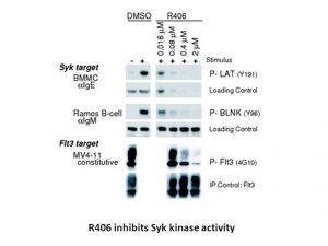
Tamatinib HCl targets spleen tyrosine kinase (Syk). IC50 in biochemical kinase assay = 41 nM (at ATP concentration corresponding to its Km). Ki for ATP competition = 30 nM [1]. Also inhibits Flt3 (≈5‑fold less potent than Syk in cells), Jak, Lck, and adenosine A3 receptor (IC50 = 0.081 μM), adenosine transporter (IC50 = 1.84 μM), monoamine transporter (IC50 = 2.74 μM) [1]. |
|---|---|
| ln Vitro |
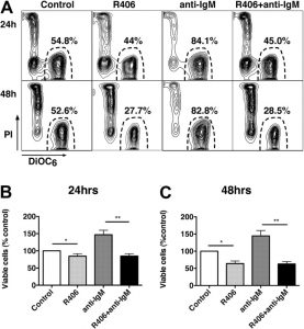
R406 is an ATP-competitive inhibitor of Syk with a Ki value of 30 nM. R406 selectively inhibits Syk-dependent signaling with EC50 values ranging from 33 nM to 171 nM, more potently than Syk-independent pathways in different cells. R406 inhibits cellular proliferation of a large panel of diffuse large B-cell lymphoma (DLBCL) cell lines at EC50 values ranging from 0.8 μM to 8.1 μM. R406 treatment (1 μM or 4 μM) induces the activation of caspases 9 and 3, but not caspase 8, leading to significant apoptosis of the majority of DLBCL cell lines. Pretreatment of R406 completely blocks the phosphorylation of SYK525/526 and the SYK-dependent phosphorylation of BLNK in R406-sensitive DLBCLs following B-cell receptor (BCR) crosslinking. R406 potently decreases MMP-9 mRNA levels by 2.8- and 4.3-fold lower than controls after 24 and 48 hours treatment, respectively, and reduces the invasive capacity of the RL cells.
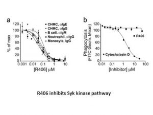
Kinase Assay: R406 is serially diluted in DMSO and then diluted to 1% DMSO in kinase buffer (20 mM HEPES, pH 7.4, 5 mM MgCl2, 2 mM MnCl2, 1 mM DTT, 0.1 mg/mL acetylated BGG). ATP and substrate in kinase buffer are added at room temperature, resulting in a final DMSO concentration on 0.2%. The kinase reactions are performed in a final volume of 20 μL containing 5 μM HS1 peptide substrate and 4 μM ATP and started by addition of 0.125 ng of Syk in kinase buffer. The reaction is allowed to proceed for 40 minutes at room temperature. The reaction is stopped by the addition of 20 μL of PTK quench mix containing EDTA/anti-phosphotyrosine antibody (1X final)/fluorescent phosphopeptide tracer (0.5X final) diluted in FP Dilution Buffer. The plate is incubated for 30 minutes in the dark at room temperature and then read on a Polarion fluorescence polarization plate reader. Data are converted to amount of phosphopeptide present using a calibration curve generated by competition with the phosphopeptide competitor provided in the Tyrosine Kinase Assay Kit. For IC50 determination, R406 is tested at eleven concentrations in duplicate and curve-fitting is performed by non-linear regression analysis using Prism GraphPad Software. Cell Assay: DLBCL cell lines are treated with serial dilutions of R406 (0.3, 0.6, 1.25, 2.5, or 5 μM) for 72 or 96 hours. Thereafter, cellular proliferation is determined by MTT assay, and cell apoptosis is assessed by using annexin V–FITC/propidium iodide (PI) staining. For the determination of caspase 9, 8, and 3, cells are lysed, size-fractionated by polyacrylamide gel electrophoresis (PAGE), and immunoblotted. Tamatinib HCl inhibited IgE‑mediated degranulation in cultured human mast cells with EC50 = 56 ± 20 nM (tryptase release), and also inhibited anti‑IgE‑induced LTC4, TNFα, IL‑8, GM‑CSF production (EC50 93‑158 nM). It had no effect on ionomycin‑induced degranulation (EC50 >10 μM), indicating specificity for FcεRI signaling [1]. In mast cells, Tamatinib HCl blocked phosphorylation of LAT (Tyr191) and downstream signaling molecules (PLCγ1, AKT, ERK, p38, JNK) but did not inhibit Syk phosphorylation (Tyr352) or FcRγ chain phosphorylation (Lyn‑dependent), confirming Syk as the primary target [1]. In human macrophages (primary and THP‑1), Tamatinib HCl inhibited anti‑IgG‑induced TNFα production with EC50 = 0.111 μM (primary) and 0.171 μM (THP‑1). In primary neutrophils, it inhibited anti‑IgG‑induced oxidative burst with EC50 = 33 nM. In primary B cells, it inhibited anti‑IgM‑induced CD69 up‑regulation with EC50 = 48 nM. In contrast, it was much less potent (≈10‑fold higher EC50) in LPS‑induced TNFα production (EC50 = 1.15‑2.10 μM) [1]. Tamatinib HCl did not inhibit phagocytosis of FITC‑labeled opsonized E. coli (EC50 >50 μM), oxidative burst induced by opsonized E. coli (EC50 >30 μM), chemotaxis toward fMLP (no inhibition at 10 μM), or microbicidal activity against opsonized S. aureus (no inhibition up to 20 μM). It also did not inhibit PMA‑induced oxidative burst in neutrophils (EC50 >10 μM) [1]. |
| ln Vivo |
R406 has shown efficacy in a number of animal models of immune disorders. Oral administration of R406 in mice with immune complex-mediated inflammation significantly inhibits the cutaneous reverse passive Arthus reaction by approximately 72% and 86% at 1 mg/kg and 5 mg/kg, respectively, compared with the control. R406 treatment at 10 mg/kg significantly reduces inflammation and swelling, decreases the progressive arthritis to a lower level in the passive anticollagen antibody-challenged mice, and delays the onset and reduces paw thickening and clinical arthritis by approximately 50% in the K/BxN serum transfer mice model.
In mice, oral administration of Tamatinib HCl 1 h before immune complex challenge reduced the reverse passive Arthus reaction (cutaneous edema and dye extravasation) by 72% at 1 mg/kg and 86% at 5 mg/kg (p < 0.001) [1]. In the collagen antibody‑induced arthritis (CAIA) model, Tamatinib HCl (oral b.i.d. for 14 days) delayed disease onset and reduced clinical scores; histopathology showed marked reduction of synovitis, pannus formation, and leukocyte infiltration [1]. In the K/BxN serum transfer arthritis model, Tamatinib HCl at 10 mg/kg b.i.d. reduced paw thickening and clinical arthritis by approximately 50% [1]. In a first‑in‑human study (healthy volunteers, single oral doses 80‑600 mg), Tamatinib HCl inhibited ex vivo anti‑IgE‑induced basophil degranulation (CD63 up‑regulation) in a dose‑dependent manner. The plasma concentration producing 50% inhibition was 496 ± 42 ng/mL (~1.06 μM). Inhibition was sustained for up to 24 h at higher doses [1]. |
| Enzyme Assay |
R406 is serially diluted in DMSO, diluted in kinase buffer (20 mM HEPES, pH 7.4, 5 mM MgCl2, 2 mM MnCl2, 1 mM DTT, 0.1 mg/mL acetylated BGG) and finally diluted to 1% DMSO by volume. After adding ATP and substrate to kinase buffer at room temperature, the final DMSO concentration is 0.2%. 0.125 ng of Syk is added to kinase buffer to initiate the kinase reactions, which are carried out in a final volume of 20 mL with 5 mM HS1 peptide substrate and 4 mM ATP. The reaction is left to continue at room temperature for forty minutes. 20 mL of PTK quench mix containing EDTA, anti-phosphotyrosine antibody (1X final), and fluorescent phosphopeptide tracer (0.5X final) diluted in FP Dilution Buffer is added to stop the reaction. A Polarion fluorescence polarization plate reader is used to read the plate after it has been incubated for 30 minutes at room temperature in the dark. Through competition with the phosphopeptide competitor included in the Tyrosine Kinase Assay Kit, a calibration curve is created that is used to convert data into the amount of phosphopeptide present. Non-linear regression analysis is used to fit the curve and test R406 at eleven different concentrations in order to determine the IC50.
Syk kinase activity was measured using a fluorescence polarization assay. For Ki determination, reactions were set up at eight different ATP concentrations (2‑fold serial dilutions from 200 μM) with R406 at 7.8‑125 nM. Reaction rates were plotted against ATP concentration to determine apparent Km and Vmax, then apparent Km (or Km/Vmax) plotted against inhibitor concentration to determine Ki = 30 nM [1]. Protein crystallography: The kinase domain of Syk (Ile358‑Asn635, E440Q mutation) was crystallized, soaked with R406. Data collected to 2.3 Å resolution. Structure showed R406 binding in the ATP pocket in a U‑shape" conformation, with pyrimidine N1 and linker N2 forming key hydrogen bonds with the hinge region [1]." |
| Cell Assay |
Cultured human mast cells (CHMC) were derived from cord blood CD34+ progenitors. Cells were preincubated with Tamatinib HCl for 30 min, then stimulated with anti‑IgE (0.25‑2 mg/mL) or anti‑IgG. Degranulation measured as tryptase release (luminescence of peptide substrate). LTC4 and cytokines measured by Luminex multiplex. For Western blotting, cells were preincubated with compound for 40 min, stimulated with anti‑IgE for 5 min, lysed, and analyzed with phospho‑specific antibodies [1].
Primary human macrophages were derived from CD14+ PBMC with GM‑CSF. Cells were stimulated by immobilized human IgG for 16‑20 h, TNFα measured by Luminex. THP‑1 cells were primed with IFN‑γ for 6 days before stimulation. Neutrophils isolated from peripheral blood were preincubated with compound for 20 min, primed with TNFα (20 ng/mL), stimulated with anti‑IgG (0.4 μg/mL), and oxidative burst measured by dihydrorhodamine 123 fluorescence. Primary B cells isolated by CD19+ selection were preincubated with compound for 60 min, stimulated with anti‑IgM (5 μg/mL) for 6 h, and CD69 expression analyzed by flow cytometry [1]. Phagocytosis assay: Heparinized whole blood incubated with compound for 30 min, then FITC‑labeled opsonized E. coli added; phagocytosis measured by flow cytometry. Microbicidal activity: purified neutrophils pretreated with compound (or DMSO) for 1 h at 4°C, mixed with opsonized S. aureus (1:1), rotated at 37°C, aliquots plated on blood agar and colony counts determined [1]. |
| Animal Protocol |
Formulated in DMSO and diluted in saline containing 35% TPGS, 60% PEG 400, and 5% propylene glycol; 10 mg/kg; Oral gavage
Female C57BL/6 mice challenged intravenously with 1% ovalbumin (OVA) in saline (10 mg/kg) containing 1% Evans blue dye, female Balb/c mice with the anticollagen antibody-induced arthritis, and female C57BL/6 mice with arthritis induced by intraperitoneal Reverse passive Arthus reaction: Female C57BL/6 mice were challenged i.v. with ovalbumin (OVA, 10 mg/kg) containing Evans blue dye. Ten minutes later, anti‑OVA IgG (50 μg) injected intradorsally. Tamatinib HCl (or vehicle, 67% PEG400) was administered orally 1 h before antibody challenge. Four hours later, skin punch biopsies were incubated in formamide at 80°C overnight, and extravasated Evans blue measured at OD610 [1]. CAIA model: Female Balb/c mice received i.v. collagen antibody blend on day 0, then i.p. LPS (25 μg) on day 2. Tamatinib HCl was administered orally b.i.d. for 14 days starting 4 h after antibody challenge. Clinical scores assessed daily. Joint histopathology: Safranin O staining [1]. K/BxN serum transfer model: C57BL/6 mice received i.p. K/BxN serum on days 0 and 2. Tamatinib HCl or vehicle (35% TPGS, 60% PEG400, 5% propylene glycol) was administered orally 1 h before serum injection and then b.i.d. for 13 days. Ankle thickness and arthritis scores measured daily [1]. Human study: Double‑blind, placebo‑controlled, ascending single dose (80, 250, 400, 500, 600 mg) in healthy male volunteers. Blood collected at various time points; plasma R406 concentration measured by LC‑MS/MS. Basophil activation: whole blood stimulated ex vivo with anti‑IgE, CD63 expression on basophils measured by flow cytometry. Platelet aggregation induced by collagen or ADP in platelet‑rich plasma [1]. |
| ADME/Pharmacokinetics |
In mice, after oral administration of Tamatinib HCl, rapid metabolism was observed (supplemental figure). In humans, R406 was highly bioavailable. Plasma concentration increased dose‑proportionally up to 400 mg then plateaued. Tmax ≈1.2‑1.3 h; half‑life ≈15 h. Protein binding in human plasma >98% [1].
|
| Toxicity/Toxicokinetics |
In the human study, no significant effects on hematologic or chemistry safety parameters were observed. At 600 mg, subjects complained of postural dizziness more frequently (5/6 vs. 1/8 placebo, 4/21 lower doses). R406 at high doses caused a transient reduction in circulating CD14+ mononuclear cells 4 h post‑dose, reversed by 20 h. No inhibition of collagen‑ or ADP‑induced platelet aggregation at any dose. In mice, R406 up to 100 mg/kg (achieving plasma exposures up to 25 μM) did not prolong bleeding times, whereas aspirin (100 mg/kg) prolonged by 88% [1].
|
| References |
J Pharmacol Exp Ther.2006 Dec;319(3):998-1008;Blood.2008 Feb 15;111(4):2230-7.
|
| Additional Infomation |
Tamatinib HCl (R406) is a potent, ATP‑competitive Syk inhibitor that blocks Fc receptor and B‑cell receptor signaling. It has demonstrated efficacy in multiple animal models of immune complex‑mediated inflammation and in human ex vivo basophil activation. The compound is orally bioavailable and has a favorable safety profile in a first‑in‑human study. It is being developed for autoimmune and allergic diseases. R406 structure: N4‑(2,2‑dimethyl‑3‑oxo‑4H‑pyrid[1,4]oxazin‑6‑yl)‑5‑fluoro‑N2‑(3,4,5‑trimethoxyphenyl)‑2,4‑pyrimidinediamine [1].
|
| Related CAS # |
841290-81-1;841290-80-0;R406
|
|---|---|
| Appearance |
Typically exists as solid at room temperature
|
| HS Tariff Code |
2934.99.9001
|
| Storage |
Powder -20°C 3 years 4°C 2 years In solvent -80°C 6 months -20°C 1 month |
| Shipping Condition |
Room temperature (This product is stable at ambient temperature for a few days during ordinary shipping and time spent in Customs)
|
| Solubility (In Vitro) |
May dissolve in DMSO (in most cases), if not, try other solvents such as H2O, Ethanol, or DMF with a minute amount of products to avoid loss of samples
|
|---|---|
| Solubility (In Vivo) |
Note: Listed below are some common formulations that may be used to formulate products with low water solubility (e.g. < 1 mg/mL), you may test these formulations using a minute amount of products to avoid loss of samples.
Injection Formulations
Injection Formulation 1: DMSO : Tween 80: Saline = 10 : 5 : 85 (i.e. 100 μL DMSO stock solution → 50 μL Tween 80 → 850 μL Saline)(e.g. IP/IV/IM/SC) *Preparation of saline: Dissolve 0.9 g of sodium chloride in 100 mL ddH ₂ O to obtain a clear solution. Injection Formulation 2: DMSO : PEG300 :Tween 80 : Saline = 10 : 40 : 5 : 45 (i.e. 100 μL DMSO → 400 μLPEG300 → 50 μL Tween 80 → 450 μL Saline) Injection Formulation 3: DMSO : Corn oil = 10 : 90 (i.e. 100 μL DMSO → 900 μL Corn oil) Example: Take the Injection Formulation 3 (DMSO : Corn oil = 10 : 90) as an example, if 1 mL of 2.5 mg/mL working solution is to be prepared, you can take 100 μL 25 mg/mL DMSO stock solution and add to 900 μL corn oil, mix well to obtain a clear or suspension solution (2.5 mg/mL, ready for use in animals). View More
Injection Formulation 4: DMSO : 20% SBE-β-CD in saline = 10 : 90 [i.e. 100 μL DMSO → 900 μL (20% SBE-β-CD in saline)] Oral Formulations
Oral Formulation 1: Suspend in 0.5% CMC Na (carboxymethylcellulose sodium) Oral Formulation 2: Suspend in 0.5% Carboxymethyl cellulose Example: Take the Oral Formulation 1 (Suspend in 0.5% CMC Na) as an example, if 100 mL of 2.5 mg/mL working solution is to be prepared, you can first prepare 0.5% CMC Na solution by measuring 0.5 g CMC Na and dissolve it in 100 mL ddH2O to obtain a clear solution; then add 250 mg of the product to 100 mL 0.5% CMC Na solution, to make the suspension solution (2.5 mg/mL, ready for use in animals). View More
Oral Formulation 3: Dissolved in PEG400 (Please use freshly prepared in vivo formulations for optimal results.) |
Calculation results
Working concentration: mg/mL;
Method for preparing DMSO stock solution: mg drug pre-dissolved in μL DMSO (stock solution concentration mg/mL). Please contact us first if the concentration exceeds the DMSO solubility of the batch of drug.
Method for preparing in vivo formulation::Take μL DMSO stock solution, next add μL PEG300, mix and clarify, next addμL Tween 80, mix and clarify, next add μL ddH2O,mix and clarify.
(1) Please be sure that the solution is clear before the addition of next solvent. Dissolution methods like vortex, ultrasound or warming and heat may be used to aid dissolving.
(2) Be sure to add the solvent(s) in order.
|
|---|
|
|
Products are for research use only; We do not sell to patients
Copyright 2020 InvivoChem LLC | All Rights Reserved