| Size | Price | Stock | Qty |
|---|---|---|---|
| 5mg |
|
||
| 10mg |
|
||
| 25mg |
|
||
| 50mg |
|
||
| 100mg |
|
||
| 250mg |
|
||
| 500mg |
|
||
| Other Sizes |
|
Purity: ≥98%
SB408124 HCl is a potent, novel, selective, non-peptide antagonist for OX1 receptor with Ki of 57 nM and 27 nM in both whole cell and membrane, respectively, it exhibits 50-fold selectivity over OX2 receptor. In primary astrocyte cultures from the rat cerebral cortex, pretreatment with SB408124 significantly reduced the orexin A a stimulating effect on basal-induced cAMP production and forskolin.
| Targets |
OX1 Receptor ( Ki = 57 nM ); OX1 Receptor ( Ki = 27 nM )
|
||
|---|---|---|---|
| ln Vitro |
|
||
| ln Vivo |
|
||
| Enzyme Assay |
SB-408124 is a non-peptide antagonist that shows 50-fold selectivity over OX2 receptor and has a Ki of 57 nM and 27 nM in whole cell and membrane, respectively, for the OX1 receptor.
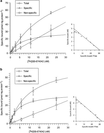
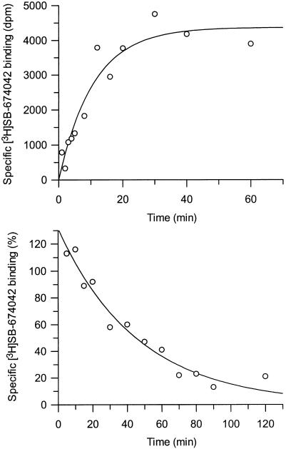
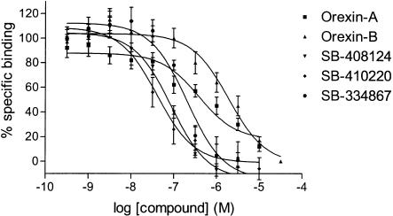
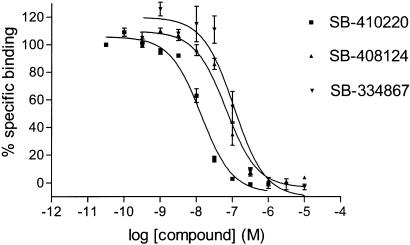
[3H]SB-674042 whole cell binding assays[1] After overnight culture in 96-well Packard Cultur plates, the medium was discarded and cells were incubated in buffer containing 150 mM NaCl, 20 mM HEPES and 0.5% bovine serum albumin (pH 7.4) for 60 min at 25°C. Saturation studies were carried out by incubating cells with a range of concentrations of [3H]SB-674042 (0.2–24 nM); the total assay volume was 250 μl. Protein content was assayed by lysing cells with 0.1 M NaOH and using the Bradford method (Bradford, 1976) with bovine serum albumin (BSA) as a standard. Association kinetic studies were performed by measuring the specific binding of [3H]SB-674042 (3 nM) at 1–60 min after addition of [3H]SB-674042. For dissociation studies, cells were first incubated with [3H]SB-674042 (3 nM) for 60 min. Specific binding was then measured at 2–120 min after the addition of 3 μM SB-408124. Competition studies were performed by incubating cells with [3H]SB-674042 (3 nM) and a range of concentrations of the test compound. All assays were terminated by washing the cells three times with 250 μl ice-cold phosphate-buffered saline. A volume of 100 μl of Microscint 40 was added to each well and the plate was left at room temperature for 2 h. Cell-associated radioactivity was then measured using a Packard Topcount, with a count time of 2 min well−1. [3H]SB-674042 membrane-based SPA binding assays[1] CHO-K1_OX1 cell membranes (75 μg ml−1) were precoupled by shaking with wheatgerm-agglutinin polyvinyltoluene (WGA-PVT) scintillation proximity assay (SPA) beads (5 mg ml−1) in buffer containing 25 mM HEPES, 2.5 mM MgCl2, 0.5 mM EDTA and 0.025% bacitracin (pH 7.4) at 4°C for 1 h. The bead-membrane suspension was centrifuged at 300 × g and resuspended in the same volume of room temperature assay buffer. A volume of 100 μl of bead-membrane suspension was incubated with [3H]SB-674042 (5 nM) in a total assay volume of 200 μl in a 96-well Packard Optiplate to give a final protein concentration of 7.5 μg well−1. Nonspecific binding was measured as that remaining in the presence of 3 μM SB-408124. Assay plates were shaken for 10 min and then incubated at room temperature for 4 h before being counted on a Packard TopCount scintillation counter (count time 2 min well−1). Saturation studies were carried out by incubating bead-membranes (equivalent to 7.5 μg protein well−1 and 2.5 mg beads ml−1) with a range of concentrations of [3H]SB-674042 (0.1–20 nM). Protein content was assayed using the Bradford method (Bradford, 1976) using bovine serum albumin as a standard. Association kinetic studies were performed by measuring specific binding of [3H]SB-674042 (5 nM) at 1–30 min after addition of bead-membranes (equivalent to 7.5 μg protein well−1 and 2.5 mg beads ml−1). For dissociation studies, bead-membranes were first incubated with [3H]SB-674042 (5 nM) for 30 min. Specific binding was then measured at 2–120 min after the addition of 3 μM SB-408124. Competition studies were performed by incubating bead-membranes (equivalent to 7.5 μg protein well−1 and 2.5 mg beads ml−1) with [3H]SB-674042 (5 nM) and a range of concentrations of the test compound. |
||
| Cell Assay |
SB-408124 has a pKi of 7.57 when it comes to binding the hypocretin type 1 receptor (HcrtR1). According to studies on calcium mobilization, SB-408124 functions as a functional antagonist of the OX1 receptor and has an affinity that is roughly 50 times more selective than the OX2 receptor. According to a recent study, the stimulatory action of Orexin A on basal and forskolin-acivated cAMP production was significantly reduced when primary cultures of rat astrocytes were pretreated with SB-401824 prior to Orexin A administration.
Measurement of orexin A-induced AVP mRNA expression in cultured brain neurons.[3] Primary neuronal cultures from the hypothalamus were incubated with vehicle control or differing concentrations of orexin A (10 nM, 100 nM, 1 μM, or 10 μM) in DMEM-HSPS for 6 h. The culture medium was removed, and cells were washed with cold PBS, collected, and subjected to RNA isolation. Real-time PCR was performed to measure mRNA levels of AVP. To test which orexin receptor mediates the AVP increase induced by orexin, we coincubated neuronal cultures with 1 μM orexin A with or without the OX1R antagonist SB-408124 (100 µM) or OX2Ra TCS-OX2-29 (100 µM) for 6 h. The mRNA level of AVP was determined by real-time quantitative PCR. Each set of experiments was performed using 3 culture wells in a 24-well plate, and all cDNA samples were assayed in duplicate. The whole experiment was repeated two to three times. Data were normalized to GAPDH mRNA. |
||
| Animal Protocol |
|
||
| References |
|
||
| Additional Infomation |
The orexin system is involved in arginine vasopressin (AVP) regulation, and its overactivation has been implicated in hypertension. However, its role in salt-sensitive hypertension (SSHTN) is unknown. Here, we tested the hypothesis that hyperactivity of the orexin system in the paraventricular nucleus (PVN) contributes to SSHTN via enhancing AVP signaling. Eight-week-old male Dahl salt-sensitive (Dahl S) and age- and sex-matched Sprague-Dawley (SD) rats were placed on a high-salt (HS; 8% NaCl) or normal-salt (NS; 0.4% NaCl) diet for 4 wk. HS intake did not alter mean arterial pressure (MAP), PVN mRNA levels of orexin receptor 1 (OX1R), or OX2R but slightly increased PVN AVP mRNA expression in SD rats. HS diet induced significant increases in MAP and PVN mRNA levels of OX1R, OX2R, and AVP in Dahl S rats. Intracerebroventricular infusion of orexin A (0.2 nmol) dramatically increased AVP mRNA levels and immunoreactivity in the PVN of SD rats. Incubation of cultured hypothalamus neurons from newborn SD rats with orexin A increased AVP mRNA expression, which was attenuated by OX1R blockade. In addition, increased cerebrospinal fluid Na+ concentration through intracerebroventricular infusion of NaCl solution (4 µmol) increased PVN OX1R and AVP mRNA levels and immunoreactivity in SD rats. Furthermore, bilateral PVN microinjection of the OX1R antagonist SB-408124 resulted in a greater reduction in MAP in HS intake (-16 ± 5 mmHg) compared with NS-fed (-4 ± 4 mmHg) anesthetized Dahl S rats. These results suggest that elevated PVN OX1R activation may contribute to SSHTN by enhancing AVP signaling.NEW & NOTEWORTHY To our best knowledge, this study is the first to investigate the involvement of the orexin system in salt-sensitive hypertension. Our results suggest that the orexin system may contribute to the Dahl model of salt-sensitive hypertension by enhancing vasopressin signaling in the hypothalamic paraventricular nucleus.[3]
1. This study characterises the binding of a novel nonpeptide antagonist radioligand, [(3)H]SB-674042 (1-(5-(2-fluoro-phenyl)-2-methyl-thiazol-4-yl)-1-((S)-2-(5-phenyl-(1,3,4)oxadiazol-2-ylmethyl)-pyrrolidin-1-yl)-methanone), to the human orexin-1 (OX(1)) receptor stably expressed in Chinese hamster ovary (CHO) cells in both a whole cell assay and in a cell membrane-based scintillation proximity assay (SPA) format. 2. Specific binding of [(3)H]SB-674042 was saturable in both whole cell and membrane formats. Analyses suggested a single high-affinity site, with K(d) values of 3.76+/-0.45 and 5.03+/-0.31 nm, and corresponding B(max) values of 30.8+/-1.8 and 34.4+/-2.0 pmol mg protein(-1), in whole cell and membrane formats, respectively. Kinetic studies yielded similar K(d) values. 3. Competition studies in whole cells revealed that the native orexin peptides display a low affinity for the OX(1) receptor, with orexin-A displaying a approximately five-fold higher affinity than orexin-B (K(i) values of 318+/-158 and 1516+/-597 nm, respectively). 4. SB-334867, SB-408124 (1-(6,8-difluoro-2-methyl-quinolin-4-yl)-3-(4-dimethylamino-phenyl)-urea) and SB-410220 (1-(5,8-difluoro-quinolin-4-yl)-3-(4-dimethylamino-phenyl)-urea) all displayed high affinity for the OX(1) receptor in both whole cell (K(i) values 99+/-18, 57+/-8.3 and 19+/-4.5 nm, respectively) and membrane (K(i) values 38+/-3.6, 27+/-4.1 and 4.5+/-0.2 nm, respectively) formats. 5. Calcium mobilisation studies showed that SB-334867, SB-408124 and SB-410220 are all functional antagonists of the OX(1) receptor, with potencies in line with their affinities, as measured in the radioligand binding assays, and with approximately 50-fold selectivity over the orexin-2 receptor. 6. These studies indicate that [(3)H]SB-674042 is a specific, high-affinity radioligand for the OX(1) receptor. The availability of this radioligand will be a valuable tool with which to investigate the physiological functions of OX(1) receptors.[1] |
| Molecular Formula |
C19H19CLF2N4O
|
|
|---|---|---|
| Molecular Weight |
392.83
|
|
| Exact Mass |
392.121
|
|
| CAS # |
1431697-90-3
|
|
| Related CAS # |
SB-408124; 288150-92-5
|
|
| PubChem CID |
71576692
|
|
| Appearance |
White to gray solid powder
|
|
| Hydrogen Bond Donor Count |
3
|
|
| Hydrogen Bond Acceptor Count |
5
|
|
| Rotatable Bond Count |
3
|
|
| Heavy Atom Count |
27
|
|
| Complexity |
484
|
|
| Defined Atom Stereocenter Count |
0
|
|
| InChi Key |
DIHXPSGMLOETTI-UHFFFAOYSA-N
|
|
| InChi Code |
InChI=1S/C19H18F2N4O.ClH/c1-11-8-17(15-9-12(20)10-16(21)18(15)22-11)24-19(26)23-13-4-6-14(7-5-13)25(2)3;/h4-10H,1-3H3,(H2,22,23,24,26);1H
|
|
| Chemical Name |
1-(6,8-difluoro-2-methylquinolin-4-yl)-3-[4-(dimethylamino)phenyl]urea;hydrochloride
|
|
| Synonyms |
|
|
| HS Tariff Code |
2934.99.9001
|
|
| Storage |
Powder -20°C 3 years 4°C 2 years In solvent -80°C 6 months -20°C 1 month Note: Please store this product in a sealed and protected environment, avoid exposure to moisture. |
|
| Shipping Condition |
Room temperature (This product is stable at ambient temperature for a few days during ordinary shipping and time spent in Customs)
|
| Solubility (In Vitro) |
|
|||
|---|---|---|---|---|
| Solubility (In Vivo) |
Note: Listed below are some common formulations that may be used to formulate products with low water solubility (e.g. < 1 mg/mL), you may test these formulations using a minute amount of products to avoid loss of samples.
Injection Formulations
Injection Formulation 1: DMSO : Tween 80: Saline = 10 : 5 : 85 (i.e. 100 μL DMSO stock solution → 50 μL Tween 80 → 850 μL Saline)(e.g. IP/IV/IM/SC) *Preparation of saline: Dissolve 0.9 g of sodium chloride in 100 mL ddH ₂ O to obtain a clear solution. Injection Formulation 2: DMSO : PEG300 :Tween 80 : Saline = 10 : 40 : 5 : 45 (i.e. 100 μL DMSO → 400 μLPEG300 → 50 μL Tween 80 → 450 μL Saline) Injection Formulation 3: DMSO : Corn oil = 10 : 90 (i.e. 100 μL DMSO → 900 μL Corn oil) Example: Take the Injection Formulation 3 (DMSO : Corn oil = 10 : 90) as an example, if 1 mL of 2.5 mg/mL working solution is to be prepared, you can take 100 μL 25 mg/mL DMSO stock solution and add to 900 μL corn oil, mix well to obtain a clear or suspension solution (2.5 mg/mL, ready for use in animals). View More
Injection Formulation 4: DMSO : 20% SBE-β-CD in saline = 10 : 90 [i.e. 100 μL DMSO → 900 μL (20% SBE-β-CD in saline)] Oral Formulations
Oral Formulation 1: Suspend in 0.5% CMC Na (carboxymethylcellulose sodium) Oral Formulation 2: Suspend in 0.5% Carboxymethyl cellulose Example: Take the Oral Formulation 1 (Suspend in 0.5% CMC Na) as an example, if 100 mL of 2.5 mg/mL working solution is to be prepared, you can first prepare 0.5% CMC Na solution by measuring 0.5 g CMC Na and dissolve it in 100 mL ddH2O to obtain a clear solution; then add 250 mg of the product to 100 mL 0.5% CMC Na solution, to make the suspension solution (2.5 mg/mL, ready for use in animals). View More
Oral Formulation 3: Dissolved in PEG400 (Please use freshly prepared in vivo formulations for optimal results.) |
| Preparing Stock Solutions | 1 mg | 5 mg | 10 mg | |
| 1 mM | 2.5456 mL | 12.7282 mL | 25.4563 mL | |
| 5 mM | 0.5091 mL | 2.5456 mL | 5.0913 mL | |
| 10 mM | 0.2546 mL | 1.2728 mL | 2.5456 mL |
*Note: Please select an appropriate solvent for the preparation of stock solution based on your experiment needs. For most products, DMSO can be used for preparing stock solutions (e.g. 5 mM, 10 mM, or 20 mM concentration); some products with high aqueous solubility may be dissolved in water directly. Solubility information is available at the above Solubility Data section. Once the stock solution is prepared, aliquot it to routine usage volumes and store at -20°C or -80°C. Avoid repeated freeze and thaw cycles.
Calculation results
Working concentration: mg/mL;
Method for preparing DMSO stock solution: mg drug pre-dissolved in μL DMSO (stock solution concentration mg/mL). Please contact us first if the concentration exceeds the DMSO solubility of the batch of drug.
Method for preparing in vivo formulation::Take μL DMSO stock solution, next add μL PEG300, mix and clarify, next addμL Tween 80, mix and clarify, next add μL ddH2O,mix and clarify.
(1) Please be sure that the solution is clear before the addition of next solvent. Dissolution methods like vortex, ultrasound or warming and heat may be used to aid dissolving.
(2) Be sure to add the solvent(s) in order.
 |
|---|
 |
  |