| Size | Price | Stock | Qty |
|---|---|---|---|
| 50mg |
|
||
| 100mg |
|
||
| 250mg |
|
||
| 500mg | |||
| 1g | |||
| Other Sizes |
Purity: ≥98%
| Targets |
VHL (von Hippel-Lindau E3 ubiquitin ligase) – binding IC50 = 0.54 ± 0.06 μM (determined by fluorescence polarization assay) [1]
HaloTag7 fusion proteins (e.g., GFP-HaloTag7, HaloTag7-ERK1, HaloTag7-MEK1) – degradation DC50 for GFP-HaloTag7 = 19 ± 1 nM, maximal degradation = 90 ± 1% at 625 nM [1] Protein degrader 1 TFA targets the Von Hippel-Lindau (VHL) protein, which is the substrate-recognition component of an E3 ubiquitin ligase complex. By binding to VHL, it serves as a "handle" to recruit the ubiquitin-proteasome system to a protein of interest when incorporated into a PROTAC molecule. It is specifically designed to degrade HaloTag7 fusion proteins. |
|---|---|
| ln Vitro |
Protein degrader 1 TFA (HaloPROTAC3) at 625 nM for 24 h induced 90 ± 1% degradation of stably expressed GFP-HaloTag7 in HEK293 cells, with DC50 = 19 ± 1 nM. [1]
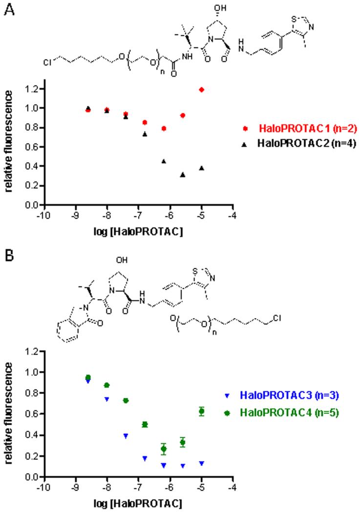
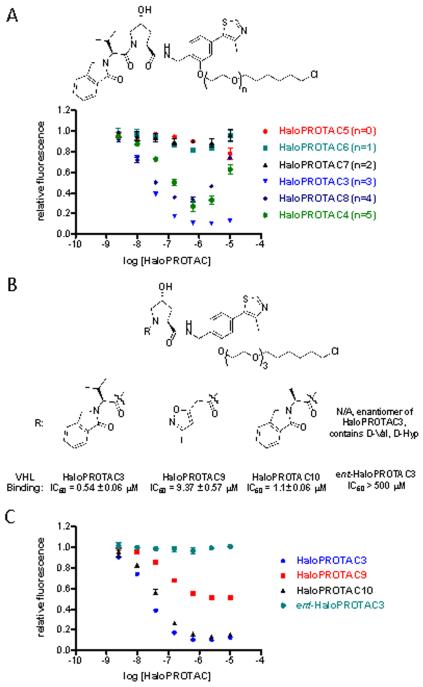
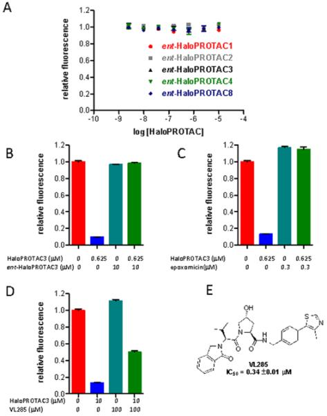
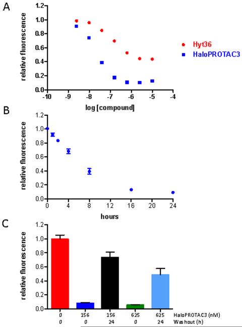
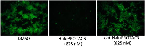
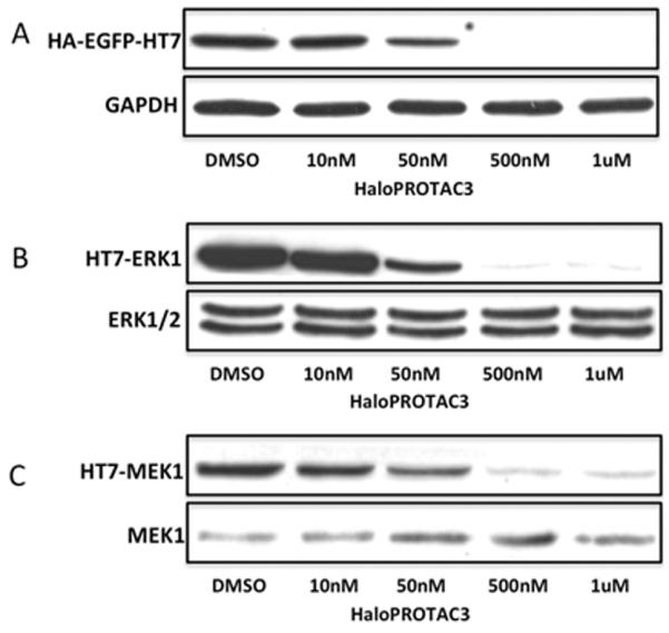
Linker length optimization showed that three ethylene glycol units (as in HaloPROTAC3) were optimal; shorter linkers (HaloPROTAC5-7) resulted in <20% degradation, while longer linkers (HaloPROTAC4,8) showed significant knockdown but less efficacy. [1] Reducing VHL binding affinity via isoxazole substitution (HaloPROTAC9) attenuated degradation to 45% maximal; alanine substitution (HaloPROTAC10) gave similar maximal degradation but slightly reduced potency (DC50 = 36 ± 4 nM). [1] ent-HaloPROTAC3 (enantiomer with D-amino acids, no VHL binding) did not induce significant degradation, confirming VHL dependence. [1] Pre-treatment with excess ent-HaloPROTAC3 blocked HaloPROTAC3-mediated degradation, indicating competitive binding to HaloTag7. [1] Pre-treatment with the proteasome inhibitor epoxomicin (300 nM, 4 h) completely prevented degradation, confirming proteasome dependency. [1] Co-treatment with excess free VHL ligand VL285 significantly reduced degradation to ~50%, further implicating VHL. [1] HaloPROTAC3 was more potent and efficacious than the hydrophobic tag HyT36 (DC50 = 134 ± 7 nM, max degradation 56 ± 1% for HyT36). [1] Kinetics: 50% degradation of GFP-HaloTag7 occurred between 4 and 8 h of treatment. [1] Washout experiment: After 24 h treatment with HaloPROTAC3, a 24 h washout led to significant recovery of GFP-HaloTag7 protein levels. [1] HaloPROTAC3 (500 nM, 24 h) induced nearly complete degradation of HaloTag7-ERK1 and HaloTag7-MEK1 in HEK293T cells, while endogenous ERK and MEK were unaffected. [1] Fluorescence microscopy confirmed drastic loss of GFP fluorescence after 24 h treatment with 625 nM HaloPROTAC3, but not with ent-HaloPROTAC3. [1] HaloPROTAC3 showed low toxicity (Supplemental Figure 3) and did not affect endogenous HIF (hypoxia-inducible factor) stability, indicating selectivity. [1] Protein degrader 1 TFA does not exhibit direct enzymatic inhibitory activity or receptor modulation. Its in vitro activity is defined by its ability to bind to the VHL protein with high affinity. When used as HaloPROTAC3, it induces the degradation of HaloTag7 fusion proteins in a concentration-dependent manner. |
| ln Vivo |
Protein degrader 1 TFA is not a therapeutic agent and does not have direct in vivo biological activity. Its in vivo activity is determined by the PROTAC molecules that incorporate this ligand. When used as HaloPROTAC3, it can induce the degradation of target proteins in vivo.
|
| Enzyme Assay |
VHL binding affinity was determined by fluorescence polarization assay as previously described (Buckley et al., 2012). The assay measured the ability of Protein degrader 1 TFA (HaloPROTAC3) to displace a fluorescently labeled HIF-1α peptide from VHL. The IC50 was calculated to be 0.54 ± 0.06 μM. [1]
In vitro binding assays for Protein degrader 1 TFA typically involve surface plasmon resonance (SPR) or isothermal titration calorimetry (ITC) to measure its affinity for the VHL protein. The VHL protein is immobilized, and varying concentrations of the ligand are passed over the surface. The KD is calculated from the binding isotherm. |
| Cell Assay |
For flow cytometry: HEK293 Flp-In cells stably expressing GFP-HaloTag7 were treated with Protein degrader 1 TFA (HaloPROTAC3) for 24 h. Cells were detached with trypsin, resuspended in DMEM, and intracellular EGFP fluorescence was measured on the FL1 channel of a FACSCalibur. Mean fluorescence intensity was quantified using CellQuest software, normalized to DMSO control (set to 1), and DC50 values were determined using Prism 5.0. All experiments were repeated 3-5 times. [1]
For immunoblotting: Cells were lysed in RIPA buffer (1% Triton X-100, 0.1% SDS, 0.5% sodium deoxycholate, 150 mM NaCl, 25 mM Tris pH 8) with protease inhibitors. Lysates were normalized by BCA assay, and 20 μg of protein per sample was loaded on 10% Bis-Tris gels for SDS-PAGE. Proteins were transferred to nitrocellulose membranes, blocked with 5% milk/TBST, and probed with anti-ERK1 (1:2000), anti-MEK-1 (1:1000), or anti-HA (1:5000) antibodies. [1] For washout experiments: After treatment, media was removed and replaced with fresh compound-free media for 30 min, then media was replaced again to remove residual compound. [1] In vitro cell-based assays for Protein degrader 1 TFA are performed using cells expressing HaloTag7 fusion proteins. Cells are cultured in appropriate media and treated with Protein degrader 1 TFA at various concentrations. The degradation of the HaloTag7 fusion protein is assessed by Western blot. Cell viability is measured by MTT assays. |
| Animal Protocol |
In vivo animal studies for Protein degrader 1 TFA are conducted using the PROTAC molecules that incorporate this ligand, rather than the ligand alone. Typical protocols involve administering the PROTAC to mice via intravenous, intraperitoneal, or oral routes. Target protein degradation is assessed in tissues by Western blot.
|
| ADME/Pharmacokinetics |
Specific pharmacokinetic properties of Protein degrader 1 TFA are not extensively characterized, as it is a research tool rather than a therapeutic agent. It is soluble in DMSO and is typically formulated for in vivo administration as part of a PROTAC.
|
| Toxicity/Toxicokinetics |
The study reports that Protein degrader 1 TFA (HaloPROTAC3) has low toxicity (as shown in Supplemental Figure 3), but no quantitative toxicity data (e.g., LD50, specific cytotoxicity values) are provided. Additionally, it does not affect the stability of endogenous HIF (hypoxia-inducible factor), an endogenous target of VHL (Supplemental Figure 4), indicating selectivity. [1]
Comprehensive toxicological data for Protein degrader 1 TFA are not widely available in public literature. As a research compound, it is intended for laboratory use only and is not for human therapeutic use. Standard safety precautions should be followed when handling this compound. It is supplied with a purity of ≥98%. |
| References |
ACS Chem Biol.2015 Aug 21;10(8):1831-7.
|
| Additional Infomation |
Protein degrader 1 TFA (HaloPROTAC3) is a chemical genetic tool that enables conditional knockdown of HaloTag7 fusion proteins with high potency and selectivity. It functions as an orthogonal approach to the Shield1 system (which stabilizes mutant FKBP12). Unlike Shield1, HaloPROTAC3 induces degradation rather than stabilization. Plasmids containing HaloTag7 fused with over 20,000 human genes are commercially available, making this system suitable for large-scale screening applications. HaloPROTAC3 does not affect endogenous VHL targets such as HIF, further supporting its specificity. [1]
Protein degrader 1 TFA is a small molecule ligand for VHL. It is also known as VH032-NH2 TFA, VHL ligand 1 TFA, and HaloPROTAC3. It enables conditional knockdown of HaloTag7 fusion proteins. This product is for research use only. |
| Molecular Formula |
C24H31F3N4O5S
|
|
|---|---|---|
| Molecular Weight |
544.586955308914
|
|
| Exact Mass |
544.196
|
|
| Elemental Analysis |
C, 52.93; H, 5.74; F, 10.47; N, 10.29; O, 14.69; S, 5.89
|
|
| CAS # |
1631137-51-3
|
|
| Related CAS # |
1448297-52-6;1631137-51-3 (TFA);
|
|
| PubChem CID |
133053568
|
|
| Appearance |
Solid powder
|
|
| Hydrogen Bond Donor Count |
4
|
|
| Hydrogen Bond Acceptor Count |
11
|
|
| Rotatable Bond Count |
6
|
|
| Heavy Atom Count |
37
|
|
| Complexity |
701
|
|
| Defined Atom Stereocenter Count |
3
|
|
| SMILES |
CC1=C(SC=N1)C2=CC=C(C=C2)CNC(=O)[C@@H]3C[C@H](CN3C(=O)[C@H](C(C)(C)C)N)O.C(=O)(C(F)(F)F)O
|
|
| InChi Key |
SGNZARGJXDPTDJ-MSSRUXLCSA-N
|
|
| InChi Code |
InChI=1S/C22H30N4O3S.C2HF3O2/c1-13-18(30-12-25-13)15-7-5-14(6-8-15)10-24-20(28)17-9-16(27)11-26(17)21(29)19(23)22(2,3)4;3-2(4,5)1(6)7/h5-8,12,16-17,19,27H,9-11,23H2,1-4H3,(H,24,28);(H,6,7)/t16-,17+,19-;/m1./s1
|
|
| Chemical Name |
|
|
| Synonyms |
|
|
| HS Tariff Code |
2934.99.9001
|
|
| Storage |
Powder -20°C 3 years 4°C 2 years In solvent -80°C 6 months -20°C 1 month |
|
| Shipping Condition |
Room temperature (This product is stable at ambient temperature for a few days during ordinary shipping and time spent in Customs)
|
| Solubility (In Vitro) |
|
|||
|---|---|---|---|---|
| Solubility (In Vivo) |
Note: Listed below are some common formulations that may be used to formulate products with low water solubility (e.g. < 1 mg/mL), you may test these formulations using a minute amount of products to avoid loss of samples.
Injection Formulations
Injection Formulation 1: DMSO : Tween 80: Saline = 10 : 5 : 85 (i.e. 100 μL DMSO stock solution → 50 μL Tween 80 → 850 μL Saline)(e.g. IP/IV/IM/SC) *Preparation of saline: Dissolve 0.9 g of sodium chloride in 100 mL ddH ₂ O to obtain a clear solution. Injection Formulation 2: DMSO : PEG300 :Tween 80 : Saline = 10 : 40 : 5 : 45 (i.e. 100 μL DMSO → 400 μLPEG300 → 50 μL Tween 80 → 450 μL Saline) Injection Formulation 3: DMSO : Corn oil = 10 : 90 (i.e. 100 μL DMSO → 900 μL Corn oil) Example: Take the Injection Formulation 3 (DMSO : Corn oil = 10 : 90) as an example, if 1 mL of 2.5 mg/mL working solution is to be prepared, you can take 100 μL 25 mg/mL DMSO stock solution and add to 900 μL corn oil, mix well to obtain a clear or suspension solution (2.5 mg/mL, ready for use in animals). View More
Injection Formulation 4: DMSO : 20% SBE-β-CD in saline = 10 : 90 [i.e. 100 μL DMSO → 900 μL (20% SBE-β-CD in saline)] Oral Formulations
Oral Formulation 1: Suspend in 0.5% CMC Na (carboxymethylcellulose sodium) Oral Formulation 2: Suspend in 0.5% Carboxymethyl cellulose Example: Take the Oral Formulation 1 (Suspend in 0.5% CMC Na) as an example, if 100 mL of 2.5 mg/mL working solution is to be prepared, you can first prepare 0.5% CMC Na solution by measuring 0.5 g CMC Na and dissolve it in 100 mL ddH2O to obtain a clear solution; then add 250 mg of the product to 100 mL 0.5% CMC Na solution, to make the suspension solution (2.5 mg/mL, ready for use in animals). View More
Oral Formulation 3: Dissolved in PEG400 (Please use freshly prepared in vivo formulations for optimal results.) |
| Preparing Stock Solutions | 1 mg | 5 mg | 10 mg | |
| 1 mM | 1.8362 mL | 9.1812 mL | 18.3624 mL | |
| 5 mM | 0.3672 mL | 1.8362 mL | 3.6725 mL | |
| 10 mM | 0.1836 mL | 0.9181 mL | 1.8362 mL |
*Note: Please select an appropriate solvent for the preparation of stock solution based on your experiment needs. For most products, DMSO can be used for preparing stock solutions (e.g. 5 mM, 10 mM, or 20 mM concentration); some products with high aqueous solubility may be dissolved in water directly. Solubility information is available at the above Solubility Data section. Once the stock solution is prepared, aliquot it to routine usage volumes and store at -20°C or -80°C. Avoid repeated freeze and thaw cycles.
Calculation results
Working concentration: mg/mL;
Method for preparing DMSO stock solution: mg drug pre-dissolved in μL DMSO (stock solution concentration mg/mL). Please contact us first if the concentration exceeds the DMSO solubility of the batch of drug.
Method for preparing in vivo formulation::Take μL DMSO stock solution, next add μL PEG300, mix and clarify, next addμL Tween 80, mix and clarify, next add μL ddH2O,mix and clarify.
(1) Please be sure that the solution is clear before the addition of next solvent. Dissolution methods like vortex, ultrasound or warming and heat may be used to aid dissolving.
(2) Be sure to add the solvent(s) in order.
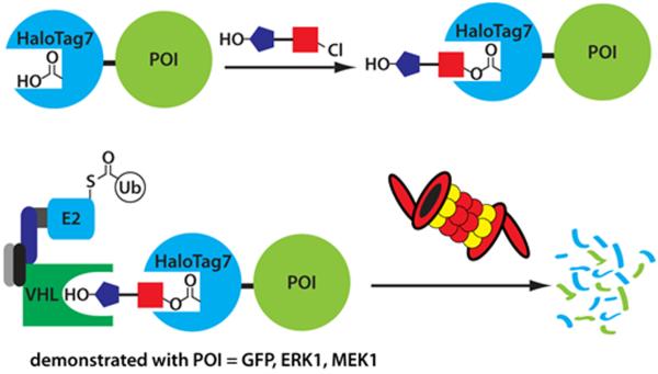
Schematic depiction of a bifunctional HaloPROTAC containing chloroalkane (which binds HaloTag7 fusion proteins) and a hydroxyproline derivative which binds VHL.
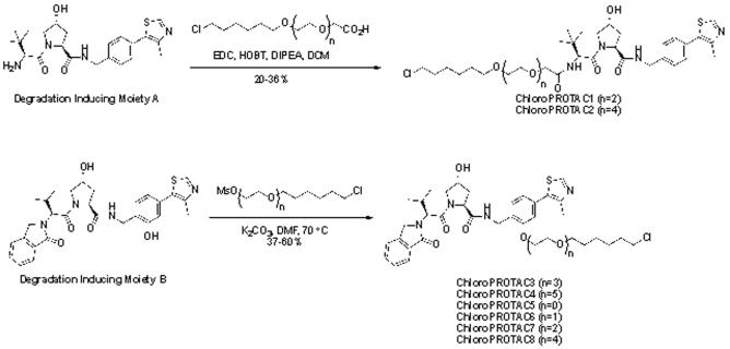
Synthesis of HaloPROTACs containing Degradation Inducing Moiety A and Degradation Inducing Moiety B.ACS Chem Biol.2015 Aug 21;10(8):1831-7. |
The average fluorescence per cell compared to vehicle control was measured by flow cytometry after 24 hour treatment with the indicated compounds and concentrations.ACS Chem Biol.2015 Aug 21;10(8):1831-7. |
A) A study of linker length with Degradation Inducing Moiety B shows that three ethylene glycol units are optimal for the degradation of GFP-HaloTag7. B) Structures of HaloPROTACs that have weaker affinity for VHL. C) Reducing the affinity for VHL attenuates their ability to induce degradation of GFP-HaloTag7, although the effect is not necessarily linear.ACS Chem Biol.2015 Aug 21;10(8):1831-7. |
A) The enantiomers of HaloPROTACs (containing D-amino acid residues) which do not bind VHL do not induce degradation of GFP-HaloTag7, supporting the necessity of VHL binding for activity. B) Pre-treatment with excessent-HaloPROTAC3 (1 hour) prevents degradation of GFP-HaloTag7 by HaloPROTAC3 after 24 hours. C) Pre-treatment with epoxomicin (4 hours) prevents degradation of GFP-HaloTag7 by HaloPROTAC3 after 20 hours. D)Treatment with VL285 attenuates the ability of HaloPROTAC3 to induce the degradation of GFP-HaloTag7. E) Structure of VL285. All error bars depict SEM.ACS Chem Biol.2015 Aug 21;10(8):1831-7. |
A) Comparison of HaloPROTAC3 (quintuplicate) to Hyt36 (triplicate) shows that HaloPROTAC3 is significantly more potent and efficacious. B) HaloPROTAC3 leads to 50% degradation of GFP-HaloTag7 within 4 to 8 hours. C) Significant recovery from 24 hour treatment with HaloPROTAC3 is observed after a 24 hour washout.ACS Chem Biol.2015 Aug 21;10(8):1831-7. |
Immunoblotting confirms that nearly complete degradation of A) GFP-HaloTag7 is observed after 24 hour treatment with 500 nM HaloPROTAC3, with significant degradation at 50 nM HaloPROTAC3. HaloPROTAC3 can lead to degradation of other HaloTag7 fusion proteins such as B) HaloTag7-ERK1 and HaloTag7-MEK1. As expected, endogenous ERK and MEK are not degraded.ACS Chem Biol.2015 Aug 21;10(8):1831-7. |
Products are for research use only; We do not sell to patients
Copyright 2020 InvivoChem LLC | All Rights Reserved


 COA
COA