
| Size | Price | |
|---|---|---|
| 500mg | ||
| 1g | ||
| Other Sizes |


| Targets |
PPAR-γ/NF-κB signaling pathway; NLRP3/caspase-1 signaling pathway; NLRP3, ASC, caspase-1 p20, and GSDMD-N. No IC50, Ki, EC50, or DC50 reported. [1]
NKTR (natural killer cell triggering receptor); AKT phosphorylation. No IC50, Ki, EC50, or DC50 reported. [2] AKT; NFkB; NLRP3 inflammasome. Pull-down indicated AKT binding; AKT molecular weight approximately 60 kDa. No IC50, Ki, EC50, or DC50 reported. [3] Gut microbiota-bile acid-FXR axis; intestinal FXR-FGF15 pathway; hepatic CYP7A1; BSEP. No IC50, Ki, EC50, or DC50 reported. [4] |
|---|---|
| ln Vitro |
In LPS-induced NR8383 alveolar macrophages, ACT001 (10, 20, and 40 µM pretreatment for 4 h before 10 µg/ml LPS for 24 h) dose-dependently decreased secretion and mRNA expression of TNF-α, IL-6, IL-1β, and IL-18 and increased IL-10. ACT001 reduced pyroptosis, shown by lower double-positive caspase-1/PI cells, and downregulated NLRP3, ASC, caspase-1 p20, and GSDMD-N. ACT001 upregulated PPAR-γ and inhibited phosphorylation of IκB-α and NF-κB p65; the PPAR-γ inhibitor T0070907 reversed these protective effects. [1]
In NSCLC cell lines, ACT001 inhibited proliferation with 72-h IC50 values: H1703 9.21 µM, H1975 14.42 µM, EBC-1 58.75 µM, H226 91.58 µM, PC-9 53.62 µM, LTEP-A2 70.93 µM, and HCC827 52.79 µM. ACT001 (0, 10, and 20 µM) inhibited colony formation and induced G1/S arrest, with decreased cyclin D1 and CDK6. RNA-seq showed NKTR upregulation; ACT001 increased NKTR mRNA/protein. NKTR knockdown promoted proliferation and reversed ACT001 effects, increased p-AKT; ACT001 inhibited AKT phosphorylation without changing total AKT. [2] In BV2 cells, ACT001 safe range was up to 10 µM; in primary mouse/rat microglia up to 5 µM; in HT22 up to 10 µM; in bEnd.3 up to 5 µM. ACT001 (1-10 µM) suppressed LPS-induced CD16, CD86, iNOS, IL-1β, TNF-α, and IFN-γ and increased Arg1, CD206, TGF-β, and IL-10; it reduced NO and CD68+ microglia. In co-culture, ACT001 (1-2 µM) reduced microglia-induced HT22 apoptosis, improved bEnd.3 tube formation, decreased VEGF, and restored ZO-1 and Occludin. ACT001 decreased p-AKT, p-IKKα/β, p-NFkB, NLRP3, ASC, cleaved-caspase-1, and IL-1β and inhibited NFkB nuclear translocation. [3] No in vitro cell data reported. [4] |
| ln Vivo |
H1703 xenograft in BALB/c nude mice; 200 mg/kg ACT001 orally daily significantly reduced tumor volume versus saline; HE of liver and kidney showed no detectable hepatic or renal toxicity; IHC showed increased NKTR and reduced Ki67. [2]
Controlled cortical impact TBI model in C57BL/6 mice; ACT001 100 mg/kg oral gavage daily from surgery to 7 days reduced blood-brain barrier damage, lesion volume (decreased by 5.36±1.41% at 7 days, P<0.001), Evans blue extravasation at 7 days, microglial activation, and neuronal apoptosis, and improved tight junction proteins and neurobehavioral recovery at 14 days. Delayed microglia depletion with PLX5622 attenuated efficacy. [3] HFD and MCD MASLD models; ACT001 200 mg/kg/day by gavage for 20 weeks (HFD) or 6 weeks (MCD) reduced liver injury, hepatic lipid accumulation, restored intestinal barrier, rebalanced gut microbiota, increased fecal 23-DCA, LCA-3S, 6-ketoLCA, and ILCA, decreased conjugated BAs, downregulated intestinal FXR-FGF15, and increased hepatic CYP7A1 and BSEP (MCD). [4] |
| Enzyme Assay |
AKT phosphorylation was assessed in cells by Western blot. [2]
Pull-down assay using active biotin-ACT001 and inactive biotin-S-ACT001 probes in rat primary microglia and BV2 cells; proteins precipitated by the active probe were visualized by silver staining, showing bands at approximately 70-55 kDa corresponding to AKT (60 kDa); Western blot confirmed AKT was precipitated by biotin-ACT001 but not biotin-S-ACT001. [3] No enzyme assay reported. [4] |
| Cell Assay |
NR8383 rat alveolar macrophages were cultured in Ham's F-12K medium with 10% FBS at 37°C and 5% CO2. Cell viability was measured by MTS after treatment with LPS or ACT001. For inflammatory cytokine measurement, cells were pretreated with ACT001 for 4 h and then stimulated with 10 µg/ml LPS for 24 h; TNF-α, IL-1β, IL-6, IL-18, and IL-10 in supernatant were measured by ELISA, and mRNA levels were measured by qRT-PCR. Pyroptosis was assessed by flow cytometry using FAM-FLICA caspase-1 and PI double staining. Western blot measured NLRP3, ASC, caspase-1 p20, GSDMD-N, PPAR-γ, IκB-α, p-IκB-α, NF-κB p65, and p-NF-κB p65 with GAPDH as internal control. [1]
NSCLC cells were cultured in standard medium with 10% FBS and penicillin/streptomycin. MTT assay was used to measure proliferation and IC50. Colony formation assay seeded 500 cells/well in six-well plates, treated after adhesion, counted clones after 10-14 days, fixed, and stained. Cell cycle was analyzed by flow cytometry using a DNA content quantitation kit after 70% cold ethanol fixation, RNase treatment, and PI staining. RNA was extracted, reverse transcribed, and qRT-PCR performed with GAPDH normalization. Western blot measured NKTR, cyclin D1, CDK6, AKT, and phosphorylated AKT. NKTR was knocked down by lentiviral shRNA in H1703 and H1975 cells. [2] BV2, primary mouse/rat microglia, HT22, and bEnd.3 cells were cultured in DMEM with 10% FBS and penicillin/streptomycin at 37°C and 5% CO2. Cell viability was measured by CCK-8. Microglial activation was induced by 100 or 500 ng/ml LPS. Pro-inflammatory and anti-inflammatory cytokines were measured by Real-time PCR; NO was measured by Griess assay. Immunofluorescence used Iba1/CD68, NeuN/TUNEL, CD31/ZO-1, and CD31/Occludin. Apoptosis was assessed by TUNEL and Annexin V-FITC/PI flow cytometry. Tube formation was performed with bEnd.3 cells on Matrigel. ELISA measured VEGF. Western blot measured p-AKT, total AKT, p-IKKα/β, total IKKβ, p-NFkB, total NFkB, NLRP3, ASC, cleaved-caspase-1, pro-caspase-1, IL-1β, pro-IL-1β, and GAPDH. [3] |
| Animal Protocol |
H1703 cells (1×10^7) suspended in 100 µl sterile PBS were inoculated subcutaneously into the groin of 5-6-week-old BALB/c nude mice. Tumor volume was measured every two days. When average tumor volume reached 100 mm^3, mice received 200 mg/kg ACT001 or sterile saline orally every day. After 12 days of administration, mice were killed under anesthesia; tumors, livers, and kidneys were collected and fixed in 10% formalin. [2]
Adult male C57BL/6 mice (8-12 weeks old, 20-25 g) underwent controlled cortical impact TBI. ACT001 was dissolved in 0.1 mmol PBS to 20 mg/ml and administered daily by oral gavage at 100 mg/kg from the day of CCI surgery until 7 days after surgery. For microglia depletion, mice were fed PLX5622-containing diet at 1200 ppm for 2 weeks before CCI. Brain tissues were collected at indicated times after injury. [3] Female C57BL/6J mice (8 weeks old, 20±2 g) were used. For HFD model, mice were fed normal diet or high-fat diet for 20 weeks; groups received saline, 200 mg/kg/day ACT001, or 150 mg/kg/day PPC by gavage. For MCD model, mice were fed methionine-choline-supplement or methionine-choline-deficient diet for 6 weeks; groups received saline, 200 mg/kg/day ACT001, or 150 mg/kg/day PPC by gavage. ACT001 was dissolved in saline. Body weights were recorded weekly; at the end, blood, liver, ileum, and cecal feces were collected. [4] |
| ADME/Pharmacokinetics |
Molecular formula C17H27NO3·C4H4O4; molecular weight 409.47 Da. No PK parameters reported. [2]
ACT001 displays higher plasma stability, more sustained release, superior efficacy, increased oral bioavailability, and can cross the blood-brain barrier; no numerical PK parameters reported. [3] ACT001 is synthesized from MCL by Michael addition and can slowly and continuously release MCL in plasma, resulting in longer drug potency and more stable pharmacokinetic properties; no numerical PK parameters reported. [4] |
| Toxicity/Toxicokinetics |
In NR8383 alveolar macrophages, ACT001 alone at 2.5, 5, 10, 20, and 40 µM did not significantly alter cell viability compared with no addition; higher concentrations were not used in subsequent experiments. [1]
In H1703 xenograft mice, HE staining of liver and kidney showed no detectable hepatic or renal toxicity after ACT001 treatment. [2] ACT001 is described as having low systemic and local toxicity; no specific toxicokinetic parameters reported. [3] Previous study in C57BL/6J mice observed no obvious toxicity; the MASLD study did not evaluate off-target or systemic effects. [4] |
| References |
|
| Additional Infomation |
ACT001 is a derivative of michelolide from the natural product parthenolide, extracted from Tanacetum parthenium. It has been investigated in tumors, intracranial diseases, and fibrotic diseases, but its role in acute lung injury was less known. The study concluded ACT001 ameliorated inflammation and pyroptosis via PPAR-γ/NF-κB in LPS-induced NR8383 alveolar macrophages. [1]
ACT001 is a novel sesquiterpene lactone derivative with anticancer and anti-inflammatory activities. It is an FDA orphan drug for glioblastoma and is in phase II clinical trials. It can pass the blood-brain barrier. This study suggested NKTR may be the target of ACT001 in NSCLC. [2] ACT001 (dimethylamino-micheliolide, DMAMCL) is a fumarate salt form of DMAMCL, a sesquiterpene lactone derivative. It is an FDA orphan drug for GBM and can cross the blood-brain barrier. The study concluded ACT001 attenuated microglia-mediated neuroinflammation after TBI via AKT/NFkB/NLRP3 pathway. [3] ACT001 is a novel anti-inflammatory and anticancer agent derived from parthenolide. It has been approved as an orphan drug for human cerebral glioma by FDA and has clinical trials. This study concluded ACT001 alleviated MASLD potentially via gut microbiota-bile acid-FXR axis. [4] |
| Molecular Formula |
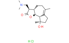
C17H28CLNO3
|
|---|---|
| Molecular Weight |
329.86
|
| CAS # |
1403357-80-1
|
| Appearance |
Light yellow to yellow solid
|
| SMILES |
Cl.O1C([C@@H](CN(C)C)[C@@H]2CCC(C)=C3CC[C@](C)([C@@H]3[C@@H]12)O)=O
|
| Synonyms |
Dimethylaminomicheliolide hydrochloride; DMAMCL hydrochloride
|
| HS Tariff Code |
2934.99.9001
|
| Storage |
Powder -20°C 3 years 4°C 2 years In solvent -80°C 6 months -20°C 1 month |
| Shipping Condition |
Room temperature (This product is stable at ambient temperature for a few days during ordinary shipping and time spent in Customs)
|
| Solubility (In Vitro) |
May dissolve in DMSO (in most cases), if not, try other solvents such as H2O, Ethanol, or DMF with a minute amount of products to avoid loss of samples
|
|---|---|
| Solubility (In Vivo) |
Note: Listed below are some common formulations that may be used to formulate products with low water solubility (e.g. < 1 mg/mL), you may test these formulations using a minute amount of products to avoid loss of samples.
Injection Formulations
Injection Formulation 1: DMSO : Tween 80: Saline = 10 : 5 : 85 (i.e. 100 μL DMSO stock solution → 50 μL Tween 80 → 850 μL Saline)(e.g. IP/IV/IM/SC) *Preparation of saline: Dissolve 0.9 g of sodium chloride in 100 mL ddH ₂ O to obtain a clear solution. Injection Formulation 2: DMSO : PEG300 :Tween 80 : Saline = 10 : 40 : 5 : 45 (i.e. 100 μL DMSO → 400 μLPEG300 → 50 μL Tween 80 → 450 μL Saline) Injection Formulation 3: DMSO : Corn oil = 10 : 90 (i.e. 100 μL DMSO → 900 μL Corn oil) Example: Take the Injection Formulation 3 (DMSO : Corn oil = 10 : 90) as an example, if 1 mL of 2.5 mg/mL working solution is to be prepared, you can take 100 μL 25 mg/mL DMSO stock solution and add to 900 μL corn oil, mix well to obtain a clear or suspension solution (2.5 mg/mL, ready for use in animals). View More
Injection Formulation 4: DMSO : 20% SBE-β-CD in saline = 10 : 90 [i.e. 100 μL DMSO → 900 μL (20% SBE-β-CD in saline)] Oral Formulations
Oral Formulation 1: Suspend in 0.5% CMC Na (carboxymethylcellulose sodium) Oral Formulation 2: Suspend in 0.5% Carboxymethyl cellulose Example: Take the Oral Formulation 1 (Suspend in 0.5% CMC Na) as an example, if 100 mL of 2.5 mg/mL working solution is to be prepared, you can first prepare 0.5% CMC Na solution by measuring 0.5 g CMC Na and dissolve it in 100 mL ddH2O to obtain a clear solution; then add 250 mg of the product to 100 mL 0.5% CMC Na solution, to make the suspension solution (2.5 mg/mL, ready for use in animals). View More
Oral Formulation 3: Dissolved in PEG400 (Please use freshly prepared in vivo formulations for optimal results.) |
| Preparing Stock Solutions | 1 mg | 5 mg | 10 mg | |
| 1 mM | 3.0316 mL | 15.1579 mL | 30.3159 mL | |
| 5 mM | 0.6063 mL | 3.0316 mL | 6.0632 mL | |
| 10 mM | 0.3032 mL | 1.5158 mL | 3.0316 mL |
*Note: Please select an appropriate solvent for the preparation of stock solution based on your experiment needs. For most products, DMSO can be used for preparing stock solutions (e.g. 5 mM, 10 mM, or 20 mM concentration); some products with high aqueous solubility may be dissolved in water directly. Solubility information is available at the above Solubility Data section. Once the stock solution is prepared, aliquot it to routine usage volumes and store at -20°C or -80°C. Avoid repeated freeze and thaw cycles.
Calculation results
Working concentration: mg/mL;
Method for preparing DMSO stock solution: mg drug pre-dissolved in μL DMSO (stock solution concentration mg/mL). Please contact us first if the concentration exceeds the DMSO solubility of the batch of drug.
Method for preparing in vivo formulation::Take μL DMSO stock solution, next add μL PEG300, mix and clarify, next addμL Tween 80, mix and clarify, next add μL ddH2O,mix and clarify.
(1) Please be sure that the solution is clear before the addition of next solvent. Dissolution methods like vortex, ultrasound or warming and heat may be used to aid dissolving.
(2) Be sure to add the solvent(s) in order.
Products are for research use only; We do not sell to patients
Copyright 2020 InvivoChem LLC | All Rights Reserved


