| Size | Price | Stock | Qty |
|---|---|---|---|
| 5mg |
|
||
| 10mg |
|
||
| 25mg |
|
||
| 50mg |
|
||
| 100mg |
|
||
| 250mg |
|
||
| 500mg |
|
||
| Other Sizes |
| Targets |
Necrosis; MLKL/mixed lineage kinase domain-like protein
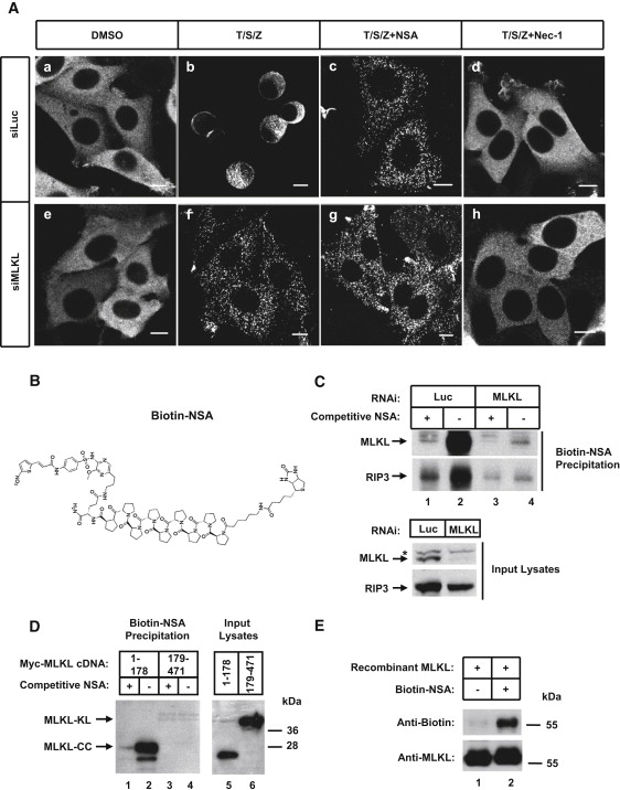
Mixed Lineage Kinase Domain-like protein (MLKL) [2][3]. The compound targets the N-terminal coiled-coil (CC) domain of human MLKL, specifically covalently modifying the Cys86 residue [2]. It does not inhibit mouse MLKL due to a tryptophan residue at the corresponding position [2]. |
|---|---|
| ln Vitro |
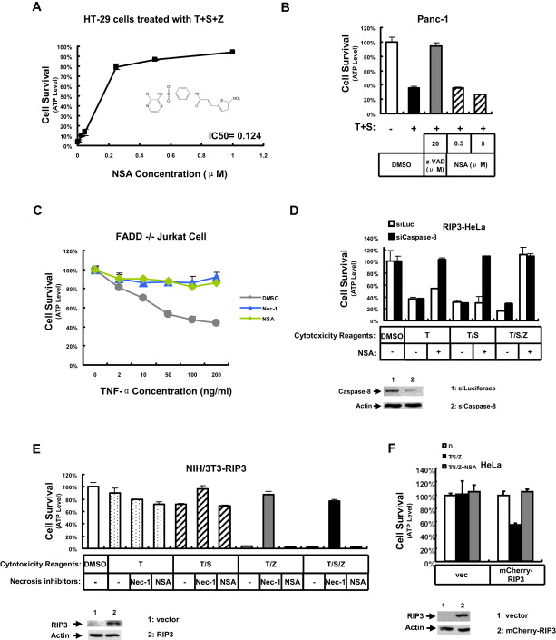
Necrosulfonamide inhibits MLKL-mediated Necrosis by blocking its N-terminal CC domain function. Following RIP3 activation, it prevents necrosis. Even at a concentration of 5 μM , necrosulfonamide has no impact on the apoptosis induced by TNF-α plus Smac mimetic in Panc-1 cells deficient in RIP3. In human cells, necrosulfonamide effectively inhibits necrosis, but not in mouse cells. The cysteine at residue 86 in human MLKL that necrosulfonamide covalently modifies is replaced by a tryptophan residue in mouse MLKL (mixed lineage kinase domain-like protein), which accounts for necrosulfonamide's species specificity[2].
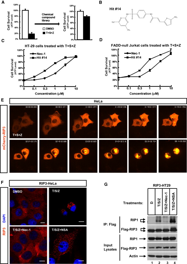
- Necrosulfonamide (NSA) blocked necrosis in human colon cancer HT-29 cells and FADD-null human T cell leukemia Jurkat cells with an IC50 of less than 1 μM, showing higher potency than necrostatin-1. A derivative, 3-methoxypyrazin-2-yl, had an IC50 of less than 0.2 μM [2]. - In a HeLa cell line expressing RIP3, NSA efficiently blocked necrosis induced by TNF-α plus Smac mimetic [2]. - Necrosis in mouse 3T3 cells expressing mouse RIP3 was insensitive to NSA inhibition [2]. - Live-cell imaging showed that NSA arrested necrosis at a step where RIP3 formed discrete punctae that failed to enlarge [2]. - In RIP3-expressing HeLa cells, NSA did not block RIP1 and RIP3 interaction but enhanced their interaction and phosphorylation [2]. - NSA treatment blocked necroptosis measured by membrane leakage assay but did not block the phospho-MLKL signal [3]. - In a cell-free liposome leakage assay, purified recombinant phosphomimic MLKL (T357E/S358D) caused over 50% Tb3+ release from liposomes containing 5% PI(4,5)P2 and close to 90% from liposomes containing 15% cardiolipin. The wild-type MLKL caused about 30% and 60% release, respectively. Necrosulfonamide (2 μM) significantly blocked this leakage from both types of liposomes [3]. - In HT-29 cells, NSA prevented phospho-MLKL from shifting to the detergent phase in a Triton X-114 phase separation experiment, indicating it blocked phospho-MLKL targeting to membranes [3]. - In an Alzheimer's disease rat model (AlCl3-induced), treatment with NSA significantly lowered the hippocampal level of phosphorylated MLKL (p-MLKL, Ser358) by 87.24% compared to the AD group, without affecting the elevated hippocampal levels of p-RIP1 (Ser166) and p-RIP3 (Ser232) [4]. - NSA treatment in the AD rat model significantly decreased hippocampal β-amyloid and BACE1 levels by 67.4% and 69.6%, respectively, and reduced p-tau (Ser404) and GSK-3β by 74.2% and 74.3%, respectively, compared to the AD group [4]. |
| ln Vivo |
Necrosulfonamide (NSA) is a small molecule that targets MLKL, the final executor of necroptosis, to specifically inhibit necroptosis.
- In a rat model of Alzheimer's disease (induced by 17 mg/kg AlCl3 for 6 weeks), treatment with Necrosulfonamide (1.65 mg/kg/day, i.p. for 6 weeks) resulted in significant improvement in spatial learning and memory deficits as demonstrated by Morris water maze (decreased escape latency, increased time spent in target quadrant) and Y-maze (increased spontaneous alternation percentage, decreased same arm returns) behavioral tests [4]. - In the same AD rat model, NSA treatment significantly reduced β-amyloid plaque deposition in the hippocampal CA1 region by almost 50% as shown by Congo red staining [4]. - NSA administration to AD rats significantly restored the hippocampal level of acetylcholine (2.6-fold increase) and decreased acetylcholinesterase activity (63.3% decline) compared to the AD group [4]. - Histological examination of the hippocampus (CA1, CA3, and dentate gyrus) in AD rats treated with NSA showed significantly higher numbers of intact neurons (e.g., 89.8% increase in CA1 region) compared to the AD group [4]. |
| Enzyme Assay |
RIP1 and RIP3 were immunoprecipitated with an anti-Flag antibody. The Flag beads were incubated with 2 μCi of [32P]γ-ATP at 37°C for 1 hour with the artificial substrate MBP or purified recombinant MLKL after being washed three times with kinase buffer (50 mM HEPES, pH 7.5, 10 mM MgCl2, 50 mM NaCl, 0.02% BSA, 150 μM ATP, and 1 mM DTT). Then SDS-PAGE and autoradiography were applied to the reaction mixtures. We describe the discovery of a small molecule known as (E)-N-(4-(N-(3-methoxypyrazin-2-yl)sulfamoyl)phenyl)-3-(5-nitrothiophene-2-yl)acrylamide, also known as necrosulfonamide, which specifically inhibits necrosis downstream of RIP3 activation. The mixed lineage kinase domain-like protein (MLKL) was identified as the interacting target by coimmunoprecipitation with anti-RIP3 antibodies and an affinity probe made from necrosulfonamide. The threonine 357 and serine 358 residues on MLKL were phosphorylated by RIP3 and these phosphorylation events were essential for necrosis.
- In vitro kinase assay: RIP1 and RIP3 were immunopurified from HeLa cells using anti-Flag antibody. The Flag beads were washed three times with kinase buffer (50 mM HEPES [pH 7.5], 10 mM MgCl2, 50 mM NaCl, 0.02% BSA, 150 μM ATP, and 1 mM DTT). The beads were then incubated with 2 μCi of [γ-32P]-ATP at 37°C for 1 hour with the artificial substrate myelin basic protein (MBP) or purified recombinant MLKL. The reaction mixtures were then subjected to SDS-PAGE followed by autoradiography. This assay showed that RIP3, but not RIP1, could phosphorylate MLKL [2]. - Liposome leakage assay: Liposomes containing Tb3+ ions and different lipid compositions (e.g., 5% PI(4,5)P2 or 15% cardiolipin) were prepared. Purified recombinant MLKL proteins (wild-type, phospho-dead, or phosphomimic) were incubated with these liposomes in the presence of dipicolinic acid (DPA). The released fluorescence from the Tb3+-DPA chelate was recorded using a plate reader. To test NSA's effect, the phosphomimic MLKL was pre-incubated with 2 μM Necrosulfonamide or DMSO at room temperature for 2 hours before the assay [3]. |
| Cell Assay |
Necrosis inhibitors induce diverse effects on MLKL phosphorylation. T/S/Z is applied to HT-29 cells for either 12 or 8 hours, with or without necrosis inhibitors. By monitoring released protease activity in the culture medium, the quantity of dead cells is calculated. The whole-cell extracts are made, and western blotting is used to analyze them. Final concentrations of 1 or 10 μM necrosulfonamide or necrostatin-1 inhibit necrosis.
- Screen Assay Design: 2,000 HT-29 cells were seeded per well in a 384-well plate. The next day, necrosis was induced by adding TNF-α (20 ng/ml), Smac mimetic (100 nM), and z-VAD (20 μM). Concurrently, compounds from a library (~200,000) were added at a final concentration of 10 μM. Cell viability was determined after 24 hrs by measuring ATP levels using Cell Titer-Glo assay [1]. - Cell Survival Assay: Cell survival was performed using the CellTiter-Glo Luminescent Cell Viability Assay kit according to the manufacturer's instructions. Luminescence was recorded with a plate reader [1][2]. - Biotinylated Necrosulfonamide Precipitation: Cells were harvested and lysed. Biotinylated NSA (20 nmol) was pre-incubated with 20 μl streptavidin agarose for 2 hrs at 4°C. The beads were washed two times in lysis buffer. The NSA-bound agarose was then incubated with cell lysates overnight at 4°C. Non-biotinylated NSA was included as a binding competitor. The beads were washed four times with lysis buffer, then directly boiled in 1X SDS loading buffer [1][2]. - Gene Knockdown and Rescue: RIP3-HeLa cells were plated in 96-well plates and treated with doxycycline. The next day, each well was transfected with 2.5 pmol of siRNA against MLKL or luciferase (control) along with 0.05 μg of a cDNA bearing silent mutations (siRNA-resistant wild-type MLKL or C86S mutant) or vector plasmid. The following day, cells were treated with necrosis inducers for 24 hrs, and cell viability was determined by measuring ATP levels [2]. |
| Animal Protocol |
Male Wistar rats
1.65 mg/kg i.p. Rats were randomly allocated into four groups (8 rats/group). Group 1 (Control group) comprised normal vehicle-treated rats. Group 2 (AlCl3 group; AD group) comprised rats that were treated with AlCl3, dissolved in distilled water, orally at a dose of 17 mg/kg daily for 6 consecutive weeks, and represented the AD group. Group 3 (AlCl3 + necrosulfonamide (NSA) group) comprised rats that were treated with AlCl3, as in group 2, concomitantly with necrosulfonamide (NSA), dissolved in dimethyl sulfoxide, intraperitoneally at a dose of 1.65 mg/kg daily for 6 weeks. Group 4 (necrosulfonamide (NSA) group) comprised normal rats that were treated with NSA dissolved in dimethyl sulfoxide at a dose of 1.65 mg/kg/day intraperitoneally for 6 weeks. The dose of NSA was selected based on a pilot experiment conducted prior to the main study. In this preliminary study, the dose efficacy was evaluated based on histological examination of the hippocampus for amyloid plaque deposits and neuronal degeneration, learning and memory evaluation by Morris water maze and Y-maze tests, and analysis of hippocampal p-MLKL, p-tau, and β-amyloid levels, in AlCl3 + NSA-treated rats compared to AlCl3-treated rats.[4] - Alzheimer's Disease Rat Model: Adult male Wistar rats (180-220 g) were randomly allocated into groups (n=8). Alzheimer's disease was induced by oral administration of aluminum chloride (AlCl3) dissolved in distilled water at a dose of 17 mg/kg daily for 6 consecutive weeks. Necrosulfonamide (NSA) was dissolved in dimethyl sulfoxide (DMSO) and administered intraperitoneally at a dose of 1.65 mg/kg daily for 6 weeks, either alone or concurrently with AlCl3. Behavioral tests (Morris water maze and Y-maze) were conducted five days prior to the termination of the experiment [4]. |
| References | |
| Additional Infomation |
Necrosulfonamide (NSA) is a sulfonamide drug, a 3-methoxypyrazine-2-yl derivative of (E)-N-(4-(N-(4,6-dimethylpyrimidin-2-yl)sulfonyl)phenyl)-3-(5-nitrothiophene-2-yl)acrylamide. SSA specifically blocks the activation of downstream necrosis by RIP3 (receptor-interacting serine/threonine kinase 3), a key signaling molecule in the programmed necrosis (apoptosis) pathway. It acts as an inhibitor of necroptosis and a neuroprotective agent. It is a sulfonamide compound belonging to the pyrazine and thiophene classes. Through high-throughput screening of 200,000 compounds and subsequent structure-activity relationship (SAR) studies, we found that SSA is a potent small-molecule inhibitor that inhibits necroptosis induced by the combined action of TNF-α, Smac mimics, and z-VAD-fmk (T/S/Z). We used a forward chemogenetic approach and NSA-based chemical probes to further reveal that NSA selectively targets mixed lineage kinase domain-like protein (MLKL) to block the formation of necrosomes. [1]
Receptor-interacting serine/threonine kinase 3 (RIP3) is a key signaling molecule in the programmed necrosis (necrotizing apoptosis) pathway. This pathway plays an important role in a variety of physiological and pathological conditions, including development, tissue damage response and antiviral immunity. This article reports the identification of a small molecule compound called (E)-N-(4-(N-(3-methoxypyrazin-2-yl)sulfonyl)phenyl)-3-(5-nitrothiophene-2-yl)acrylamide (hereinafter referred to as necrotizing sulfonamide), which specifically blocks the downstream necrosis process activated by RIP3. The mixed lineage kinase domain-like protein (MLKL) was identified as the interaction target by both affinity probes derived from necrotizing sulfonamide and co-immunoprecipitation experiments with anti-RIP3 antibodies. RIP3 phosphorylates MLKL at threonine 357 and serine 358, and these phosphorylation events are crucial for necrosis. Treatment of cells with necrotizing sulfonamide or knockdown of MLKL expression arrests the necrosis process at a specific step in which RIP3 forms discrete spots within the cell. These results suggest that MLKL is a key mediator of downstream necrosis signaling of RIP3 kinases. [2] Programmed necrotizing cell death induced by the tumor necrosis factor α (TNF-α) cytokine family depends on a kinase cascade consisting of receptor-interacting kinases RIP1 and RIP3. How these kinase activities lead to cell death is unclear. MLKL, a mixed-lineage kinase domain-like protein, is a functional substrate of RIP3 that binds to RIP3 through its kinase-like domain but lacks kinase activity itself. RIP3 phosphorylates MLKL at T357 and S358. This article reports the development of a monoclonal antibody that specifically recognizes phosphorylated MLKL in cells that die through this pathway and in liver biopsy samples from patients with drug-induced liver injury. Phosphorylated MLKL forms oligomers that can bind to phosphatidylinositol and cardiolipin. This property allows MLKL to migrate from the cytosol to the plasma membrane and intracellular membrane, where it directly disrupts membrane integrity, leading to cell death. [3] Alzheimer's disease (AD) is a progressive neurodegenerative disease for which there is currently no effective treatment. Existing treatments can only alleviate symptoms and have limited efficacy. Necrophage is a controlled form of cell death that has been found to be associated with the pathogenesis of various neurodegenerative diseases in recent years. This study investigated the role of necroptosis in the pathogenesis of AD and evaluated the potential therapeutic effect of the necroptosis inhibitor sulfamethoxazole (NSA) in an AD rat model. AD was induced by oral administration of aluminum chloride (AlCl3, 17 mg/kg/day) for 6 consecutive weeks. Intraperitoneal injection of NSA (1.65 mg/kg/day) for 6 weeks significantly improved AlCl3-induced spatial learning and memory impairment, as evidenced by enhanced performance in the Morris water maze and Y maze in rats. NSA can reduce the abnormally high expression of tumor necrosis factor-α (TNF-α), β-amyloid precursor protein lyase 1 (BACE1), β-amyloid protein, glycogen synthase kinase-3β (GSK-3β), phosphorylated tau protein, and acetylcholinesterase in the hippocampus, accompanied by acetylcholine supplementation. The improvement of Alzheimer's disease-related disorders by NSA is associated with its inhibition of phosphorylation of the key necrotizing apoptosis executive factor, mixed lineage kinase domain-like protein (MLKL). Histopathological changes support the above biochemical results. In conclusion, NSA therapy is a promising approach to Alzheimer's disease by targeting MLKL-dependent necrotizing apoptosis to alleviate the neuropathology of AD. [4] - Necrosulfonamide (NSA) blocks necroptosis by selectively targeting the mixed lineage kinase domain-like protein (MLKL) [2]. It was identified from a high-throughput screen of ~200,000 compounds using a necrosis model in HT-29 cells induced by TNF-α, Smac mimetic, and z-VAD-fmk [2]. - The compound binds to the N-terminal coiled-coil (CC) domain of human MLKL through covalent bonding, likely as a Michael acceptor with its α,β-unsaturated enone moiety, targeting the Cys86 residue [2]. - Its mechanism of action is downstream of RIP3 activation, as it does not prevent RIP1-RIP3 interaction or RIP3 phosphorylation but blocks the membrane translocation and oligomerization of phosphorylated MLKL [2][3]. - Necrosulfonamide shows species specificity, effectively inhibiting necroptosis in human cells but not in mouse cells [2]. - The compound has been used as a pharmacological tool to demonstrate that MLKL phosphorylation is a key commitment step for necroptosis execution and has been used to detect necroptosis in human disease, such as in liver biopsy samples from patients with drug-induced liver injury, using a phospho-specific MLKL antibody [3]. - This is the first study to report the ameliorative effect of NSA against Alzheimer's disease-associated neuropathologies in an AlCl3-induced rat model, suggesting MLKL as a potential therapeutic target for AD [4]. |
| Molecular Formula |
C18H15N5O6S2
|
|---|---|
| Molecular Weight |
461.4716
|
| Exact Mass |
461.046
|
| Elemental Analysis |
C, 46.85; H, 3.28; N, 15.18; O, 20.80; S, 13.89
|
| CAS # |
432531-71-0
|
| Related CAS # |
Necrosulfonamide;1360614-48-7
|
| PubChem CID |
1566236
|
| Appearance |
Light yellow to yellow solid powder
|
| Density |
1.6±0.1 g/cm3
|
| Index of Refraction |
1.695
|
| LogP |
4.08
|
| Hydrogen Bond Donor Count |
2
|
| Hydrogen Bond Acceptor Count |
10
|
| Rotatable Bond Count |
7
|
| Heavy Atom Count |
31
|
| Complexity |
760
|
| Defined Atom Stereocenter Count |
0
|
| SMILES |
COC1=NC=CN=C1NS(=O)(=O)C2=CC=C(C=C2)NC(=O)C=CC3=CC=C(S3)[N+](=O)[O-]
|
| InChi Key |
FNPPHVLYVGMZMZ-XBXARRHUSA-N
|
| InChi Code |
InChI=1S/C18H15N5O6S2/c1-29-18-17(19-10-11-20-18)22-31(27,28)14-6-2-12(3-7-14)21-15(24)8-4-13-5-9-16(30-13)23(25)26/h2-11H,1H3,(H,19,22)(H,21,24)/b8-4+
|
| Chemical Name |
(E)-N-[4-[(3-methoxypyrazin-2-yl)sulfamoyl]phenyl]-3-(5-nitrothiophen-2-yl)prop-2-enamide
|
| Synonyms |
Necrosulfonamide, MLKL inhibitor; Necrosome Inhibitor II; Necrosis Inhibitor III; Necrosulfonamide; 1360614-48-7; 432531-71-0; (E)-N-(4-(N-(3-methoxypyrazin-2-yl)sulfamoyl)phenyl)-3-(5-nitrothiophen-2-yl)acrylamide; CHEBI:63770; (E/Z)-Necrosulfonamide; (2E)-N-{4-[(3-methoxypyrazin-2-yl)sulfamoyl]phenyl}-3-(5-nitrothiophen-2-yl)prop-2-enamide; (E)-N-[4-[(3-methoxypyrazin-2-yl)sulfamoyl]phenyl]-3-(5-nitrothiophen-2-yl)prop-2-enamide;
Mixed Lineage Kinase Domain-Like Protein Inhibitor
|
| HS Tariff Code |
2934.99.9001
|
| Storage |
Powder -20°C 3 years 4°C 2 years In solvent -80°C 6 months -20°C 1 month |
| Shipping Condition |
Room temperature (This product is stable at ambient temperature for a few days during ordinary shipping and time spent in Customs)
|
| Solubility (In Vitro) |
DMSO: ~92 mg/mL (~199.4 mM)
|
|---|---|
| Solubility (In Vivo) |
Note: Listed below are some common formulations that may be used to formulate products with low water solubility (e.g. < 1 mg/mL), you may test these formulations using a minute amount of products to avoid loss of samples.
Injection Formulations
Injection Formulation 1: DMSO : Tween 80: Saline = 10 : 5 : 85 (i.e. 100 μL DMSO stock solution → 50 μL Tween 80 → 850 μL Saline)(e.g. IP/IV/IM/SC) *Preparation of saline: Dissolve 0.9 g of sodium chloride in 100 mL ddH ₂ O to obtain a clear solution. Injection Formulation 2: DMSO : PEG300 :Tween 80 : Saline = 10 : 40 : 5 : 45 (i.e. 100 μL DMSO → 400 μLPEG300 → 50 μL Tween 80 → 450 μL Saline) Injection Formulation 3: DMSO : Corn oil = 10 : 90 (i.e. 100 μL DMSO → 900 μL Corn oil) Example: Take the Injection Formulation 3 (DMSO : Corn oil = 10 : 90) as an example, if 1 mL of 2.5 mg/mL working solution is to be prepared, you can take 100 μL 25 mg/mL DMSO stock solution and add to 900 μL corn oil, mix well to obtain a clear or suspension solution (2.5 mg/mL, ready for use in animals). View More
Injection Formulation 4: DMSO : 20% SBE-β-CD in saline = 10 : 90 [i.e. 100 μL DMSO → 900 μL (20% SBE-β-CD in saline)] Oral Formulations
Oral Formulation 1: Suspend in 0.5% CMC Na (carboxymethylcellulose sodium) Oral Formulation 2: Suspend in 0.5% Carboxymethyl cellulose Example: Take the Oral Formulation 1 (Suspend in 0.5% CMC Na) as an example, if 100 mL of 2.5 mg/mL working solution is to be prepared, you can first prepare 0.5% CMC Na solution by measuring 0.5 g CMC Na and dissolve it in 100 mL ddH2O to obtain a clear solution; then add 250 mg of the product to 100 mL 0.5% CMC Na solution, to make the suspension solution (2.5 mg/mL, ready for use in animals). View More
Oral Formulation 3: Dissolved in PEG400 (Please use freshly prepared in vivo formulations for optimal results.) |
| Preparing Stock Solutions | 1 mg | 5 mg | 10 mg | |
| 1 mM | 2.1670 mL | 10.8349 mL | 21.6699 mL | |
| 5 mM | 0.4334 mL | 2.1670 mL | 4.3340 mL | |
| 10 mM | 0.2167 mL | 1.0835 mL | 2.1670 mL |
*Note: Please select an appropriate solvent for the preparation of stock solution based on your experiment needs. For most products, DMSO can be used for preparing stock solutions (e.g. 5 mM, 10 mM, or 20 mM concentration); some products with high aqueous solubility may be dissolved in water directly. Solubility information is available at the above Solubility Data section. Once the stock solution is prepared, aliquot it to routine usage volumes and store at -20°C or -80°C. Avoid repeated freeze and thaw cycles.
Calculation results
Working concentration: mg/mL;
Method for preparing DMSO stock solution: mg drug pre-dissolved in μL DMSO (stock solution concentration mg/mL). Please contact us first if the concentration exceeds the DMSO solubility of the batch of drug.
Method for preparing in vivo formulation::Take μL DMSO stock solution, next add μL PEG300, mix and clarify, next addμL Tween 80, mix and clarify, next add μL ddH2O,mix and clarify.
(1) Please be sure that the solution is clear before the addition of next solvent. Dissolution methods like vortex, ultrasound or warming and heat may be used to aid dissolving.
(2) Be sure to add the solvent(s) in order.
Products are for research use only; We do not sell to patients
Copyright 2020 InvivoChem LLC | All Rights Reserved