
| Size | Price | |
|---|---|---|
| 500mg | ||
| 1g | ||
| Other Sizes |
| ADME/Pharmacokinetics |
Absorption, Distribution and Excretion
This study aimed to characterize the intestinal transport of β-phenylethylamine (PEA). Caco-2 cells exhibited Na+-independent uptake of [(14)C]PEA, but were strongly stimulated by the extracellular H+ gradient. At extracellular pH 7.5, concentration-dependent uptake of PEA reached saturation, with kinetic parameters of 2.6 mM (K(t)) and 96.2 nmol/min/mg protein (V(max)). Various biogenic amines, such as halamine and N-methylphenylethylamine, as well as cationic drugs such as phenelzine, transphenylcyclopropionate, d,l-phenylpropionate, methadone, chlorpheniramine, diphenhydramine, and promethazine, strongly inhibited [(14)C]PEA uptake, with K(i) values of approximately 1 mM. Tetraethylammonium, N-methyl-4-phenylpyridine, and choline had no effect. We also investigated bidirectional transepithelial transport of [(14)C]PEA in cell monolayers cultured on permeable membranes. The net transepithelial flux of [(14)C]PEA from apex to basolateral was 5 times that from basolateral to apex. We conclude that PEA is transported into Caco-2 cells via a highly active, saturable, H(+)-dependent (reverse transport) process. This transport characteristic is inconsistent with the known characteristics of SLC22, SLC44, SLC47, and other families of organic cationic carriers. Phenylethylamine was detected in the dorsal and ventral horns of the rat cervical spinal cord, the intermediate zone, and the dorsal and ventral horns of the lumbar spinal cord at concentrations ranging from 114 to 238 pg/mg protein. It was also detected in the caudate nucleus (218 pg/mg) and cerebellum (73 pg/mg). Levels of this substance increased in the brain and spinal cord after 10 consecutive days of amphetamine treatment. The specific binding of tritium-labeled β-phenylethylamine to the rat anterior meninges is saturable; the apparent dissociation constant is 55 nmol, and the binding site density is approximately 1078 pmol/mg protein. The highest binding rates were observed in the hypothalamus and striatum. In human subjects, the urinary excretion rate of endogenous amphetamine-like β-phenylethylamine significantly increased after the first skydive. The increase in urinary excretion rate was delayed in most subjects and was independent of changes in urinary pH or creatinine excretion. The data suggest that β-phenylethylamine plays a role in the stress response. Normal urine contains β-phenylethylamine (approximately 30 μg/L). Metabolism/Metabolites In adults and children with phenylketonuria (PKU), the excretion of free β-phenylethylamine is significantly increased even with normal or slightly restricted phenylalanine intake. Urinary β-phenylethylamine levels are also significantly elevated in children with PKU receiving a low-phenylalanine diet. The excretion of conjugated β-phenylethylamine was not found to be higher than normal. β-Phenylethylamine is an endogenous component of the human brain, involved in brain signal transmission. It is also present in certain foods and may have toxic side effects in susceptible individuals. The metabolism of 2-phenylethylamine to phenylacetaldehyde is mainly catalyzed by monoamine oxidase, while the oxidation of active aldehydes to phenylglyoxylic acid derivatives is mainly catalyzed by aldehyde dehydrogenase, and possibly also by aldehyde oxidase; the catalytic effect of xanthine oxidase is minimal. This study investigated the metabolism of 2-phenylethylamine to phenylacetaldehyde in liver slices and compared the relative activities of aldehyde oxidase, xanthine oxidase, and aldehyde dehydrogenase in the oxidation of phenylacetaldehyde under conditions of presence and absence of enzyme-specific inhibitors. In liver slices, phenylacetaldehyde was rapidly converted to phenylacetic acid. Phenylacetic acid is the main metabolite of 2-phenylethylamine, and its metabolic pathway is phenylacetaldehyde. Disulfiram (an aldehyde dehydrogenase-specific inhibitor) completely inhibits the formation of phenylacetic acid, while isovanillin (an aldehyde oxidase-specific inhibitor) has a weak inhibitory effect on phenylacetic acid formation, and allopurinol (a xanthine oxidase-specific inhibitor) has almost no effect. Therefore, in liver sections, phenylacetaldehyde is rapidly oxidized primarily by aldehyde dehydrogenase and aldehyde oxidase, with little or no effect from xanthine oxidase. We report a case of extremely high phenylethylamine levels in the urine of a neonate with phenylketonuria (PKU), and differences in phenylethylamine levels in urine among PKU patients with similar phenylalanine levels. Phenethylamine is a highly toxic metabolite of phenylalanine and is rapidly degraded by monoamine oxidase type B (an enzyme with very low activity in neonates). Therefore, these results are consistent with the hypothesis that monoamine oxidase type B acts as a modifying gene in PKU. Incubation of phenylethylamine with rabbit liver microsomes, divalent micronuclei, and an NADPH-generating system produces azo-2-phenylethane (a mutagen). 10 μmol of phenylethylamine yields 38 nmol of azo-2-phenylethane during a 30-minute incubation. The metabolism of 2-phenylethylamine to azo-2-aniline appears to be entirely dependent on the presence of divalent MN. For more complete data on the metabolism/metabolites of 2-phenylethylamine (9 metabolites in total), please visit the HSDB record page. The plasma pharmacokinetics of PEA can be described using first-order kinetics, with an estimated t/2 of approximately 5–10 minutes. |
|---|---|
| Toxicity/Toxicokinetics |
Toxicity Summary
Identification and Uses: 2-Phenylethylamine (PEA) is a colorless to pale yellow liquid with a fishy odor. It is used in organic synthesis, as a laboratory reagent, and in scintillation counters (carbon dioxide absorbers). Human Studies: PEA poisoning can be severe in young, healthy individuals. The most common reported symptoms include anxiety and hallucinations (49%), dilated pupils and headache (41%), tachycardia (40%), and hypertension (15%). Complications such as seizures (7%), cardiac arrest (5%), toxic myocarditis (1%), and hemorrhagic stroke (1%) have also been observed. PEA is teratogenic at concentrations observed in patients with phenylketonuria (PKU). Animal Studies: This study investigated the behavioral effects of PEA (a drug believed to act on both serotonin and dopaminergic brain mechanisms) in Wistar, Sprague-Dawley, and Long-Evans rats. Behavioral indicators assessed included forepaw thumping, head shaking, and hindlimb abduction, behaviors considered hallmarks of central serotonin stimulation. Following injection of 50 mg/kg PEA, Wistar rats were more active and responsive than Long-Evans and Sprague-Dawley rats. Wistar rats showed significantly different responses to central monoaminergic stimulation compared to Long-Evans and Sprague-Dawley strains. This study also examined the behavioral consequences of daily PEA injections for 6 weeks. Rats exhibited symptoms of serotonin-related behavioral syndrome after a single injection of PEA (50 mg/kg) or 7 consecutive days of daily injections of PEA (25 mg/kg). This syndrome peaked after 3 weeks of treatment. Following a single PEA injection, male rats showed reduced 24-hour food intake, and no tolerance developed during the 4-week treatment period. During this period, PEA-induced weight gain decreased in a dose-dependent manner. The anorexic effect of PEA appeared to be behavior-specific, affecting only food intake, as it did not simultaneously inhibit water drinking or diuresis. In anesthetized rats, PEA induces myoclonic contractions of the mandibular muscle, and epileptiform myoclonic discharges are observed in electrocorticography recordings. PEA-induced stereotyped behaviors in rodents are thought to mimic psychotic symptoms of schizophrenia. PEA is metabolized to azo-2-phenylethane (a mutagen). In whole embryo culture, exposing D9 (neurotube-forming) mouse embryos to 0.01 to 1 mM phenylethylamine (PEA) for 24 hours showed a neural tube closure defect rate of 0.01 mM, 67% at 0.1 mM, and embryonic maldevelopment at 1 mM. Histological analysis also showed increased neuroepithelial cell death in embryos exposed to 0.1 mM and 1 mM PEA. D8 (headfold-forming) embryos exposed to 0.1 mM PEA exhibited similar neural tube closure defects (89%) and craniofacial deformities (67%) to D9 embryos; furthermore, 28% of the embryos also showed abnormal cardiac circumduction. In summary, phenylalanine has low teratogenicity, while its metabolite PEA causes malformations similar to those in offspring with phenylketonuria (PKU). Furthermore, the headfold stage appears to be more sensitive than the neurogenesis stage. Interactions The effects of long-term administration of antidepressants and 2-phenylethylamine on β-adrenergic receptor function were assessed. Male Sprague-Dawley rats were administered monoamine oxidase inhibitors [phenelzine sulfate, 5 or 10 mg/kg/day, and (-)-deprani hydrochloride, 1 mg/kg/day] and 2-phenylethylamine hydrochloride (10 mg/kg/day) via an Alzet osmotic pump. The motor-inhibitory effect of the β-adrenergic receptor agonist salbutamol sulfate (3 mg/kg, 15 minutes after intraperitoneal injection) was assessed on days 21 and 22 as an indicator of β-adrenergic receptor sensitivity. On day 28, animals were sacrificed, and brain tissue was collected to determine monoamine oxidase activity and the concentration of 2-phenylethylamine (an endogenous amine and a metabolite of phenethylhydrazine). Daily administration of 10 mg/kg phenethylhydrazine sulfate (instead of 5 mg/kg daily) and the combination of (-)-deprazole with 2-phenylethylamine both resulted in a decreased response to salbutamol. These treatments also led to a significant increase in 2-phenylethylamine concentration in brain tissue. Each phenethylhydrazine treatment resulted in the same degree of inhibition of monoamine oxidase activity in brain tissue. These results support the view that 2-phenylethylamine may at least partially mediate the effects of phenethylhydrazine on β-adrenergic receptor function. Systemic administration of β-phenylethylamine induced a behavioral syndrome in rats consistent with central nervous system 5-HT receptor activation. Restoration of endogenous serotonin (5-HT) levels did not prevent this syndrome. The 5-HT receptor antagonists mesimergot and mianserin blocked the effects of β-phenylethylamine. β-Phenethylamine produces serotonergic effects through direct 5-HT agonist action. In rats, β-phenylethylamine (50-100 mg/g, intraperitoneal injection) induces stereotyped behaviors and disrupts polyribosomes. Haloperidol and chlorpromazine antagonize these effects. This study investigated the cerebrovascular effects of phenethylamine (an amine associated with the pathogenesis of migraine) in 16 anesthetized baboons. The effects of monoaminergic blockers and monoamine oxidase-specific inhibitors on the cerebral circulation and metabolism of phenethylamine were examined. The reduction in cerebral blood flow (28%) and cerebral oxygen consumption (31%) induced by internal carotid artery injection of phenethylamine (2 × 10⁻⁶ mol/kg/min) was not affected by prior intravenous administration of phenoxybenzamide (1.5 mg/kg) or pimozide (0.5 mg/kg). Phenoxybenzamide or pimozide alone did not significantly affect cerebral blood flow or cerebral oxygen consumption. Migraine sufferers have a reduced capacity to oxidize deaminated phenethylamine during an attack. In this study, intravenous administration of the monoamine oxidase type B inhibitor selegiline (1 mg/kg) did not cause significant changes in cerebral blood flow or cerebral oxygen consumption. However, administration of phenethylamine (4 × 10⁻⁸ mol/kg/min) after depravide, a concentration that had no effect in normal animals, significantly reduced cerebral blood flow. This article explores several possible mechanisms affecting cerebral circulation sensitivity to phenethylamine and their relationship with migraine. For more complete data on interactions of 2-phenylethylamines (9 in total), please visit the HSDB record page. Non-human toxicity values Subcutaneous LD50 in mice: 320 mg/kg Intravenous LD50 in mice: 100 mg/kg Intracervical LD50 in mice: 39 mg/kg |
| Additional Infomation |
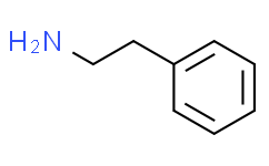
2-Phenylacetamine is a phenylethylamine compound with a phenyl substituent at the 2-position. It is found in humans, E. coli, and mice as a metabolite. It is a phenylethylamine, an aralkylamine, and an alkaloid, and is also the conjugate base of 2-phenylethylamineonium. Phenylacetamine is a metabolite found or produced in E. coli (K12 strain, MG1655 strain). It has been reported in Sedum berlandieri, Sedum lydium, and other organisms with relevant data. Phenylacetamine is a metabolite found or produced in Saccharomyces cerevisiae. See also: Phenylalanine (note moved here).
|
| Molecular Formula |
C8H11N
|
|---|---|
| Molecular Weight |
121.1796
|
| Exact Mass |
121.089
|
| CAS # |
64-04-0
|
| Related CAS # |
156-28-5 (hydrochloride);5471-08-9 (sulfate[2:1]);71750-39-5 (sulfate)
|
| PubChem CID |
1001
|
| Appearance |
Colorless to slightly yellow liquid
Liquid |
| LogP |
1.4
|
| Hydrogen Bond Donor Count |
1
|
| Hydrogen Bond Acceptor Count |
1
|
| Rotatable Bond Count |
2
|
| Heavy Atom Count |
9
|
| Complexity |
65
|
| Defined Atom Stereocenter Count |
0
|
| InChi Key |
BHHGXPLMPWCGHP-UHFFFAOYSA-N
|
| InChi Code |
InChI=1S/C8H11N/c9-7-6-8-4-2-1-3-5-8/h1-5H,6-7,9H2
|
| Chemical Name |
2-phenylethanamine
|
| HS Tariff Code |
2934.99.9001
|
| Storage |
Powder -20°C 3 years 4°C 2 years In solvent -80°C 6 months -20°C 1 month |
| Shipping Condition |
Room temperature (This product is stable at ambient temperature for a few days during ordinary shipping and time spent in Customs)
|
| Solubility (In Vitro) |
May dissolve in DMSO (in most cases), if not, try other solvents such as H2O, Ethanol, or DMF with a minute amount of products to avoid loss of samples
|
|---|---|
| Solubility (In Vivo) |
Note: Listed below are some common formulations that may be used to formulate products with low water solubility (e.g. < 1 mg/mL), you may test these formulations using a minute amount of products to avoid loss of samples.
Injection Formulations
Injection Formulation 1: DMSO : Tween 80: Saline = 10 : 5 : 85 (i.e. 100 μL DMSO stock solution → 50 μL Tween 80 → 850 μL Saline)(e.g. IP/IV/IM/SC) *Preparation of saline: Dissolve 0.9 g of sodium chloride in 100 mL ddH ₂ O to obtain a clear solution. Injection Formulation 2: DMSO : PEG300 :Tween 80 : Saline = 10 : 40 : 5 : 45 (i.e. 100 μL DMSO → 400 μLPEG300 → 50 μL Tween 80 → 450 μL Saline) Injection Formulation 3: DMSO : Corn oil = 10 : 90 (i.e. 100 μL DMSO → 900 μL Corn oil) Example: Take the Injection Formulation 3 (DMSO : Corn oil = 10 : 90) as an example, if 1 mL of 2.5 mg/mL working solution is to be prepared, you can take 100 μL 25 mg/mL DMSO stock solution and add to 900 μL corn oil, mix well to obtain a clear or suspension solution (2.5 mg/mL, ready for use in animals). View More
Injection Formulation 4: DMSO : 20% SBE-β-CD in saline = 10 : 90 [i.e. 100 μL DMSO → 900 μL (20% SBE-β-CD in saline)] Oral Formulations
Oral Formulation 1: Suspend in 0.5% CMC Na (carboxymethylcellulose sodium) Oral Formulation 2: Suspend in 0.5% Carboxymethyl cellulose Example: Take the Oral Formulation 1 (Suspend in 0.5% CMC Na) as an example, if 100 mL of 2.5 mg/mL working solution is to be prepared, you can first prepare 0.5% CMC Na solution by measuring 0.5 g CMC Na and dissolve it in 100 mL ddH2O to obtain a clear solution; then add 250 mg of the product to 100 mL 0.5% CMC Na solution, to make the suspension solution (2.5 mg/mL, ready for use in animals). View More
Oral Formulation 3: Dissolved in PEG400 (Please use freshly prepared in vivo formulations for optimal results.) |
| Preparing Stock Solutions | 1 mg | 5 mg | 10 mg | |
| 1 mM | 8.2522 mL | 41.2609 mL | 82.5219 mL | |
| 5 mM | 1.6504 mL | 8.2522 mL | 16.5044 mL | |
| 10 mM | 0.8252 mL | 4.1261 mL | 8.2522 mL |
*Note: Please select an appropriate solvent for the preparation of stock solution based on your experiment needs. For most products, DMSO can be used for preparing stock solutions (e.g. 5 mM, 10 mM, or 20 mM concentration); some products with high aqueous solubility may be dissolved in water directly. Solubility information is available at the above Solubility Data section. Once the stock solution is prepared, aliquot it to routine usage volumes and store at -20°C or -80°C. Avoid repeated freeze and thaw cycles.
Calculation results
Working concentration: mg/mL;
Method for preparing DMSO stock solution: mg drug pre-dissolved in μL DMSO (stock solution concentration mg/mL). Please contact us first if the concentration exceeds the DMSO solubility of the batch of drug.
Method for preparing in vivo formulation::Take μL DMSO stock solution, next add μL PEG300, mix and clarify, next addμL Tween 80, mix and clarify, next add μL ddH2O,mix and clarify.
(1) Please be sure that the solution is clear before the addition of next solvent. Dissolution methods like vortex, ultrasound or warming and heat may be used to aid dissolving.
(2) Be sure to add the solvent(s) in order.
Products are for research use only; We do not sell to patients
Copyright 2020 InvivoChem LLC | All Rights Reserved


