
| Size | Price | Stock | Qty |
|---|---|---|---|
| 100mg |
|
||
| 500mg |


| Targets |
RA-9's primary targets are proteasome-associated deubiquitinating enzymes (DUBs), specifically the 19S regulatory particle (19S RP)-associated DUBs including USP14 and UCHL5. It also inhibits other 19S RP-associated DUBs such as USP8, UCH-L1, and UCH-L3. By inhibiting these DUBs, RA-9 blocks the removal of ubiquitin chains from proteasome substrates, thereby preventing ubiquitin-dependent protein degradation. Importantly, RA-9 does not affect the 20S proteasome's catalytic core activity, distinguishing it from bortezomib and other classical proteasome inhibitors.
|
|---|---|
| ln Vitro |
The growth of primary cultures and cell lines of ovarian cancer is inhibited by RA-9 (10-30 μM; 48 hours) [1]. In ovarian cancer cells, RA-9 (1.25-5 μM; 18 hours) induces caspase-mediated apoptosis and cell cycle arrest [1]. In ovarian cancer cells, RA-9 (5 μM; 0-24 hours) causes endoplasmic reticulum stress [1]. As early as 8 hours after treatment, RA-9 (5 μM; over 24 hours) causes a time-dependent buildup of PARP cleavage formation [1].
In vitro, RA-9 inhibits proteasomal DUB activity at 20 μM while sparing all three 20S proteasome catalytic activities. It selectively induces apoptosis in ovarian cancer cell lines and primary cultures derived from donors. RA-9 triggers ER stress responses and activates the unfolded protein response (UPR) in ovarian cancer cells. It has demonstrated efficacy against bortezomib-resistant multiple myeloma cells, enabling resistance mechanism studies. The compound shows favorable toxicity profile and potent anticancer activity. |
| ln Vivo |
In a mouse model of ovarian cancer, RA-9 (5 mg/kg; intraperitoneally; one day on, two days off) prolongs survival in vivo and suppresses the development of human ovarian cancer cells [1].
In vivo, RA-9 has been validated in ES-2 ovarian cancer xenograft models with an intraperitoneal dosing regimen of 5 mg/kg administered on a one-day-on, two-days-off schedule. The compound has demonstrated therapeutic potential for ovarian cancer treatment in preclinical studies. Its mechanism of action involves selective apoptosis induction in cancer cells through the inhibition of 19S RP-associated DUB activity and the subsequent activation of ER stress responses. |
| Enzyme Assay |
The in vitro activity of RA-9 is assessed using cell-free DUB enzyme activity assays. In these assays, purified 19S proteasome-associated DUBs (e.g., USP14 or UCHL5) are incubated with a ubiquitin-AMC (7-amino-4-methylcoumarin) substrate in the presence of varying concentrations of RA-9. The cleavage of the ubiquitin-AMC substrate by the DUB releases free AMC, which produces a fluorescent signal measurable at excitation/emission wavelengths of 380/460 nm. The IC50 is determined as the concentration of RA-9 required to inhibit 50% of the DUB activity. Importantly, the selectivity of RA-9 for DUBs over 20S proteasome catalytic activities is confirmed by parallel assays measuring chymotrypsin-like, trypsin-like, and caspase-like activities using fluorogenic peptide substrates (e.g., Suc-LLVY-AMC, Boc-LRR-AMC, and Z-LLE-AMC) in the presence of the compound.
|
| Cell Assay |
Cell Viability Assay[1]
Cell Types: Cisplatin-sensitive ovarian cancer cell lines TOV-21G and ES-2, Cisplatin-resistant ovarian cancer cell lines HEY and OVCAR-3, primary ovarian cancer cells Tested Concentrations: 10, 20, 30 μM Incubation Duration: 48 hrs (hours) Experimental Results: Compromised the viability of ovarian cancer cells in a dose-dependent fashion. Cell Cycle Analysis[1] Cell Types: ES-2 cells Tested Concentrations: 1.25, 5 μM Incubation Duration: 18 hrs (hours) Experimental Results: Resulted in a dose -dependent increase in the fraction of ES-2 cells in the G2-M cell cycle phase. Western Blot Analysis[1] Cell Types: ES-2, SKOV-3 and TOV-21G ovarian cancer cells Tested Concentrations: 5 μM Incubation Duration: 0-24 h Experimental Results: Caused a time-dependent increase in the steady levels of the early ER-stress marker GRP-78, as well as the late ER-stress markers IRE1-α and Ero1L-α. For cellular assays, various cancer cell lines including ovarian cancer cells (e.g., ES-2, Caov-3, A2780) and bortezomib-resistant multiple myeloma cells are cultured in appropriate media (e.g., RPMI-1640 or DMEM with 10% fetal bovine serum). Cells are treated with various concentrations of RA-9 (typically ranging from 0.1 to 50 μM) for different time periods (e.g., 24-72 hours). Cell viability is assessed using the MTT or CellTiter-Glo assay. Apoptosis is evaluated by measuring caspase-3/7 activity, by flow cytometry using Annexin V/PI staining, or by Western blotting for cleaved caspases and PARP. The accumulation of ubiquitinated proteins is assessed by Western blotting using an anti-ubiquitin antibody. ER stress markers such as BiP, CHOP, and phosphorylated eIF2α are analyzed by Western blotting. |
| Animal Protocol |
Animal/Disease Models: Sixweeks old female immunodeficient (NCr nu/nu) mice[1]
Doses: 5 mg/kg Route of Administration: Ip; one-day on, two-days off Experimental Results: Significant reduction in tumor burden at day 12. In vivo, RA-9 is typically administered to mice bearing human tumor xenografts via intraperitoneal injection. The compound is formulated in a suitable vehicle (e.g., DMSO or a mixture of DMSO and saline) and administered at a dose of 5 mg/kg on a one-day-on, two-days-off schedule. Tumor growth is monitored by measuring tumor volume over time. At the end of the study, tumors are excised and analyzed for ubiquitinated protein accumulation, apoptosis markers, and ER stress markers to confirm target engagement and mechanism of action. Pharmacodynamic studies can be performed by collecting tumor tissue at various time points post-administration. |
| ADME/Pharmacokinetics |
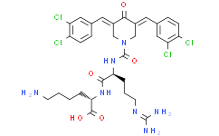
RA-9 has a molecular weight of 741.49 g/mol and a molecular formula of C32H37Cl4N7O5. It is soluble in DMSO and should be stored in a dry, dark place at 0-4°C for short-term storage (days to weeks) or at -20°C for long-term storage (months to years). The compound is available as a solid powder. For in vivo administration, it can be formulated in appropriate vehicles.
|
| Toxicity/Toxicokinetics |
RA-9 exhibits a favorable toxicity profile in preclinical studies. Unlike bortezomib and other classical proteasome inhibitors that directly target 20S proteasome catalytic activity and can cause significant toxicity, RA-9 selectively inhibits 19S RP-associated DUBs while preserving 20S proteasome proteolytic activity. This selectivity may contribute to a more favorable safety profile. In vitro studies have shown that RA-9 selectively induces apoptosis in cancer cells while sparing normal cells. Comprehensive toxicological studies are required to establish its full safety profile.
|
| References | |
| Additional Infomation |
RA-9 is a research compound developed as a selective inhibitor of 19S proteasome-associated deubiquitinating enzymes (DUBs). Its unique mechanism of action—blocking ubiquitin-dependent protein degradation without affecting 20S proteasome catalytic activity—distinguishes it from both classical proteasome inhibitors (e.g., bortezomib) and broad-spectrum DUB inhibitors. RA-9 has demonstrated therapeutic potential for ovarian cancer and has shown efficacy against bortezomib-resistant multiple myeloma cells. It is not approved for clinical use and is intended for research purposes only. Its development has provided valuable insights into the role of 19S RP-associated DUBs in cancer biology and the potential of targeting these enzymes for cancer therapy.
|
| Molecular Formula |
C32H37CL4N7O5
|
|---|---|
| Molecular Weight |
741.492083311081
|
| Exact Mass |
739.161
|
| CAS # |
1262295-74-8
|
| Appearance |
Light yellow to yellow solid powder
|
| SMILES |
ClC1=C(C=CC(=C1)/C=C1/C(/C(=C/C2C=CC(=C(C=2)Cl)Cl)/CN(C(N[C@H](C(N[C@H](C(=O)O)CCCCN)=O)CCC/N=C(\N)/N)=O)C/1)=O)Cl
|
| Synonyms |
RA9; RA 9; RA-9
|
| HS Tariff Code |
2934.99.9001
|
| Storage |
Powder -20°C 3 years 4°C 2 years In solvent -80°C 6 months -20°C 1 month |
| Shipping Condition |
Room temperature (This product is stable at ambient temperature for a few days during ordinary shipping and time spent in Customs)
|
| Solubility (In Vitro) |
May dissolve in DMSO (in most cases), if not, try other solvents such as H2O, Ethanol, or DMF with a minute amount of products to avoid loss of samples
|
|---|---|
| Solubility (In Vivo) |
Note: Listed below are some common formulations that may be used to formulate products with low water solubility (e.g. < 1 mg/mL), you may test these formulations using a minute amount of products to avoid loss of samples.
Injection Formulations
Injection Formulation 1: DMSO : Tween 80: Saline = 10 : 5 : 85 (i.e. 100 μL DMSO stock solution → 50 μL Tween 80 → 850 μL Saline)(e.g. IP/IV/IM/SC) *Preparation of saline: Dissolve 0.9 g of sodium chloride in 100 mL ddH ₂ O to obtain a clear solution. Injection Formulation 2: DMSO : PEG300 :Tween 80 : Saline = 10 : 40 : 5 : 45 (i.e. 100 μL DMSO → 400 μLPEG300 → 50 μL Tween 80 → 450 μL Saline) Injection Formulation 3: DMSO : Corn oil = 10 : 90 (i.e. 100 μL DMSO → 900 μL Corn oil) Example: Take the Injection Formulation 3 (DMSO : Corn oil = 10 : 90) as an example, if 1 mL of 2.5 mg/mL working solution is to be prepared, you can take 100 μL 25 mg/mL DMSO stock solution and add to 900 μL corn oil, mix well to obtain a clear or suspension solution (2.5 mg/mL, ready for use in animals). View More
Injection Formulation 4: DMSO : 20% SBE-β-CD in saline = 10 : 90 [i.e. 100 μL DMSO → 900 μL (20% SBE-β-CD in saline)] Oral Formulations
Oral Formulation 1: Suspend in 0.5% CMC Na (carboxymethylcellulose sodium) Oral Formulation 2: Suspend in 0.5% Carboxymethyl cellulose Example: Take the Oral Formulation 1 (Suspend in 0.5% CMC Na) as an example, if 100 mL of 2.5 mg/mL working solution is to be prepared, you can first prepare 0.5% CMC Na solution by measuring 0.5 g CMC Na and dissolve it in 100 mL ddH2O to obtain a clear solution; then add 250 mg of the product to 100 mL 0.5% CMC Na solution, to make the suspension solution (2.5 mg/mL, ready for use in animals). View More
Oral Formulation 3: Dissolved in PEG400 (Please use freshly prepared in vivo formulations for optimal results.) |
| Preparing Stock Solutions | 1 mg | 5 mg | 10 mg | |
| 1 mM | 1.3486 mL | 6.7432 mL | 13.4864 mL | |
| 5 mM | 0.2697 mL | 1.3486 mL | 2.6973 mL | |
| 10 mM | 0.1349 mL | 0.6743 mL | 1.3486 mL |
*Note: Please select an appropriate solvent for the preparation of stock solution based on your experiment needs. For most products, DMSO can be used for preparing stock solutions (e.g. 5 mM, 10 mM, or 20 mM concentration); some products with high aqueous solubility may be dissolved in water directly. Solubility information is available at the above Solubility Data section. Once the stock solution is prepared, aliquot it to routine usage volumes and store at -20°C or -80°C. Avoid repeated freeze and thaw cycles.
Calculation results
Working concentration: mg/mL;
Method for preparing DMSO stock solution: mg drug pre-dissolved in μL DMSO (stock solution concentration mg/mL). Please contact us first if the concentration exceeds the DMSO solubility of the batch of drug.
Method for preparing in vivo formulation::Take μL DMSO stock solution, next add μL PEG300, mix and clarify, next addμL Tween 80, mix and clarify, next add μL ddH2O,mix and clarify.
(1) Please be sure that the solution is clear before the addition of next solvent. Dissolution methods like vortex, ultrasound or warming and heat may be used to aid dissolving.
(2) Be sure to add the solvent(s) in order.
Products are for research use only; We do not sell to patients
Copyright 2020 InvivoChem LLC | All Rights Reserved


