| Size | Price | Stock | Qty |
|---|---|---|---|
| 50mg |
|
||
| 100mg |
|
||
| 250mg |
|
||
| 500mg | |||
| Other Sizes |


| Targets |
CYP51; fungicidal
|
|---|---|
| ln Vitro |
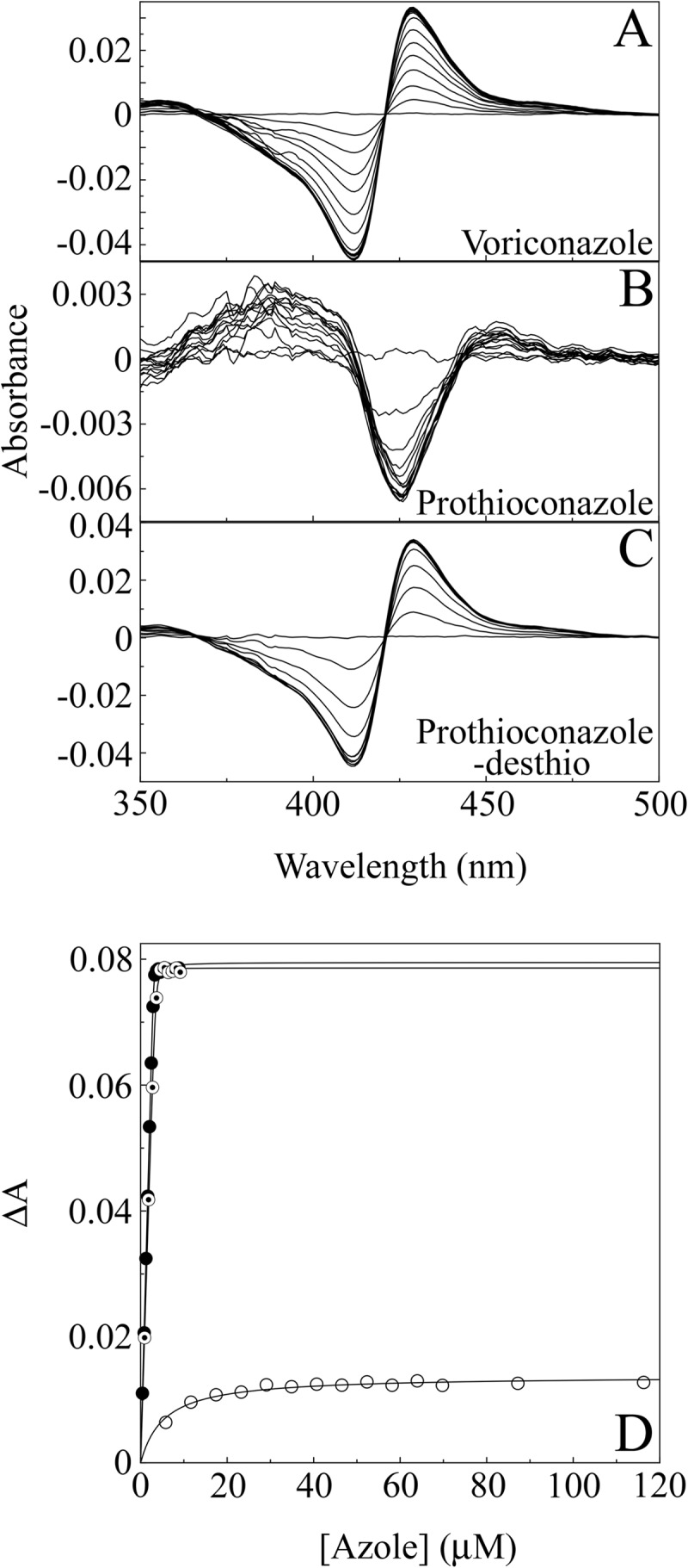
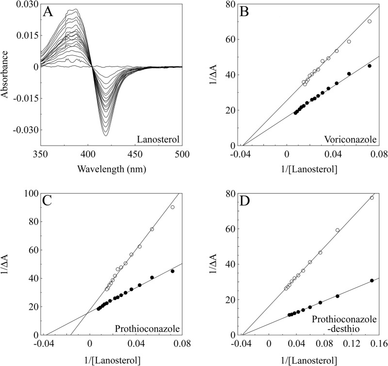
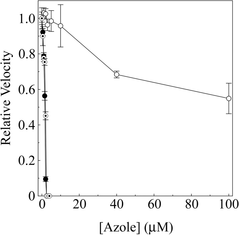
Prothioconazole is a new triazolinthione fungicide used in agriculture. We have used Candida albicans CYP51 (CaCYP51) to investigate the in vitro activity of prothioconazole and to consider the use of such compounds in the medical arena. Treatment of C. albicans cells with prothioconazole, prothioconazole-desthio, and voriconazole resulted in CYP51 inhibition, as evidenced by the accumulation of 14α-methylated sterol substrates (lanosterol and eburicol) and the depletion of ergosterol. We then compared the inhibitor binding properties of prothioconazole, prothioconazole-desthio, and voriconazole with CaCYP51. We observed that prothioconazole-desthio and voriconazole bind noncompetitively to CaCYP51 in the expected manner of azole antifungals (with type II inhibitors binding to heme as the sixth ligand), while prothioconazole binds competitively and does not exhibit classic inhibitor binding spectra. Inhibition of CaCYP51 activity in a cell-free assay demonstrated that prothioconazole-desthio is active, whereas prothioconazole does not inhibit CYP51 activity. Extracts from C. albicans grown in the presence of prothioconazole were found to contain prothioconazole-desthio. We conclude that the antifungal action of prothioconazole can be attributed to prothioconazole-desthio [1].
|
| Enzyme Assay |
Antifungal binding determinations. [1]
The chemical structures of the azoles used in this study are shown in Fig. 1. Binding of azole to CaCYP51 was performed as previously described. A stock 2 mg · ml−1 solution of Prothioconazole, a 0.2 mg · ml−1 solution of prothioconazole-desthio, and a 0.1 mg · ml−1 solution of voriconazole were prepared in DMF. Azoles were progressively titrated against 5 μM CaCYP51 in 0.1 M Tris-HCl (pH 8.1) and 25% (wt/vol) glycerol, with the difference spectra between 500 and 350 nm determined after each addition. Azole binding determinations were performed in triplicate for each compound. Binding saturation curves were constructed from ΔApeak-trough against the azole concentration. A rearrangement of the Morrison equation was used to determine the dissociation constant (Kd) values when ligand binding was “tight.” Tight binding is observed when the Kd for azole is similar to or lower than the concentration of CYP51 present. The Michaelis-Menten equation was used when the ligand binding was not tight. The Kd values reported are the mean values from three replicates along with the associated standard deviations. Substrate binding studies. [1] A 0.1% (wt/vol) aqueous solution of lanosterol in 0.5% (vol/vol) Tween 80 was prepared as previously described. Lanosterol was progressively titrated against 10 μM CaCYP51 in the sample cuvette with equivalent amounts of 0.5% (vol/vol) Tween 80 added to the P450-containing reference cuvette. The absorbance difference spectrum between 500 and 350 nm was determined after each incremental addition of lanosterol, and binding saturation curves were constructed from the ΔA385–419, including corrections for changes in sample volume. The substrate binding constant (Ks) was determined by nonlinear regression (Levenberg-Marquardt algorithm) using the Michaelis-Menten equation. Ks values reported for lanosterol are the mean values from three replicates along with the associated standard deviations. The spin-state change of CaCYP51 was calculated from the ΔA385–419 value using an extinction coefficient of 118 mM−1 · cm−1 derived for the type I difference spectrum of CYP164A2 which was modulated from 100% low spin to nearly 100% high spin by physicochemical means. Lanosterol binding difference spectra were determined with 10 μM CaCYP51 in the presence and absence of 4 μM voriconazole, 100 μM Prothioconazole, and 4 μM prothioconazole-desthio. Negative-control determinations were made in the presence of 1.25% (vol/vol) DMF. Determinations were performed in triplicate, and Lineweaver-Burk plots were constructed from resultant CaCYP51 substrate binding spectra. CYP51 reconstitution assays and IC50 determinations. [1] The CYP51 enzyme reconstitution system previously described containing 2.5 μM CaCYP51 and 10 μM truncated yeast cytochrome P450 reductase was used. The reaction was terminated by the addition of 2 ml 15% (wt/vol) KOH in ethanol followed by incubation at 85°C for 90 min. IC50 determinations were performed by adding various concentrations of voriconazole, Prothioconazole, and prothioconazole-desthio in 5 μl of DMF prior to incubation at 37°C and the addition of β-NADPH-Na4. Sterol substrates and products were extracted and analyzed by GC/MS as described below. |
| Cell Assay |
Antifungal treatment of cells. [1]
C. albicans cells were grown overnight in RPMI 1640 l-glutamine. A starting concentration of 1 × 10³ cells/ml was used to inoculate RPMI medium containing 4 μg · ml−1 Prothioconazole, 4 μg · ml−1 prothioconazole-desthio, or 1 μg · ml−1 voriconazole (all with a final concentration of 1% DMSO) and a control containing 1% DMSO (untreated). Cultures were incubated at 37°C and 200 rpm overnight and cells harvested and washed twice with deionized water prior to sterol extraction. Solid-phase extraction of antifungals from Candida albicans cells and growth media. [1] A 100-μl aliquot of 1 × 10⁷ cells/ml from overnight cultures of C. albicans (ATCC SC5314) was subcultured into 250 ml RPMI l-glutamine and yeast extract-peptone-dextrose (YPD) broth containing 8 μg · ml−1 Prothioconazole with a final concentration of 1% (vol/vol) DMSO (and controls without any prothioconazole) and incubated at 37°C and 200 rpm for 24 h. Cells were harvested and washed three times with deionized water before sonication (60-s bursts and 60-s rests for 10 min) in 99:1 methanol:acetic acid. Samples were then dried in a vacuum centrifuge and resuspended in 20% (vol/vol) methanol. Sep-Pak cartridges (Vac 3 cc, 200 mg) were prepared by washing with 6 ml of methanol and subsequently 6 ml of 20% (vol/vol) methanol. Extracts were each applied to a cartridge which was then washed with 4 sequential 1-ml volumes of 40%, 60%, 80%, and 100% (vol/vol) methanol. The eluent was collected for each wash. Extracts from the media used for the growth of C. albicans with prothioconazole, control media containing Prothioconazole and no cells, and sterile deionized water with prothioconazole and no cells were prepared in a manner similar to that described for the cell extractions. Methanol was added to samples to reach a final concentration of 20% (vol/vol) (standards were diluted in 20% [vol/vol] methanol) prior to solid-phase extraction. Identification of Prothioconazole and prothioconazole-desthio. [1] Samples were analyzed by ESI-MS and -MS/MS on a Q-Tof Ultima spectrometer in positive-ion mode. Samples were electrosprayed from nano-ESI metal-coated capillaries, and collision-induced dissociation was achieved with argon gas. The following instrument parameters were used: capillary voltage, 1.8 kV; collision voltage, 8 V for MS (25 V for MS/MS); m/z range, 50 to 1,000; scan time, 2.4 s/scan; MCP detector voltage, 2,100 V. The instrument was calibrated immediately prior to use with 1 pmol/μl Glu-1-Fibrinopeptide B. The data obtained were processed using the MassLynx V4.1 software package. Prothioconazole and prothioconazole-desthio were identified by their measured mass and by their fragmentation patterns obtained through MS/MS. Both prothioconazole and prothioconazole-desthio standards were found to be most abundant in the 80% (vol/vol) methanol eluent; therefore, analysis of extractions was performed by comparing the 80% (vol/vol) methanol eluents of all samples. |
| ADME/Pharmacokinetics |
Absorption, Distribution and Excretion
Following a single oral administration of a low-dose prothioconazole, the absorption rate (triazole-labeled) in male rats was approximately 94%. Extrapolating from the 48-hour excretion process of the triazole-labeled prothioconazole, the 48-hour absorption rate of the phenyl-labeled prothioconazole was estimated to be approximately 90%. Plasma radioactivity time-course data showed rapid absorption after a single oral administration of the low dose, with peak plasma concentrations in both male and female rats occurring between 0.33 and 0.66 hours post-administration. After a single oral administration of a high dose, peak plasma concentrations in both male and female rats occurred between 0.66 and 1.00 hours post-administration. The absorption rate of phenyl-labeled prothioconazole was slightly faster; after a single oral administration of a low dose in male rats, peak plasma concentrations occurred between 0.16 and 0.33 hours; after repeated oral administration of the low dose in both male and female rats, peak plasma concentrations occurred at 0.16 hours. The fluctuations in plasma concentration curves indicate that the radioactive material underwent enterohepatic circulation. This effect was more pronounced in female rats. Absorption was slightly delayed in female rats compared to male rats. 168 hours after a single oral low-dose administration, residual radioactivity was low in rats. For the triazole-labeled substance, 1.5% of the administered dose was recovered in tissues and carcasses of male rats, and 0.4% in female rats. The highest tissue concentrations were found in the liver, carcass, and gastrointestinal tract. Residual radioactivity levels ranged from 0.0004% to 0.07% in all other tissues examined. For the phenyl-labeled substance (used only in males), 5.8% of the administered dose was recovered in tissues and carcasses. The highest levels of radioactivity were found in the gastrointestinal tract, liver, and carcass. Residual radioactivity levels ranged from 0.0001% to 0.05% in all other tissues examined. 48 hours after repeated oral administration of the low-dose, residual radioactivity was also low in rats, with 3.8% of the administered dose recovered in tissues and carcasses of male rats and 0.8% in female rats. The highest levels of radioactivity were found in the liver, gastrointestinal tract, and carcass. In all other tissues examined, residual radioactivity levels ranged from 0.0002% to 0.05%. Systemic and liver cumulative doses were consistently higher in male rats than in female rats. 168 hours after a single oral high-dose administration, residual radioactivity in rats was also low, with 0.11% of the administered dose recovered from the cadaver and tissues of both male and female rats. Systemic radioactivity cumulative doses were roughly the same in male and female rats, but liver radioactivity cumulative doses were higher in male rats. The primary route of excretion for both markers and in both sexes was feces. Following a single oral low-dose (triazole-labeled) administration, the overall recovery rate in both sexes was approximately 94-95% of the administered dose, with 10% excreted in urine in male rats and 16% in female rats, and 84% in feces in male rats and 78% in female rats. In the phenyl-labeled group (male rats only), 5% of the administered dose was excreted in urine and 85% in feces, with an overall recovery rate of approximately 90%. In the triazole-labeled group (males only), approximately 90% of the administered dose was excreted in bile within 24–48 hours. In the phenyl-labeled group (males only), approximately 81% of the administered dose was excreted in bile after 24 hours, and 93% after 48 hours. In whole-body autoradiography distribution studies, peak concentrations were reached 1 hour after administration in males and continued to decline until sacrifice at 168 hours. Absorption was slightly delayed in females, with some tissues reaching peak concentrations at 8 hours after administration. The highest drug concentrations were found in the liver (up to 1.78 g/g in males and 0.97 μg/g in females), followed by the kidneys (renal medulla, up to 0.64 g/g), brown/perirhinal fat (up to 0.36 μg/g), thyroid gland (up to 0.23 μg/g), and adrenal glands (up to 0.27 μg/g). Peak concentrations in all other tissues were below 0.13 μg/g. Radioactivity levels decreased rapidly within 24 to 168 hours after administration, indicating sustained clearance of the drug from tissues. Metabolisms/Metabolites Following oral administration of prothioconazole, extensive metabolism occurred in rats. Eighteen metabolites and the parent compound were identified in urine, feces, and bile. Biotransformation of prothioconazole primarily involved three reaction types: desulfurization, phenyl oxidative hydroxylation, and glucuronide conjugation. Metabolite identification rates ranged from 26% to 63% of the administered dose. Higher metabolite separation and identification rates were not achievable due to difficulties in fecal extraction, as 67% to 79% of the administered dose remained in unextractable solid residues. The primary route of excretion for prothioconazole was feces, accounting for 22% to 53% of the administered dose. The parent compound, prothioconazole, was most abundant in feces (1% to 22% of the administered dose), followed by prothioconazole desulfurization metabolites (3% to 16% of the administered dose). All other fecal metabolites were present at less than 7% of the administered dose. No 1,2,4-triazole metabolites were detected in feces. The main urinary metabolite is albendazole-S- or O-glucuronide (0.1-8% of the administered dose), and it is primarily excreted in women. Following a single oral low- or/or high-dose administration, 1,2,4-triazole metabolites account for 0.8-2.3% of the administered dose. The remaining urinary metabolites account for 0-1.4% of the administered dose. Albendazole-S- or O-glucuronide is the most abundant metabolite in bile, accounting for approximately 46% of the administered dose. This metabolite is excreted only in the urine of women, but it has also been found in the bile of men. Glucuronide metabolites in bile account for 8-10% of the administered dose. The parent compound accounts for 3-5% of the administered dose. No 1,2,4-triazole metabolites were detected in bile. In a metabolic study, the absorption, distribution, metabolism, and excretion of albendazole desulfurization were investigated. Radioactive test material was absorbed from the gastrointestinal tract as early as 4 minutes after administration. The maximum concentration observed at 1.5 hours was 0.052 μg/g. The highest radioactivity was observed in the liver and gastrointestinal tract, likely due to continuous enterohepatic circulation. Radioactivity levels in other tissues were less than 1%. Over 90% of the administered radioactive dose was excreted via bile and urine. Very little radioactive material was recovered in exhaled carbon dioxide. Most of the administered radioactive dose was excreted via feces, with a small amount excreted via urine. Only trace amounts of recovered radioactive material were found in the skin, while the carcass and gastrointestinal tract contained up to 4% and 2.25% radioactive material, respectively. At 48 hours, excretion of the radioactive material may not be complete, as the total residual radioactivity in the body at this time is 5% to 6% of the administered dose. The elimination half-life was 44.3 hours, and the mean residence time was 48.2 hours. These observations indicate a slow redistribution of radioactive material into the plasma before elimination, consistent with a total clearance of 10.9 mL/min/kg body weight and a renal clearance of 1.4 mL/min/kg body weight. In bile duct cannulated animals, 84% to 85% of the administered dose was detected in bile 24 and 48 hours after intraduodenal administration. In these animals, urinary excretion accounted for nearly 6% of the administered dose after 48 hours, while fecal excretion accounted for 2%. Eighteen radioactive HPLC peaks were observed in mixed bile samples, representing 84.3% of the administered dose. Five compounds were isolated and identified, while the remaining 13 metabolites were unidentified, representing 44.7% of the administered dose. /Prothioconazole Desulfurization/ Rapid absorption of the test substance was confirmed in whole-body autoradiography. Absorption was not complete after one hour. Autoradiography also showed lower blood concentrations than in adipose tissue, suggesting that prothioconazole desulfurization and its metabolites may be lipophilic. Radioactivity was observed in the gastric mucosa throughout the observation period, considered an indication of the absorbed radioactive material being secreted back into the gastric lumen via bile. Mineral fractions of muscle, heart, lungs, brain, thyroid, and bones showed trace amounts of radioactivity. The radioactivity distribution pattern in the testes indicates the presence of blood circulation within the organ. Moderate levels of radioactivity were observed in some glandular organs, including the prepuce and adrenal glands. Increased radioactivity was also observed in the gingiva, but its physiological significance is unclear. The radioactivity distribution observed at 1 hour remained largely consistent over 48 hours, although it decreased due to excretion. The radioactivity content in the renal cortex was much higher than that in the renal pelvis, indicating that the radioactive material was reabsorbed in the duodenum. Furthermore, the absorbed radioactive material may not have been converted into metabolites of sufficiently high polarity to be excreted through the kidneys. This resulted in the radioactive test material passing more through the liver. /Prothioconazole Desulfurization/ For more metabolite/metabolite (complete) data on prothioconazole (6 metabolites in total), please visit the HSDB record page. Biological Half-Life The elimination half-life is 44.3 hours. ... /Prothioconazole Desulfurization/ |
| Toxicity/Toxicokinetics |
Non-Human Toxicity Values
Oral LD50 in rats (male and female) ≥ 6200 mg/kg (prothioconazole technical grade) Rainbow trout LC50 1.83 mg/L/96 hours Inhalation LC50 in rats > 4990 mg/m³ / Duration unspecified/ Dermal LD50 in rats (male and female) ≥ 2000 mg/kg |
| References | |
| Additional Infomation |
2-[2-(1-chlorocyclopropyl)-3-(2-chlorophenyl)-2-hydroxypropyl]-1,2-dihydro-1,2,4-triazol-3-thione is a triazole compound with the structure 1,2,4-triazol-3-thione substituted at the 2-position with 2-(1-chlorocyclopropyl)-3-(2-chlorophenyl)-2-hydroxypropyl. It belongs to the monochlorobenzene, triazole, tertiary alcohol, cyclopropane, and thiocarbonyl compounds.
Mechanism of Action Prothioconazole is a systemic demethylation inhibitor fungicide, belonging to the triazolinethion fungicide class. ...It combats susceptible fungi by inhibiting the demethylation at the 14-position of lanosterol or 24-methylenedihydrosterol, both precursors of fungal sterols; that is, it works by interfering with the biosynthesis of ergosterol (a precursor of vitamin D2 and an important component of the fungal cell wall). Compared to desulfurized prothioconazole, prothioconazole exhibits weaker inhibitory activity against CaCYP51 in vitro, prompting further investigation into its mechanism of action. Studies have found prothioconazole desulfurization in both cells and culture media of Candida albicans treated with prothioconazole. Therefore, the antifungal effect observed during prothioconazole treatment is due to the presence of its desulfurization product. Our results also indicate that desulfurization analogs were detected in YPD and RPMI media containing prothioconazole, but not in sterile deionized water containing prothioconazole after 24 hours of incubation at 37°C. Therefore, prothioconazole readily converts to its desulfurized form and exerts its antifungal effect. It can be inferred that the high efficiency of prothioconazole as an agricultural fungicide also benefits from the metabolism of the relatively inactive prothioconazole in pathogenic fungal cells into a highly active desulfurized form, as well as its metabolism in host cells. Further research on triazolinethion compounds may help develop such novel antifungal compounds for clinical treatment or as more effective crop protection compounds, and promote consideration of the value of fungicide precursors or prodrug strategies. [1] |
| Molecular Formula |
C14H15CL2N3OS
|
|---|---|
| Molecular Weight |
344.25
|
| Exact Mass |
343.031
|
| Elemental Analysis |
C, 48.85; H, 4.39; Cl, 20.60; N, 12.21; O, 4.65; S, 9.31
|
| CAS # |
178928-70-6
|
| PubChem CID |
6451142
|
| Appearance |
White to light yellow solid powder
|
| Density |
1.5±0.1 g/cm3
|
| Boiling Point |
486.7±55.0 °C at 760 mmHg
|
| Melting Point |
139.1-144.5°
|
| Flash Point |
248.2±31.5 °C
|
| Vapour Pressure |
0.0±1.3 mmHg at 25°C
|
| Index of Refraction |
1.698
|
| LogP |
1.77
|
| Hydrogen Bond Donor Count |
2
|
| Hydrogen Bond Acceptor Count |
2
|
| Rotatable Bond Count |
5
|
| Heavy Atom Count |
21
|
| Complexity |
458
|
| Defined Atom Stereocenter Count |
0
|
| InChi Key |
MNHVNIJQQRJYDH-UHFFFAOYSA-N
|
| InChi Code |
InChI=1S/C14H15Cl2N3OS/c15-11-4-2-1-3-10(11)7-14(20,13(16)5-6-13)8-19-12(21)17-9-18-19/h1-4,9,20H,5-8H2,(H,17,18,21)
|
| Chemical Name |
2-[2-(1-chlorocyclopropyl)-3-(2-chlorophenyl)-2-hydroxypropyl]-1H-1,2,4-triazole-3-thione
|
| Synonyms |
Proline 480 SC Fungicide; JAU 6476; Prothioconazole; 178928-70-6; 3H-1,2,4-Triazole-3-thione, 2-[2-(1-chlorocyclopropyl)-3-(2-chlorophenyl)-2-hydroxypropyl]-1,2-dihydro-; JAU 6476; Prothioconazole [ISO:BSI]; 2-[2-(1-Chlorocyclopropyl)-3-(2-chlorophenyl)-2-hydroxypropyl]-1,2-dihydro-3H-1,2,4-triazole-3-thione; UNII-27B9FV58IY; PROLINE 480 SC Fungicide; Prothioconazole
|
| HS Tariff Code |
2934.99.9001
|
| Storage |
Powder -20°C 3 years 4°C 2 years In solvent -80°C 6 months -20°C 1 month |
| Shipping Condition |
Room temperature (This product is stable at ambient temperature for a few days during ordinary shipping and time spent in Customs)
|
| Solubility (In Vitro) |
May dissolve in DMSO (in most cases), if not, try other solvents such as H2O, Ethanol, or DMF with a minute amount of products to avoid loss of samples
|
|---|---|
| Solubility (In Vivo) |
Note: Listed below are some common formulations that may be used to formulate products with low water solubility (e.g. < 1 mg/mL), you may test these formulations using a minute amount of products to avoid loss of samples.
Injection Formulations
Injection Formulation 1: DMSO : Tween 80: Saline = 10 : 5 : 85 (i.e. 100 μL DMSO stock solution → 50 μL Tween 80 → 850 μL Saline)(e.g. IP/IV/IM/SC) *Preparation of saline: Dissolve 0.9 g of sodium chloride in 100 mL ddH ₂ O to obtain a clear solution. Injection Formulation 2: DMSO : PEG300 :Tween 80 : Saline = 10 : 40 : 5 : 45 (i.e. 100 μL DMSO → 400 μLPEG300 → 50 μL Tween 80 → 450 μL Saline) Injection Formulation 3: DMSO : Corn oil = 10 : 90 (i.e. 100 μL DMSO → 900 μL Corn oil) Example: Take the Injection Formulation 3 (DMSO : Corn oil = 10 : 90) as an example, if 1 mL of 2.5 mg/mL working solution is to be prepared, you can take 100 μL 25 mg/mL DMSO stock solution and add to 900 μL corn oil, mix well to obtain a clear or suspension solution (2.5 mg/mL, ready for use in animals). View More
Injection Formulation 4: DMSO : 20% SBE-β-CD in saline = 10 : 90 [i.e. 100 μL DMSO → 900 μL (20% SBE-β-CD in saline)] Oral Formulations
Oral Formulation 1: Suspend in 0.5% CMC Na (carboxymethylcellulose sodium) Oral Formulation 2: Suspend in 0.5% Carboxymethyl cellulose Example: Take the Oral Formulation 1 (Suspend in 0.5% CMC Na) as an example, if 100 mL of 2.5 mg/mL working solution is to be prepared, you can first prepare 0.5% CMC Na solution by measuring 0.5 g CMC Na and dissolve it in 100 mL ddH2O to obtain a clear solution; then add 250 mg of the product to 100 mL 0.5% CMC Na solution, to make the suspension solution (2.5 mg/mL, ready for use in animals). View More
Oral Formulation 3: Dissolved in PEG400 (Please use freshly prepared in vivo formulations for optimal results.) |
| Preparing Stock Solutions | 1 mg | 5 mg | 10 mg | |
| 1 mM | 2.9049 mL | 14.5243 mL | 29.0487 mL | |
| 5 mM | 0.5810 mL | 2.9049 mL | 5.8097 mL | |
| 10 mM | 0.2905 mL | 1.4524 mL | 2.9049 mL |
*Note: Please select an appropriate solvent for the preparation of stock solution based on your experiment needs. For most products, DMSO can be used for preparing stock solutions (e.g. 5 mM, 10 mM, or 20 mM concentration); some products with high aqueous solubility may be dissolved in water directly. Solubility information is available at the above Solubility Data section. Once the stock solution is prepared, aliquot it to routine usage volumes and store at -20°C or -80°C. Avoid repeated freeze and thaw cycles.
Calculation results
Working concentration: mg/mL;
Method for preparing DMSO stock solution: mg drug pre-dissolved in μL DMSO (stock solution concentration mg/mL). Please contact us first if the concentration exceeds the DMSO solubility of the batch of drug.
Method for preparing in vivo formulation::Take μL DMSO stock solution, next add μL PEG300, mix and clarify, next addμL Tween 80, mix and clarify, next add μL ddH2O,mix and clarify.
(1) Please be sure that the solution is clear before the addition of next solvent. Dissolution methods like vortex, ultrasound or warming and heat may be used to aid dissolving.
(2) Be sure to add the solvent(s) in order.
|
|
|
Products are for research use only; We do not sell to patients
Copyright 2020 InvivoChem LLC | All Rights Reserved