| Size | Price | Stock | Qty |
|---|---|---|---|
| 5mg |
|
||
| 10mg |
|
||
| 25mg |
|
||
| 50mg |
|
||
| 100mg |
|
||
| 250mg |
|
||
| 500mg |
|
||
| Other Sizes |


Purity: ≥98%
| Targets |
Androgen receptor (AR) (Ki = 0.9 nM; EC50 for transcriptional activation = 3.6 nM, efficacy 134% relative to DHT)[1]
Mineralocorticoid receptor (MR) (Ki = 1261 nM; maximal efficacy 10% relative to aldosterone; EC50 = 3695 nM)[1] Glucocorticoid receptor (GR) (Ki = 581 nM; maximal efficacy 1% relative to dexamethasone)[1] Progesterone receptor (PR) (Ki = 136 nM; maximal efficacy 38% relative to progesterone; EC50 = 2233 nM)[1] Estrogen receptor α (ERα) (maximal efficacy 1% relative to 17β-estradiol)[1] LGD-3303 targets the androgen receptor (AR) with high affinity (Ki = 0.9 nM). It potently activates transcription through the androgen receptor with an EC50 of 3.6 nM and has 134% efficacy relative to the steroidal androgen DHT. The compound shows little or no cross-reactivity with related nuclear receptors. |
|---|---|
| ln Vitro |
LGD‐3303 is a nonsteroidal, nonaromatizable androgen receptor ligand that binds to the androgen receptor with high affinity in a radiolabeled to competitive binding assay (Ki = 0.9 nM). LGD‐3303 binds to the mineralocorticoid, glucocorticoid, and progesterone receptors with greatly reduced affinity in comparison with the androgen receptor (Ki = 1261, 581, and 136 nM, respectively). LGD‐3303 potently activates transcription through the androgen receptor (EC50 = 3.6 nM) and has 134% efficacy relative to the steroidal androgen DHT. LGD‐3303 has minimal activity on other related nuclear hormone receptors in a transcriptional activity assay. Maximal efficacy relative to the natural ligands was determined to be 10% for the mineralocorticoid receptor, 1% for the glucocorticoid receptor, 1% for the estrogen receptor α, and 38% for the progesterone receptor. Potency could only be determined for the mineralocorticoid receptor (EC50 = 3695 nM) and the progesterone receptor (EC50 = 2233 nM).
Kinase Assay: LGD‐3303 is a nonsteroidal, nonaromatizable androgen receptor ligand that binds to the androgen receptor with high affinity in a radiolabeled to competitive binding assay (Ki = 0.9 nM). LGD‐3303 binds to the mineralocorticoid, glucocorticoid, and progesterone receptors with greatly reduced affinity in comparison with the androgen receptor (Ki = 1261, 581, and 136 nM, respectively). LGD‐3303 potently activates transcription through the androgen receptor (EC50 = 3.6 nM) and has 134% efficacy relative to the steroidal androgen DHT. Cell Assay: Data were collected at 10 Hz from the load cell and the cross‐head displacement and analyzed using software designed for materials testing (TestWorks 4; MTS). Whole femurs were tested to failure in three‐point bending, and the fifth lumbar vertebral body was tested to failure in compression after removal of the endplates and spinous processes. The preparation and testing of the vertebral body have been previously described. Maximum load, stiffness, and energy absorption were measured from the load‐deformation curve for each specimen. Elastic modulus, maximum stress, and toughness were calculated based on standard engineering equations for three‐point bending or compression testing, respectively. LGD-3303 binds to the androgen receptor with high affinity (Ki = 0.9 nM) in a competitive binding assay using recombinant human androgen receptor.[1] In transcriptional activity reporter assays using CV1 cells transfected with androgen receptor and MMTV-LUC reporter, LGD-3303 potently activates transcription with an EC50 of 3.6 nM and 134% efficacy relative to DHT.[1] LGD-3303 shows minimal cross-reactivity with other nuclear receptors: it has reduced binding affinity for MR (Ki = 1261 nM), GR (Ki = 581 nM), and PR (Ki = 136 nM), and minimal transcriptional activity on MR (10% max efficacy, EC50 = 3695 nM), GR (1% max efficacy), ERα (1% max efficacy), and PR (38% max efficacy, EC50 = 2233 nM).[1] LGD-3303 binds to the androgen receptor with high affinity (Ki = 0.9 nM) and activates AR-mediated transcription with an EC50 of 3.6 nM. It has 134% efficacy relative to DHT. The compound has potent activity on levator ani muscle but is a partial agonist on the preputial gland and ventral prostate, demonstrating tissue selectivity. |
| ln Vivo |
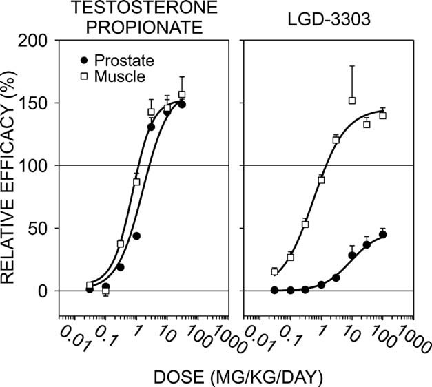
Both testosterone propionate and LGD‐3303 had anabolic activity in muscle in ORDX male rats, significantly increasing the levator ani weight in a dose‐dependent manner. Histological examination was not performed in this experiment; however, examination of H&E‐stained skeletal muscle sections in other studies showed no abnormal histological findings at doses of LGD‐3303 up to 450 mg/kg, suggesting normal muscle physiology (unpublished data). LGD‐3303 and testosterone displayed similar potency, maintaining the levator ani near eugonadal levels with a 1‐mg/kg dose. Testosterone stimulated the ventral prostate to 50% of eugonadal levels with the 1‐mg/kg dose and exceeded the eugonadal level with the 3‐mg/kg dose, indicating minimal tissue selective activity. LGD‐3303, however, had negligible activity on the ventral prostate at 1 mg/kg (<5% efficacy). At higher doses of LGD‐3303, the ventral prostate weight never reached the eugonadal level, restoring the ventral prostate to <50% of eugonadal levels at 100 mg/kg. The finding that prostate weight is maximally stimulated by LGD‐3303 to a level markedly less than testosterone, a full agonist, indicates that this compound is a partial agonist on the prostate. This partial agonist activity on the prostate occurred despite increasing plasma concentrations of compound (data not shown).
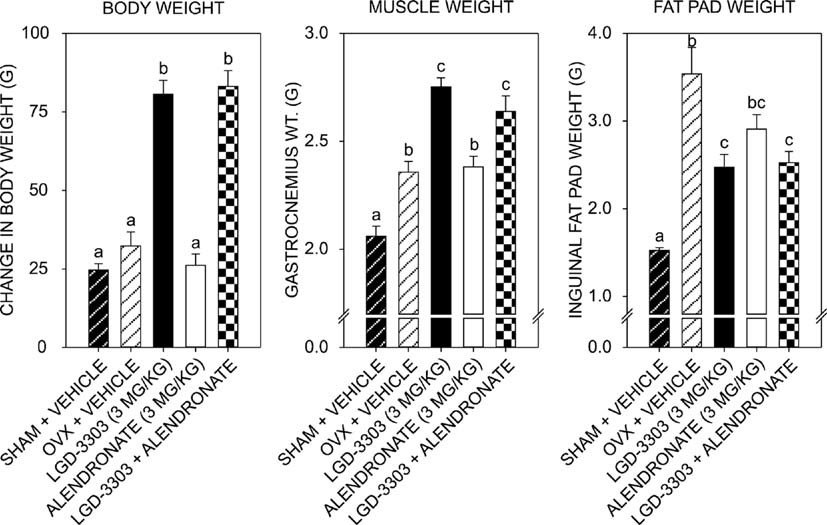
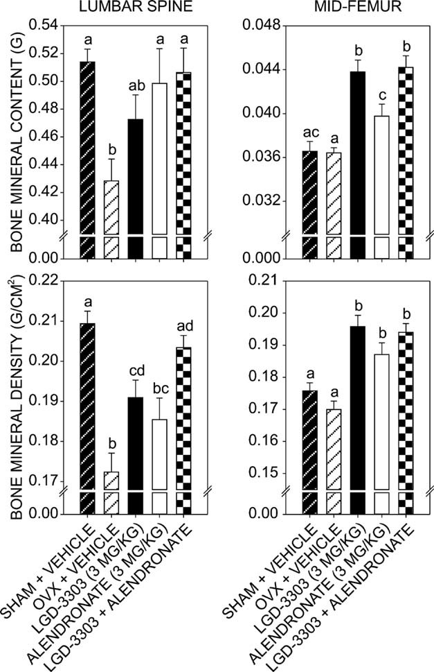
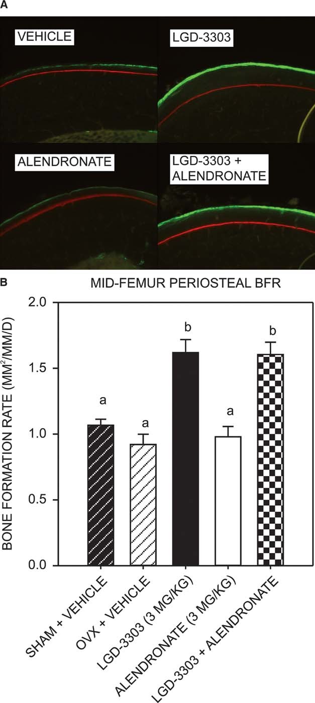
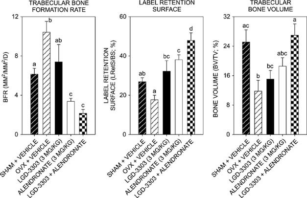
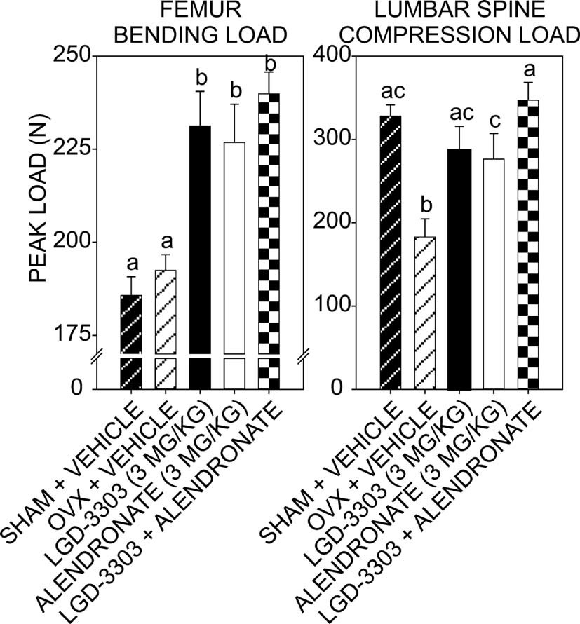
In orchidectomized (ORDX) male rats, LGD-3303 administered orally for 14 days dose-dependently increased levator ani muscle weight, restoring it to near eugonadal levels at 1 mg/kg, while having minimal effect on ventral prostate weight (never exceeding 50% of eugonadal levels even at 100 mg/kg), demonstrating tissue-selective anabolic activity on muscle.[1] In ovariectomized (OVX) female rats with established osteopenia, LGD-3303 (3 mg/kg/day orally for 12 weeks) significantly increased body weight and gastrocnemius muscle weight, and decreased inguinal fat pad weight. Alendronate co-administration did not alter these muscle anabolic effects.[1] LGD-3303 significantly reduced serum osteocalcin levels in OVX rats (55% and 59% decrease at 5 and 12 weeks, respectively), below sham-operated levels.[1] DXA scans showed that LGD-3303 increased lumbar spine BMD (cancellous bone) and midfemoral BMD and BMC (cortical bone) in OVX rats. At the midfemur, LGD-3303 was significantly more effective than alendronate at increasing BMC.[1] Histomorphometry revealed that LGD-3303 significantly increased periosteal bone formation rate at the midfemoral diaphysis (92.2% mineralizing surface, MAR 1.75 μm/d, BFR increased) compared to OVX controls, indicating anabolic activity on cortical bone. It also decreased endocortical mineralizing surface and increased baseline label retention (decreased resorption).[1] In cancellous bone (lumbar spine), LGD-3303 slightly reduced bone formation rate, increased label retention surface (indicating reduced resorption), and increased trabecular bone volume (not significant alone). Combination with alendronate significantly increased trabecular bone volume to sham levels.[1] Biomechanical testing showed that LGD-3303 significantly increased peak bending load of the femur and peak compressive load of lumbar vertebrae in OVX rats, similar to alendronate.[1] In vivo, LGD-3303 has anabolic effects on muscle and cortical bone not observed with bisphosphonates. In castrated male rats, oral administration for 14 days shows selective anabolic effects. The compound enhances male-directed sexual preference, proceptive behavior, and lordosis behavior in sexually experienced female rats. |
| Enzyme Assay |
Competitive binding assays were performed using recombinant baculovirus-expressed human androgen receptor (hAR), human glucocorticoid receptor (hGR), human mineralocorticoid receptor (hMR), or human progesterone receptor (hPR). Tritium-labeled reference ligand was used with varying concentrations of LGD-3303 as a competing ligand. Inhibition constant (Ki) values were calculated by application of the Cheng-Prusoff equation.[1]
Transcriptional activity reporter assays were performed using CV1 cells cultured in DMEM supplemented with 10% charcoal resin-stripped fetal bovine serum. Cells were seeded 48 hours before transfection in 96-well microtiter plates. Transient transfection was performed using a nonliposomal formulation, with luciferase reporter plasmids (MMTV-LUC or MTV-ERE5-LUC), a β-galactosidase expression plasmid, and expression plasmids for hAR, hGR, hMR, hPR, or human estrogen receptor α. Cells were treated with varying concentrations of LGD-3303 or reference compound for 40 hours. Normalized luciferase response was calculated as relative luciferase units divided by β-galactosidase activity. EC50 values were determined, and agonist efficacy was calculated as percent of normalized luciferase response relative to the maximum response by the reference agonist (DHT for hAR, dexamethasone for hGR, aldosterone for hMR, progesterone for hPR-B, or 17β-estradiol for hERα).[1] Cell-free AR binding assays for LGD-3303 utilize purified recombinant androgen receptor ligand-binding domain. Binding affinity is measured using radiolabeled [3H]-DHT competition binding. The compound's selectivity for AR over other nuclear receptors is assessed. Transactivation assays use AR-responsive reporter gene constructs in cell-free systems. |
| Cell Assay |
CV1 cells (American Type Culture Collection) were cultured in DMEM supplemented with 10% charcoal resin-stripped fetal bovine serum, and seeded 48 hours before transfection in 96-well microtiter plates. Cells were transiently transfected using a nonliposomal formulation (FuGENE 6 transfection reagent) with luciferase reporter plasmids MMTV-LUC or MTV-ERE5-LUC, a β-galactosidase expression plasmid coding for constitutive expression of E. coli β-galactosidase, and human androgen receptor (hAR), hGR, hMR, hPR, or hERα expression plasmids. Cells were treated with varying concentrations of LGD-3303 or reference compound for 40 hours. The normalized luciferase response was calculated as relative luciferase units divided by (β-galactosidase optical density at 415 nm divided by β-galactosidase incubation time in minutes). EC50 and agonist efficacy relative to reference agonists were determined.[1]
Cells expressing AR (e.g., C2C12 myoblasts, LNCaP prostate cancer cells) are treated with LGD-3303 at various concentrations. AR target gene expression is assessed by qRT-PCR. Muscle cell differentiation and fusion are evaluated. Androgenic effects in prostate cells are assessed as a measure of tissue selectivity. |
| Animal Protocol |
Suspended in Tween 80, polyethylene glycol-400, and 0.1% carboxy-methyl cellulose in water (0.005%:10%:89.995%); oral administration ORDX male rats
Male rat protocol (ORDX): Male Sprague-Dawley rats (7-8 weeks old, ~200 g) were orchidectomized or underwent sham surgery. After 14 days of untreated hypogonadism, rats were treated with LGD-3303 or testosterone propionate. LGD-3303 was administered in a suspension of Tween-80, polyethylene glycol-400, and 0.1% carboxymethyl cellulose in water (0.005%:10%:89.995%) by once daily oral gavage for 14 days. Testosterone propionate was dissolved in polyethylene glycol-400 and DMSO (70%:30%) and given subcutaneously once daily. Rats were then killed, and wet weights of ventral prostate and levator ani muscle were measured.[1] Female rat protocol (OVX): Female Sprague-Dawley rats (3 months old, 175-200 g) underwent ovariectomy or sham surgery. OVX animals were allowed to develop osteopenia for 7 weeks. At week -1, animals were scanned by DXA and stratified by whole femur BMC, then randomly assigned to treatment groups (n=11/group). Treatment with LGD-3303 (3 mg/kg), alendronate (3 mg/kg), or combination (both at 3 mg/kg) began at week 1 and continued once daily by oral gavage for 12 weeks. Alizarin red was injected subcutaneously at baseline DXA scan; calcein was injected subcutaneously 3 and 10 days before necropsy. After 12 weeks, animals were killed; gastrocnemius muscle and inguinal fat pad weights were measured; bones were harvested for DXA, biomechanics, and histomorphometry.[1] In vivo animal studies for LGD-3303 are conducted in castrated male rats or other rodent models. The compound is administered orally. Muscle mass (levator ani, gastrocnemius), bone density, and prostate weight are measured. Behavioral assays for sexual behavior are performed. Pharmacodynamic markers are assessed in muscle and other tissues. |
| ADME/Pharmacokinetics |
LGD-3303 is orally bioavailable and brain-penetrant. As a small molecule (MW 330.3, formula C16H14ClF3N2O), it has favorable drug-like properties. The compound is soluble in DMSO (8 mg/mL). PK parameters including Tmax, half-life, clearance, and bioavailability have been characterized in preclinical species.
|
| Toxicity/Toxicokinetics |
In orchidectomized male rats, LGD-3303 at doses up to 450 mg/kg showed no abnormal histological findings in H&E-stained skeletal muscle sections (unpublished data cited within the article).[1]
LGD-3303 is a partial agonist on the prostate, never increasing ventral prostate weight to >50% of eugonadal levels even at high doses (100 mg/kg), in contrast to full agonist testosterone.[1] In OVX female rats, the tested dose of LGD-3303 (3 mg/kg/day) was reported to be maximally effective on bone and non-toxic based on prior studies (reference to unpublished data). No adverse effects on kidney were noted at this dose; a previous rat study cited indicated that the no-observed-adverse-effect level for nephrotoxicity of alendronate was 5 mg/kg/day, but this is not directly about LGD-3303.[1] LGD-3303 has been studied for its anabolic effects on muscle and bone. As a SARM, it is designed to have a favorable safety profile compared to traditional androgens, with reduced prostate-related side effects. Standard toxicological assessments including genotoxicity, reproductive toxicity, and chronic toxicity studies would be required for development. |
| References |
J Bone Miner Res.2009 Feb;24(2):231-40.
|
| Additional Infomation |
Androgen receptor modulators.
LGD-3303 is a nonsteroidal, nonaromatizable androgen receptor ligand. It is orally available and has tissue-selective activity, preferentially stimulating skeletal muscle while sparing the prostate. The compound shows anabolic activity on muscle and cortical bone, and antiresorptive activity on cancellous bone. It is being investigated for potential treatment of osteoporosis and frailty, especially in combination with bisphosphonates such as alendronate, where additive effects were observed in an osteopenic rat model. No currently approved therapy has this combination of bone anabolic, muscle anabolic, and bone antiresorptive activity.[1] LGD-3303 (molecular formula C16H14ClF3N2O, MW 330.3) is also known as 9-Chloro-2-ethyl-3,6-dihydro-1-methyl-3-(2,2,2-trifluoroethyl)-7H-pyrrolo[3,2-f]quinolin-7-one. It is a potent, selective, orally bioavailable, non-steroidal SARM with anabolic effects on muscle and bone. The compound is intended for research use only. |
| Molecular Formula |
C16H14CLF3N2O
|
|
|---|---|---|
| Molecular Weight |
342.75
|
|
| Exact Mass |
342.075
|
|
| CAS # |
917891-35-1
|
|
| Related CAS # |
|
|
| PubChem CID |
25195253
|
|
| Appearance |
Light yellow to yellow solid powder
|
|
| LogP |
4.569
|
|
| Hydrogen Bond Donor Count |
1
|
|
| Hydrogen Bond Acceptor Count |
4
|
|
| Rotatable Bond Count |
2
|
|
| Heavy Atom Count |
23
|
|
| Complexity |
522
|
|
| Defined Atom Stereocenter Count |
0
|
|
| SMILES |
O=C1NC2C=CC3N(CC(F)(F)F)C(CC)=C(C)C=3C=2C(Cl)=C1
|
|
| InChi Key |
OMXGOGXEWUCLFI-UHFFFAOYSA-N
|
|
| InChi Code |
InChI=1S/C16H14ClF3N2O/c1-3-11-8(2)14-12(22(11)7-16(18,19)20)5-4-10-15(14)9(17)6-13(23)21-10/h4-6H,3,7H2,1-2H3,(H,21,23)
|
|
| Chemical Name |
|
|
| Synonyms |
|
|
| HS Tariff Code |
2934.99.9001
|
|
| Storage |
Powder -20°C 3 years 4°C 2 years In solvent -80°C 6 months -20°C 1 month |
|
| Shipping Condition |
Room temperature (This product is stable at ambient temperature for a few days during ordinary shipping and time spent in Customs)
|
| Solubility (In Vitro) |
DMSO: 10 mM
Water: 1 mg/mL (Insoluble) Ethanol: |
|---|---|
| Solubility (In Vivo) |
Note: Listed below are some common formulations that may be used to formulate products with low water solubility (e.g. < 1 mg/mL), you may test these formulations using a minute amount of products to avoid loss of samples.
Injection Formulations
Injection Formulation 1: DMSO : Tween 80: Saline = 10 : 5 : 85 (i.e. 100 μL DMSO stock solution → 50 μL Tween 80 → 850 μL Saline)(e.g. IP/IV/IM/SC) *Preparation of saline: Dissolve 0.9 g of sodium chloride in 100 mL ddH ₂ O to obtain a clear solution. Injection Formulation 2: DMSO : PEG300 :Tween 80 : Saline = 10 : 40 : 5 : 45 (i.e. 100 μL DMSO → 400 μLPEG300 → 50 μL Tween 80 → 450 μL Saline) Injection Formulation 3: DMSO : Corn oil = 10 : 90 (i.e. 100 μL DMSO → 900 μL Corn oil) Example: Take the Injection Formulation 3 (DMSO : Corn oil = 10 : 90) as an example, if 1 mL of 2.5 mg/mL working solution is to be prepared, you can take 100 μL 25 mg/mL DMSO stock solution and add to 900 μL corn oil, mix well to obtain a clear or suspension solution (2.5 mg/mL, ready for use in animals). View More
Injection Formulation 4: DMSO : 20% SBE-β-CD in saline = 10 : 90 [i.e. 100 μL DMSO → 900 μL (20% SBE-β-CD in saline)] Oral Formulations
Oral Formulation 1: Suspend in 0.5% CMC Na (carboxymethylcellulose sodium) Oral Formulation 2: Suspend in 0.5% Carboxymethyl cellulose Example: Take the Oral Formulation 1 (Suspend in 0.5% CMC Na) as an example, if 100 mL of 2.5 mg/mL working solution is to be prepared, you can first prepare 0.5% CMC Na solution by measuring 0.5 g CMC Na and dissolve it in 100 mL ddH2O to obtain a clear solution; then add 250 mg of the product to 100 mL 0.5% CMC Na solution, to make the suspension solution (2.5 mg/mL, ready for use in animals). View More
Oral Formulation 3: Dissolved in PEG400 (Please use freshly prepared in vivo formulations for optimal results.) |
| Preparing Stock Solutions | 1 mg | 5 mg | 10 mg | |
| 1 mM | 2.9176 mL | 14.5879 mL | 29.1758 mL | |
| 5 mM | 0.5835 mL | 2.9176 mL | 5.8352 mL | |
| 10 mM | 0.2918 mL | 1.4588 mL | 2.9176 mL |
*Note: Please select an appropriate solvent for the preparation of stock solution based on your experiment needs. For most products, DMSO can be used for preparing stock solutions (e.g. 5 mM, 10 mM, or 20 mM concentration); some products with high aqueous solubility may be dissolved in water directly. Solubility information is available at the above Solubility Data section. Once the stock solution is prepared, aliquot it to routine usage volumes and store at -20°C or -80°C. Avoid repeated freeze and thaw cycles.
Calculation results
Working concentration: mg/mL;
Method for preparing DMSO stock solution: mg drug pre-dissolved in μL DMSO (stock solution concentration mg/mL). Please contact us first if the concentration exceeds the DMSO solubility of the batch of drug.
Method for preparing in vivo formulation::Take μL DMSO stock solution, next add μL PEG300, mix and clarify, next addμL Tween 80, mix and clarify, next add μL ddH2O,mix and clarify.
(1) Please be sure that the solution is clear before the addition of next solvent. Dissolution methods like vortex, ultrasound or warming and heat may be used to aid dissolving.
(2) Be sure to add the solvent(s) in order.
In ORDX male rats, testosterone increased levator ani muscle (LA) weight at doses that have equivalent effects on ventral prostate (VP) weight, indicating a lack of tissue selectivity.J Bone Miner Res.2009 Feb;24(2):231-40. |
|---|
LGD‐3303 increases body and gastrocnemius muscle weight in OVX female rats after 12 wk of treatment.J Bone Miner Res.2009 Feb;24(2):231-40. |
LGD‐3303 increases lumbar spine and femoral BMD and BMC in OVX female rats.J Bone Miner Res.2009 Feb;24(2):231-40. |
(A) Photomicrograph of the periosteal surface of the midfemoral diaphysis viewed under epifluorescent light. (B) LGD‐3303 increases periosteal bone formation at the midfemoral diaphysis in OVX female rats. Alendronate has no activity at this site.J Bone Miner Res.2009 Feb;24(2):231-40. |
|---|
Estrogen deficiency in female rats increases lumbar spine cancellous bone formation rate.J Bone Miner Res.2009 Feb;24(2):231-40. |
Three‐point bending test of the femur and compression test of the lumbar vertebral body.J Bone Miner Res.2009 Feb;24(2):231-40. |
Products are for research use only; We do not sell to patients
Copyright 2020 InvivoChem LLC | All Rights Reserved


 COA
COA