| Size | Price | Stock | Qty |
|---|---|---|---|
| 5mg |
|
||
| 10mg |
|
||
| 25mg |
|
||
| 50mg |
|
||
| 100mg |
|
||
| 250mg | |||
| 500mg | |||
| Other Sizes |


Purity: ≥98%
| Targets |
FPH1 targets primary human hepatocytes and iPS-derived hepatocytes, promoting their expansion and differentiation. Unlike traditional small molecules that inhibit specific enzymes or receptors, FPH1 is a functional proliferation hit that enables the renewable sourcing of functional human hepatocytes. Its mechanism of action involves inducing functional proliferation of hepatocytes in vitro, leading to more pronounced hepatocyte morphologies, including polygonal cell shapes, visible nuclei, and more noticeable bile canaliculi between hepatocytes. The precise molecular target of FPH1 remains to be fully elucidated, but it is recognized as a potent inducer of hepatocyte proliferation.
|
|---|---|
| ln Vitro |
FPH1 induces functional proliferation of hepatocytes in vitro, and causes more pronounced hepatocyte morphologies, including polygonal cell shapes, visible nuclei, and more noticeable bile cannaliculi between hepatocytes.
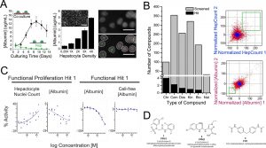
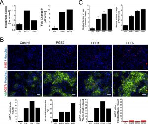
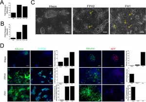
Cell Assay: FPH1 belongs to the functional proliferation hits which are screened out by their ability to permit renewable sourcing of functional human hepatocytes. This ability of FPH1 is not dependent on the donors of the hepatocytes. It has been found that FPH1 was active against the primary human hepatocytes from six cell sources of genetically diverse individuals. Besides that, FPH1 can affect the hepatocyte functions with promoting albumin secretion during the differentiation of iPS cells into iHeps. Moreover, treatment of FPH1 also resulted in the increase of CYP3A4 levels and the decrease of AFP secretion. FPH1 (BRD-6125) increased hepatocyte nuclei count and elevated the number of nuclei undergoing mitosis in primary human hepatocytes in a concentration-dependent manner (Fig. 1c, 2a). [1] After 6 days of culture with FPH1 (BRD-6125) at 20 μM, Ki67-positive nuclei colocalized with human albumin-positive hepatocytes, and quantitative image analysis showed up to a 6.6-fold increase in the area of albumin-positive colonies. [1] FPH1 (BRD-6125) treatment led to a substantial increase in hepatocyte number (up to tenfold across various FPHs, with FPH1 showing significant expansion) as measured by automated cell counter and FACS analysis. [1] FPH1 (BRD-6125) maintained normal hepatocyte morphology, stable urea synthesis, albumin secretion, CYP450 activity (CYP450 activity not compromised), and active MRP2 transport consistent with intact bile canaliculi. [1] In iHep cells, FPH1 (BRD-6125) at 20 μM for 9 days increased colony size and promoted polygonal cell shapes, visible nuclei, and more noticeable bile canaliculi. [1] Immunofluorescent staining showed that FPH1 (BRD-6125) increased albumin and CYP3A levels while largely eliminating AFP expression in iHep cells. [1] Gene expression profiling revealed that FPH1 (BRD-6125)-treated iHep cells more closely resembled adult hepatocytes, with increased expression of mature markers (e.g., ABCB11/BSEP) and decreased GSTP1. [1] FPH1 (BRD-6125) increased CYP3A4 activity by 45-fold and markedly increased CYP2A6 activity in iHep cells. [1] The maturation effect of FPH1 (BRD-6125) on iHep cells was stable for at least one week after compound removal. [1] In vitro, FPH1 induces functional proliferation of hepatocytes, causing more pronounced hepatocyte morphologies, including polygonal cell shapes, visible nuclei, and more noticeable bile canaliculi between hepatocytes. It increases the number and activity of primary human hepatocytes in vitro. FPH1 promotes the differentiation of iPS cells towards a hepatic lineage. These in vitro activities have been characterized in primary human hepatocyte cultures and iPS cell differentiation systems. The compound's ability to induce functional proliferation makes it a valuable tool for studying hepatocyte biology and for generating hepatocytes for research applications. |
| ln Vivo |
In vivo studies on FPH1 are not extensively documented, as the compound is primarily used as a research tool for in vitro applications. Its primary application is in the expansion and differentiation of hepatocytes in cell culture systems. As a small molecule that promotes hepatocyte proliferation, FPH1 has potential for in vivo applications in liver regeneration and cell therapy, but specific in vivo data are not available in standard research summaries. Further studies are needed to evaluate its in vivo efficacy and safety.
|
| Enzyme Assay |
Non-cellular assays for FPH1 typically involve cytochrome P450 inhibition studies. These assays are performed in pooled human hepatic microsomal fractions following current scientific and regulatory guidelines. Reaction conditions are linear with respect to incubation time and hepatic microsomal protein concentration. Substrates are present at concentrations equal to or less than their respective Km values. Metabolite and/or substrate concentrations are determined using specific, internal standard controlled HPLC MS/MS assays. These assays are used to characterize the compound's potential for drug-drug interactions and its effects on drug metabolism enzymes.
|
| Cell Assay |
For primary human hepatocyte expansion: Human primary hepatocytes were cocultured with growth-arrested J2-3T3 fibroblasts in collagen-coated plates. FPH1 (BRD-6125) was added to the culture medium at 20 μM on days 1 and 5 of culture. After 6–7 days, cells were fixed for immunofluorescent staining (Ki67, albumin, Hoechst) or harvested for cell counting (FACS or automated cell counter). Image analysis was performed using CellProfiler and CellProfiler Analyst. [1]
For iHep cell maturation: Induced pluripotent stem (iPS) cells were differentiated into hepatocyte-like cells (iHep) over 20 days using sequential growth factors (activin A, BMP-4, bFGF, HGF, OSM). On day 21, FPH1 (BRD-6125) at 20 μM was added to basal medium without OSM (or with OSM in some experiments) and cultured for 9 days. Cells were then analyzed by immunofluorescent staining (albumin, CYP3A, AFP), gene expression profiling (Luminex), and functional assays (albumin/AFP ELISA, CYP450 activity). [1] CYP450 activity assay: iHep cells were incubated with coumarin (for CYP2A6) or luciferin-IPA (for CYP3A4) for 4 h at 37 °C; metabolite concentrations were quantified by luminescence or fluorescence after hydrolysis of potential conjugates by β-glucuronidase/arylsulfatase. [1] In vitro cell-based assays for FPH1 are conducted using primary human hepatocytes or iPS-derived hepatocytes. Cells are cultured and treated with FPH1, and the effects on cell number, activity, and morphology are assessed. Hepatocyte proliferation is measured by counting cell numbers and assessing functional activity. Differentiation towards a hepatic lineage is evaluated by monitoring the expression of hepatocyte-specific markers and the development of characteristic hepatocyte morphologies, including polygonal cell shapes and visible bile canaliculi. These assays are essential for characterizing the compound's activity as a hepatocyte proliferation inducer. |
| Animal Protocol |
In vivo animal studies for FPH1 are not well documented, as the compound is primarily used as a research tool for in vitro applications. Its primary application is in the expansion and differentiation of hepatocytes in cell culture systems. As a small molecule that promotes hepatocyte proliferation, FPH1 has potential for in vivo applications in liver regeneration and cell therapy, but specific in vivo protocols are not available in standard research summaries. Further studies are needed to evaluate its in vivo efficacy and safety.
|
| ADME/Pharmacokinetics |
FPH1 has a molecular weight of 388.82 g/mol and a molecular formula of C16H15ClF2N2O3S. It is soluble in DMSO at 250 mg/mL (642.97 mM). It is insoluble or slightly soluble in water (< 1 mg/mL) and ethanol (< 1 mg/mL). For in vivo formulation, a mixture of 10% DMSO, 40% PEG300, 5% Tween-80, and 45% saline can be used. The compound should be stored as a powder at -20°C for up to 3 years or in solution at -80°C for up to 1 year.
|
| Toxicity/Toxicokinetics |
Comprehensive toxicological data for FPH1 are limited, as it is primarily a research compound. It is classified with GHS07 warning, indicating potential for irritation or other mild hazards. As with all research chemicals, appropriate safety precautions should be taken when handling FPH1. It is not intended for human use and is strictly a research compound for laboratory applications. Its safety profile in vivo has not been extensively characterized.
|
| References |
Nat Chem Biol.2013 Aug;9(8):514-20.
|
| Additional Infomation |
FPH1 (BRD-6125) (CAS#: 708219-39-0) has the chemical structure shown in Figure 1d (N-phenyl-2-(N-phenylmethylsulfonamido)acetamide core). Structure-activity relationship analysis identified 21 analogs of FPH1; the key driver of activity was the presence of a 5-chloro-2-methyl substitution on the sulfonamide phenyl ring and a small functional group at the para position of the phenylamide ring. [1]
FPH1 (BRD-6125) was identified from a library of 12,480 small molecules screened on primary human hepatocytes. It belongs to the functional proliferation hit class that enables both expansion of mature primary human hepatocytes and maturation of iPS-derived hepatocytes, potentially addressing the bottleneck of human hepatocyte sourcing for liver research and cell-based therapies. [1] FPH1 (BRD-6125) is a small molecule that promotes the expansion of iPS-derived hepatocytes. It was identified through a high-throughput phenotypic screening platform using primary human hepatocytes. FPH1 increases the number and activity of primary human hepatocytes in vitro and promotes the differentiation of iPS cells towards a hepatic lineage. It has the molecular formula C16H15ClF2N2O3S and a molecular weight of 388.82 g/mol. FPH1 is not approved for clinical use and is strictly a research compound for studying hepatocyte biology and regenerative medicine. |
| Molecular Formula |
C16H15CLF2N2O3S
|
|
|---|---|---|
| Molecular Weight |
388.82
|
|
| Exact Mass |
388.046
|
|
| CAS # |
708219-39-0
|
|
| Related CAS # |
|
|
| PubChem CID |
2210370
|
|
| Appearance |
White to off-white solid powder
|
|
| LogP |
4.485
|
|
| Hydrogen Bond Donor Count |
1
|
|
| Hydrogen Bond Acceptor Count |
6
|
|
| Rotatable Bond Count |
5
|
|
| Heavy Atom Count |
25
|
|
| Complexity |
562
|
|
| Defined Atom Stereocenter Count |
0
|
|
| SMILES |
CC1=C(N(S(=O)(C)=O)CC(NC2=C(F)C=CC=C2F)=O)C=C(Cl)C=C1
|
|
| InChi Key |
WAOBCCBUTHNTFO-UHFFFAOYSA-N
|
|
| InChi Code |
InChI=1S/C16H15ClF2N2O3S/c1-10-6-7-11(17)8-14(10)21(25(2,23)24)9-15(22)20-16-12(18)4-3-5-13(16)19/h3-8H,9H2,1-2H3,(H,20,22)
|
|
| Chemical Name |
2-(5-chloro-2-methyl-N-methylsulfonylanilino)-N-(2,6-difluorophenyl)acetamide
|
|
| Synonyms |
|
|
| HS Tariff Code |
2934.99.9001
|
|
| Storage |
Powder -20°C 3 years 4°C 2 years In solvent -80°C 6 months -20°C 1 month |
|
| Shipping Condition |
Room temperature (This product is stable at ambient temperature for a few days during ordinary shipping and time spent in Customs)
|
| Solubility (In Vitro) |
DMSO: 78 mg/mL (200.6 mM)
Water: 1 mg/mL (Insoluble) Ethanol: 1 mg/mL |
|---|---|
| Solubility (In Vivo) |
Note: Listed below are some common formulations that may be used to formulate products with low water solubility (e.g. < 1 mg/mL), you may test these formulations using a minute amount of products to avoid loss of samples.
Injection Formulations
Injection Formulation 1: DMSO : Tween 80: Saline = 10 : 5 : 85 (i.e. 100 μL DMSO stock solution → 50 μL Tween 80 → 850 μL Saline)(e.g. IP/IV/IM/SC) *Preparation of saline: Dissolve 0.9 g of sodium chloride in 100 mL ddH ₂ O to obtain a clear solution. Injection Formulation 2: DMSO : PEG300 :Tween 80 : Saline = 10 : 40 : 5 : 45 (i.e. 100 μL DMSO → 400 μLPEG300 → 50 μL Tween 80 → 450 μL Saline) Injection Formulation 3: DMSO : Corn oil = 10 : 90 (i.e. 100 μL DMSO → 900 μL Corn oil) Example: Take the Injection Formulation 3 (DMSO : Corn oil = 10 : 90) as an example, if 1 mL of 2.5 mg/mL working solution is to be prepared, you can take 100 μL 25 mg/mL DMSO stock solution and add to 900 μL corn oil, mix well to obtain a clear or suspension solution (2.5 mg/mL, ready for use in animals). View More
Injection Formulation 4: DMSO : 20% SBE-β-CD in saline = 10 : 90 [i.e. 100 μL DMSO → 900 μL (20% SBE-β-CD in saline)] Oral Formulations
Oral Formulation 1: Suspend in 0.5% CMC Na (carboxymethylcellulose sodium) Oral Formulation 2: Suspend in 0.5% Carboxymethyl cellulose Example: Take the Oral Formulation 1 (Suspend in 0.5% CMC Na) as an example, if 100 mL of 2.5 mg/mL working solution is to be prepared, you can first prepare 0.5% CMC Na solution by measuring 0.5 g CMC Na and dissolve it in 100 mL ddH2O to obtain a clear solution; then add 250 mg of the product to 100 mL 0.5% CMC Na solution, to make the suspension solution (2.5 mg/mL, ready for use in animals). View More
Oral Formulation 3: Dissolved in PEG400 (Please use freshly prepared in vivo formulations for optimal results.) |
| Preparing Stock Solutions | 1 mg | 5 mg | 10 mg | |
| 1 mM | 2.5719 mL | 12.8594 mL | 25.7188 mL | |
| 5 mM | 0.5144 mL | 2.5719 mL | 5.1438 mL | |
| 10 mM | 0.2572 mL | 1.2859 mL | 2.5719 mL |
*Note: Please select an appropriate solvent for the preparation of stock solution based on your experiment needs. For most products, DMSO can be used for preparing stock solutions (e.g. 5 mM, 10 mM, or 20 mM concentration); some products with high aqueous solubility may be dissolved in water directly. Solubility information is available at the above Solubility Data section. Once the stock solution is prepared, aliquot it to routine usage volumes and store at -20°C or -80°C. Avoid repeated freeze and thaw cycles.
Calculation results
Working concentration: mg/mL;
Method for preparing DMSO stock solution: mg drug pre-dissolved in μL DMSO (stock solution concentration mg/mL). Please contact us first if the concentration exceeds the DMSO solubility of the batch of drug.
Method for preparing in vivo formulation::Take μL DMSO stock solution, next add μL PEG300, mix and clarify, next addμL Tween 80, mix and clarify, next add μL ddH2O,mix and clarify.
(1) Please be sure that the solution is clear before the addition of next solvent. Dissolution methods like vortex, ultrasound or warming and heat may be used to aid dissolving.
(2) Be sure to add the solvent(s) in order.
|
|---|
|
|
Products are for research use only; We do not sell to patients
Copyright 2020 InvivoChem LLC | All Rights Reserved


 COA
COA