| Size | Price | Stock | Qty |
|---|---|---|---|
| 5mg |
|
||
| 10mg |
|
||
| 25mg |
|


Purity: =99.87%
| Targets |
sphingosine 1-phosphate (S1P)(IC50 = 0.033 nM, in K562 and NK cells); PAK1
Sphingosine 1-phosphate receptor 1 (S1P1): FTY720 binds to S1P1 on microglia. Blocking S1P1 with the antagonist W146 (250 nM) significantly reduced the suppressive effect of FTY720 on microglial TNF-α production. FTY720 binds all S1P receptor subtypes except S1P2. [1] Fingolimod phosphate is a potent agonist at four of the five sphingosine-1-phosphate (S1P) receptors: S1P1, S1P3, S1P4, and S1P5, with IC50 values of 0.2-6 nM. It shows no activity at S1P2. Upon binding, it induces receptor internalization and degradation. This functional antagonism leads to lymphocyte sequestration in lymphoid organs, preventing autoreactive lymphocytes from infiltrating the central nervous system. |
|---|---|
| ln Vitro |
Fingolimod phosphate stops autoreactive lymphocytes from entering the central nervous system and stops lymphocytes from leaving lymphoid organs [1]. Fingolimod phosphate (0, 1, 10, 100 nM) binds to the S1P1 receptor and inhibits activated microglia's production of pro-inflammatory cytokines, including interleukin-1β, interleukin-6, and tumor necrosis factor-a[1]. Additionally, microglia produce more brain-derived neurotrophic factor and glial cell-derived neurotrophic factor when exposed to (0, 1, 10, 100 nM) [1].
- S1P Receptor Expression in Microglia: Mouse primary microglia expressed all S1P receptor subtypes (S1P1, S1P2, S1P3, S1P4, S1P5), with the highest expression levels observed for S1P1. Stimulation with LPS (1 μg/ml) significantly reduced microglial expression levels of S1P2, S1P4, and S1P5. [1] - Inhibition of Pro-inflammatory Cytokine Production: LPS (1 μg/ml) enhanced microglial production of TNF-α, IL-1β, and IL-6. FTY720 (1-100 nM) reduced these effects in a dose-dependent manner. At 100 nM, FTY720 significantly decreased TNF-α, IL-1β, and IL-6 production as measured by ELISA and RT-PCR. The suppressive effect on TNF-α was significantly reduced by the S1P1 antagonist W146 (250 nM), but not by the S1P3/S1P5 antagonist suramin (10 μM) or the S1P4 antagonist CYM50358 (100 nM). [1] - Upregulation of Neurotrophic Factor Production: LPS (1 μg/ml) increased microglial expression of BDNF and GDNF. FTY720 (1-100 nM) enhanced these effects in a dose-dependent manner, as shown by RT-PCR (12h) and Western blotting (48h). At 100 nM, FTY720 significantly increased BDNF and GDNF protein levels. [1] In vitro, Fingolimod phosphate (0, 1, 10, 100 nM) binds to S1P1 receptors and downregulates activated microglial production of pro-inflammatory cytokines including tumor necrosis factor-α, interleukin-1β, and interleukin-6. It prevents lymphocytes from moving out of lymphoid organs and inhibits autoreactive lymphocyte infiltration into the central nervous system. The compound promotes the neuroprotective effects of microglia. |
| ln Vivo |
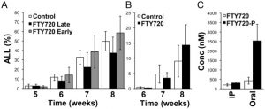
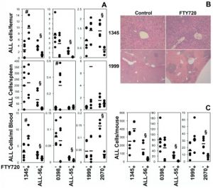
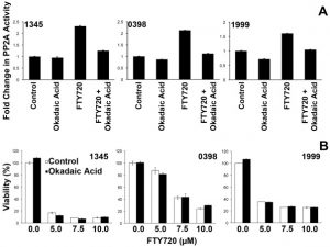
FTY720 is effective in Ph+ but not Ph- ALL xenografts using an early disease model. FTY720 produces a significant reduction in disease burden in the Ph+ ALL xenografts using an early disease model. Ph+ human ALL xenografts responds to FTY720 with an 80 % reduction in overall disease if treatment has been initiated early on. In contrast, treatment of mice with FTY720 does not result in reduced leukemia compared to controls using four separate human Ph- ALL xenografts. |
| Enzyme Assay |
For receptor binding assays, membrane preparations from cells expressing human S1P receptors are incubated with [³³P]S1P or other radiolabeled ligands at various concentrations of the test compound (typically 0.001-10 μM) in binding buffer containing BSA for 60-90 minutes at room temperature. Nonspecific binding is determined using excess unlabeled S1P. Bound radioligand is separated by filtration through GF/B filters and radioactivity is quantified by liquid scintillation counting to determine IC50 values.
|
| Cell Assay |
- Primary Microglia Culture: Mouse primary microglia were isolated from newborn C57BL/6 mice (day 14 in vitro) using the "shaking off" method. Cells were maintained in DMEM with 10% fetal calf serum, 5 μg/ml bovine insulin, and 0.2% glucose. Purity was 97-100% by Fc receptor immunostaining. [1]
- RT-PCR: Microglia were collected 12 hours after LPS stimulation. Total RNA was extracted, and first-strand cDNA was synthesized. RT-PCR was performed to amplify transcripts for S1P1-S1P5, TNF-α, IL-1β, IL-6, BDNF, GDNF, and GAPDH using specific oligonucleotide primers. [1] - Flow Cytometry: Microglia were collected 24 hours after LPS stimulation. Cells were fixed with 4% paraformaldehyde and stained with anti-S1P1, anti-S1P2, anti-S1P3, anti-S1P4, and anti-S1P5 rabbit polyclonal antibodies, followed by Alexa Fluor 488-conjugated anti-rabbit IgG. Samples were analyzed on a flow cytometer. [1] - ELISA: Microglia were collected 24 hours after LPS stimulation. Microglial production of IL-1β, IL-6, and TNF-α was assessed with specific ELISA kits according to the manufacturers' protocols. [1] - Western Blotting: Microglia were collected 48 hours after LPS stimulation. Cells were lysed in TNE buffer with protease inhibitors. 20 μg of protein from total cell lysates were separated on a 4-20% Tris-glycine SDS-polyacrylamide gradient gel, transferred to a PVDF membrane, and probed with anti-BDNF, anti-GDNF, or anti-β-actin antibodies. Signals were visualized using enhanced chemiluminescence. [1] For cytokine production assays, primary microglia or microglial cell lines are cultured and treated with Fingolimod phosphate at concentrations of 0, 1, 10, and 100 nM. Cells are stimulated with lipopolysaccharide (LPS) to induce inflammatory responses. After treatment, culture supernatants are collected and cytokine levels (TNF-α, IL-1β, IL-6) are measured by ELISA. RNA can also be extracted for qRT-PCR analysis of cytokine gene expression to confirm the anti-inflammatory effects at the transcriptional level. |
| Animal Protocol |
Dissolved in 2% ethanol or saline; 5 and 10 mg/kg; i.p. injection NOD/SCIDγc / mice bearing ALL cells. |
| ADME/Pharmacokinetics |
- Brain Penetration: FTY720 can penetrate the blood-brain barrier. [1]
- Cerebrospinal Fluid Concentration: The effective concentrations of FTY720 used in this study (1-100 nM) were approximately 100-fold higher than the concentration observed in the cerebrospinal fluid of patients treated with 0.5 mg/kg of FTY720 (the clinically relevant dose mentioned is 0.5 mg/kg, though the text states 0.3 mg/kg in one instance). [1] Fingolimod phosphate is the active metabolite formed in vivo by phosphorylation of Fingolimod (FTY720) via sphingosine kinase. The parent compound FTY720 is orally bioavailable and widely distributed throughout the body. FTY720 phosphate has a half-life of approximately 6-9 days in humans following oral administration of FTY720. It is primarily excreted in urine as inactive metabolites. The compound shows solubility in chloroform (0.5 mg/mL) and is slightly soluble in DMSO. |
| Toxicity/Toxicokinetics |
In clinical use, Fingolimod (the parent compound) has been associated with adverse effects including bradycardia, atrioventricular conduction delays, elevated liver enzymes, macular edema, and increased risk of infections. These effects are related to S1P receptor modulation. At therapeutic doses, lymphopenia is observed as a pharmacodynamic effect. The compound is contraindicated in patients with certain cardiac conditions. In research settings, the compound is handled as a hazardous substance with appropriate safety precautions.
|
| References | |
| Additional Infomation |
Fingolimod phosphate is a primary amino compound formed by the conversion of a hydroxyl group of fingolimod to its dihydrophosphate derivative. It is the active metabolite of fingolimod. It has antitumor, immunosuppressive, and sphingosine-1-phosphate receptor agonist effects. It is a monoalkyl phosphate, a primary amino compound, and a primary alcohol. Its function is related to that of fingolimod. Fingolimod phosphate (FTY720) is a sphingosine-1-phosphate (S1P) receptor agonist currently being used as a novel oral medication for the treatment of multiple sclerosis. FTY720 can prevent lymphocytes from leaving lymphoid organs and inhibit the infiltration of autoreactive lymphocytes into the central nervous system. Whether FTY720 directly affects microglia (innate immune cells of the central nervous system) is unclear. This study shows that FTY720 downregulates the production of pro-inflammatory cytokines such as tumor necrosis factor-α, interleukin-1β, and interleukin-6 by binding to the S1P1 receptor. FTY720 also upregulated the production of brain-derived neurotrophic factor (BDNF) and glial cell-derived neurotrophic factor (GNF) by microglia. These results suggest that FTY720 can directly promote the neuroprotective effect of microglia. Therefore, FTY720 may not only be an effective drug for the treatment of multiple sclerosis, but also an effective drug for the treatment of other neurological diseases related to microglia activation. [1]
- Therapeutic Implications: Because FTY720 can cross the blood-brain barrier and directly promote the neuroprotective effects of microglia, it may be a promising therapeutic agent not only for MS but also for other neurologic diseases associated with microglial activation, such as spinal cord injury, stroke, amyotrophic lateral sclerosis, Alzheimer's disease, and epilepsy. [1] Fingolimod phosphate (FTY720-P) is the active phosphorylated form of Fingolimod, an FDA-approved drug for the treatment of relapsing forms of multiple sclerosis. It functions as a sphingosine 1-phosphate (S1P) receptor modulator that induces receptor internalization and degradation, thereby functionally antagonizing S1P signaling. This leads to lymphocyte retention in lymph nodes and reduced autoimmune attack on the central nervous system. Fingolimod phosphate is available for research purposes to study S1P receptor biology and immune modulation. |
| Molecular Formula |
C19H34NO5P
|
|---|---|
| Molecular Weight |
387.45100
|
| Exact Mass |
387.217
|
| CAS # |
402615-91-2
|
| Related CAS # |
FTY720 (S)-Phosphate;402616-26-6;Fingolimod phosphate-d4;1794828-93-5
|
| PubChem CID |
9908268
|
| Appearance |
White to off-white solid powder
|
| Density |
1.2±0.1 g/cm3
|
| Boiling Point |
584.2±60.0 °C at 760 mmHg
|
| Melting Point |
122-124ºC
|
| Flash Point |
307.1±32.9 °C
|
| Vapour Pressure |
0.0±1.7 mmHg at 25°C
|
| Index of Refraction |
1.541
|
| LogP |
4.27
|
| Hydrogen Bond Donor Count |
4
|
| Hydrogen Bond Acceptor Count |
6
|
| Rotatable Bond Count |
14
|
| Heavy Atom Count |
26
|
| Complexity |
409
|
| Defined Atom Stereocenter Count |
0
|
| SMILES |
CCCCCCCCC1=CC=C(C=C1)CCC(CO)(COP(=O)(O)O)N
|
| InChi Key |
LRFKWQGGENFBFO-UHFFFAOYSA-N
|
| InChi Code |
InChI=1S/C19H34NO5P/c1-2-3-4-5-6-7-8-17-9-11-18(12-10-17)13-14-19(20,15-21)16-25-26(22,23)24/h9-12,21H,2-8,13-16,20H2,1H3,(H2,22,23,24)
|
| Chemical Name |
[2-amino-2-(hydroxymethyl)-4-(4-octylphenyl)butyl] dihydrogen phosphate
|
| Synonyms |
Fingolimod phosphate; FTY720 phosphate; 402615-91-2; FTY720 (R)-Phosphate; rac FTY720 Phosphate; 402616-23-3; FTY720-phosphate; fingolimod-P;
Fingolimod phosphate salt; 2-amino-2-(hydroxymethyl)-4-(4-octylphenyl)butyl dihydrogen phosphate; [2-amino-2-(hydroxymethyl)-4-(4-octylphenyl)butyl] dihydrogen phosphate; fingolimod-phosphate; 402615-91-2;
|
| HS Tariff Code |
2934.99.9001
|
| Storage |
Powder -20°C 3 years 4°C 2 years In solvent -80°C 6 months -20°C 1 month Note: This product requires protection from light (avoid light exposure) during transportation and storage. |
| Shipping Condition |
Room temperature (This product is stable at ambient temperature for a few days during ordinary shipping and time spent in Customs)
|
| Solubility (In Vitro) |
Chloroform: 0.5 mg/ml
DMSO: ~100 mg/mL (~258 mM) |
|---|---|
| Solubility (In Vivo) |
Solubility in Formulation 1: ≥ 2.5 mg/mL (6.45 mM) (saturation unknown) in 10% DMSO + 40% PEG300 + 5% Tween-80 + 45% Saline (add these co-solvents sequentially from left to right, and one by one), Clear solution.
For example, if 1 mL of working solution is to be prepared, you can add 100 μL of DMSO stock solution (25.0 mg/mL) to 400 μL of PEG300 and mix well; then add 50 μL of Tween-80 and mix well; finally add 450 μL of physiological saline and adjust the volume to 1 mL. Preparation of saline: Dissolve 0.9 g of sodium chloride in 100 mL ddH₂ O to obtain a clear solution. (Please use freshly prepared in vivo formulations for optimal results.) |
| Preparing Stock Solutions | 1 mg | 5 mg | 10 mg | |
| 1 mM | 2.5810 mL | 12.9049 mL | 25.8098 mL | |
| 5 mM | 0.5162 mL | 2.5810 mL | 5.1620 mL | |
| 10 mM | 0.2581 mL | 1.2905 mL | 2.5810 mL |
*Note: Please select an appropriate solvent for the preparation of stock solution based on your experiment needs. For most products, DMSO can be used for preparing stock solutions (e.g. 5 mM, 10 mM, or 20 mM concentration); some products with high aqueous solubility may be dissolved in water directly. Solubility information is available at the above Solubility Data section. Once the stock solution is prepared, aliquot it to routine usage volumes and store at -20°C or -80°C. Avoid repeated freeze and thaw cycles.
Calculation results
Working concentration: mg/mL;
Method for preparing DMSO stock solution: mg drug pre-dissolved in μL DMSO (stock solution concentration mg/mL). Please contact us first if the concentration exceeds the DMSO solubility of the batch of drug.
Method for preparing in vivo formulation::Take μL DMSO stock solution, next add μL PEG300, mix and clarify, next addμL Tween 80, mix and clarify, next add μL ddH2O,mix and clarify.
(1) Please be sure that the solution is clear before the addition of next solvent. Dissolution methods like vortex, ultrasound or warming and heat may be used to aid dissolving.
(2) Be sure to add the solvent(s) in order.
|
|---|
|
|
Products are for research use only; We do not sell to patients
Copyright 2020 InvivoChem LLC | All Rights Reserved


 COA
COA