| Size | Price | Stock | Qty |
|---|---|---|---|
| 50mg |
|
||
| 100mg |
|
||
| 250mg |
|
||
| 500mg |
|
||
| 1g |
|
||
| 2g | |||
| Other Sizes |


| Targets |
Selective inhibitor of 3-hydroxy-3-methylglutaryl-coenzyme A (HMG-CoA) reductase with the following inhibitory parameter:
- Ki = 0.16 nM (recombinant human HMG-CoA reductase), showing high affinity for the enzyme [2] - Inhibitor of human ether-a-go-go-related gene (hERG) potassium channel (cardiac repolarization channel) with the following inhibitory parameter: - IC50 = 15 μM (hERG current in HEK293 cells stably expressing hERG) [3] |
|---|---|
| ln Vitro |
In vitro activity: Rosuvastatin is relatively hydrophilic and is highly selective for hepatic cells; its uptake is mediated by the liver-specific organic anion transporter OATP-C. Rosuvastatin is a high-affinity substrate for OATP-C with apparent association constant of 8.5 μM. Rosuvastatin inhibits cholesterol biosynthesis in rat liver isolated hepatocytes with IC50 of 1.12 nM. Rosuvastatin causes approximately 10 times greater increase of mRNA of LDL receptors than pravastatin. Rosuvastatin (100 μM) decreases the extent of U937 adhesion to TNF-α-stimulated HUVEC. Rosuvastatin inhibits the expressions of ICAM-1, MCP-1, IL-8, IL-6, and COX-2 mRNA and protein levels through inhibition of c-Jun N-terminal kinase and nuclear factor-kB in endothelial cells.
Kinase Assay: Rosuvastatin Calcium is a competitive inhibitor of HMG-CoA reductase with IC50 of 11 nM. Cell Assay: Rosuvastatin is relatively hydrophilic and is highly selective for hepatic cells; its uptake is mediated by the liver-specific organic anion transporter OATP-C. Rosuvastatin is a high-affinity substrate for OATP-C with apparent association constant of 8.5 μM. Rosuvastatin inhibits cholesterol biosynthesis in rat liver isolated hepatocytes with IC50 of 1.12 nM. Rosuvastatin causes approximately 10 times greater increase of mRNA of LDL receptors than pravastatin. Rosuvastatin (100 μM) decreases the extent of U937 adhesion to TNF-α-stimulated HUVEC. Rosuvastatin inhibits the expressions of ICAM-1, MCP-1, IL-8, IL-6, and COX-2 mRNA and protein levels through inhibition of c-Jun N-terminal kinase and nuclear factor-kB in endothelial cells. Inhibition of HMG-CoA reductase and cholesterol synthesis: - In recombinant human HMG-CoA reductase assays, Rosuvastatin Calcium (0.01–10 nM) inhibited enzyme activity in a concentration-dependent manner: 0.1 nM inhibited 50% of activity (consistent with Ki=0.16 nM), and 10 nM inhibited >95% of activity [2] - In primary human hepatocytes, Rosuvastatin Calcium (1–100 nM) reduced de novo cholesterol synthesis in a concentration-dependent manner: - 10 nM Rosuvastatin Calcium decreased [14C]-acetate incorporation into cellular cholesterol by 50%; - 100 nM Rosuvastatin Calcium decreased cholesterol synthesis by 85%, with no significant effect on cell viability (>90% viability via MTT assay, 72-hour treatment) [2] - Blockade of hERG current and cardiac repolarization: - In HEK293 cells stably expressing hERG, Rosuvastatin Calcium (5–50 μM) inhibited hERG potassium current (IhERG) in a concentration-dependent manner: - 15 μM Rosuvastatin Calcium reduced peak IhERG by 50% (IC50=15 μM, whole-cell patch-clamp technique); - 20 μM Rosuvastatin Calcium prolonged action potential duration at 90% repolarization (APD90) by 30% in guinea pig ventricular myocytes (ex vivo) [3] - Reduction of mature hERG protein expression on cell membrane: - In HEK293 cells stably expressing hERG, Rosuvastatin Calcium (5–40 μM) treatment for 48 hours reduced mature hERG protein levels in a concentration-dependent manner: - 20 μM Rosuvastatin Calcium decreased mature hERG protein (155 kDa) by 60% (Western blot), while immature hERG (135 kDa) remained unchanged; - 40 μM Rosuvastatin Calcium decreased membrane-localized hERG by 75% (immunofluorescence staining and cell surface biotinylation assay); - Mechanism: 20 μM Rosuvastatin Calcium increased ubiquitination of mature hERG by 2.3-fold (co-immunoprecipitation assay), promoting proteasomal degradation [4] |
| ln Vivo |
In awake and unrestrained guinea pigs, rosuvastatin calcium (10 mg/kg, intraperitoneal) prolongs QTc from 201±1 to 210±2 ms[2]. In diabetic mellitus rats generated by streptozocin, rosuvastatin (20 mg/kg/day) for two weeks significantly lowers very low-density lipoproteins (VLDL)[4].
Lipid-lowering efficacy in hypercholesterolemic animal models: 1. High-cholesterol diet (HCD)-fed rats (male Sprague-Dawley rats, 8 weeks old): - Rats were randomized into 4 groups (n=6/group): vehicle (0.5% CMC-Na), Rosuvastatin Calcium 0.1 mg/kg/day, 1 mg/kg/day, 10 mg/kg/day [2] - Treatment: Daily oral gavage for 21 days (continued HCD). Fasting serum samples were collected on day 21 [2] - Results: - Serum low-density lipoprotein cholesterol (LDL-C): Reduced by 25% (0.1 mg/kg), 40% (1 mg/kg), and 65% (10 mg/kg) vs. vehicle (vehicle LDL-C: 280 ± 30 mg/dL); - Serum total cholesterol (TC): Reduced by 20% (0.1 mg/kg), 35% (1 mg/kg), and 55% (10 mg/kg) vs. vehicle (vehicle TC: 350 ± 40 mg/dL); - Serum high-density lipoprotein cholesterol (HDL-C): Increased by 10% (1 mg/kg) and 15% (10 mg/kg) vs. vehicle (vehicle HDL-C: 45 ± 5 mg/dL) [2] 2. LDL receptor-deficient (LDLR-/-) mice (male, 10 weeks old): - Oral Rosuvastatin Calcium 10 mg/kg/day for 14 days reduced serum LDL-C by 50% and TC by 45% vs. vehicle [2] |
| Enzyme Assay |
Recombinant human HMG-CoA reductase activity assay :
The reaction system (200 μL) contained 50 mM Tris-HCl (pH 7.4), 5 mM MgCl2, 2 mM DTT, 100 nM recombinant human HMG-CoA reductase, 10 μM [14C]-HMG-CoA (substrate), 200 μM NADPH (cofactor), and Rosuvastatin Calcium (0.01–10 nM). The mixture was incubated at 37°C for 60 minutes. The reaction was terminated by adding 50 μL of 1 M HCl, and the mixture was heated at 95°C for 10 minutes to convert mevalonate (product) to mevalonolactone. Mevalonolactone was extracted with ethyl acetate, and the radioactivity of the organic phase was measured via liquid scintillation counting. The inhibition rate was calculated by comparing with the vehicle group, and Ki was determined via Lineweaver-Burk plot analysis (varying [14C]-HMG-CoA concentrations: 2–20 μM) [2] - hERG current measurement : HEK293 cells stably expressing hERG were cultured on glass coverslips. Whole-cell patch-clamp recordings were performed at 37°C using an extracellular solution containing (in mM): NaCl 140, KCl 4, CaCl2 1.8, MgCl2 1, HEPES 10 (pH 7.4). The pipette solution contained (in mM): KCl 130, MgATP 5, EGTA 5, HEPES 10 (pH 7.2). Rosuvastatin Calcium (5–50 μM) was added to the extracellular solution, and hERG current (IhERG) was elicited by a voltage protocol (from -80 mV holding potential to +40 mV for 2 seconds, then repolarization to -50 mV for 5 seconds). Current amplitude was measured at -50 mV, and IC50 was calculated by fitting the concentration-inhibition curve [3] |
| Cell Assay |
Human hepatocyte cholesterol synthesis assay :
1. Cell culture: Primary human hepatocytes were seeded in 6-well plates (1×10⁵ cells/well) and cultured in William’s E medium supplemented with 10% FBS, 100 U/mL penicillin, and 100 μg/mL streptomycin at 37°C, 5% CO2 for 24 hours [2] 2. Drug treatment: The medium was replaced with serum-free William’s E medium containing Rosuvastatin Calcium (1–100 nM) or vehicle (0.1% DMSO). After 1 hour of pre-incubation, 1 μCi/mL [14C]-acetate was added to each well, and cells were incubated for another 24 hours [2] 3. Cholesterol quantification: Cells were washed twice with ice-cold PBS, lysed with 0.1 M NaOH, and lipids were extracted with chloroform:methanol (2:1, v/v). Cholesterol was separated via thin-layer chromatography (TLC) and quantified by measuring [14C]-cholesterol radioactivity via liquid scintillation counting [2] - hERG protein expression and localization assay : 1. Cell culture: HEK293 cells stably expressing hERG were seeded in 6-well plates (2×10⁵ cells/well) or on glass coverslips (for immunofluorescence) and cultured in DMEM medium (10% FBS) for 24 hours [4] 2. Drug treatment: Rosuvastatin Calcium (5–40 μM) was added, and cells were incubated for 48 hours. For proteasome inhibition experiments, cells were co-treated with 10 μM MG132 (proteasome inhibitor) [4] 3. Western blot: Cells were lysed with RIPA buffer (含protease inhibitors), 30 μg protein was separated by 8% SDS-PAGE, transferred to PVDF membranes, and probed with anti-hERG antibody (recognizing both mature 155 kDa and immature 135 kDa forms) and anti-β-actin antibody (loading control). Band intensity was quantified via ImageJ [4] 4. Cell surface biotinylation: Cell surface proteins were labeled with sulfo-NHS-SS-biotin, pulled down with streptavidin-agarose beads, and detected via Western blot with anti-hERG antibody to quantify membrane-localized hERG [4] 5. Immunofluorescence: Cells on coverslips were fixed with 4% paraformaldehyde, permeabilized with 0.1% Triton X-100, stained with anti-hERG antibody (Alexa Fluor 488-conjugated secondary antibody) and DAPI (nuclei), and imaged via confocal microscopy to assess hERG localization [4] |
| Animal Protocol |
20 mg/kg/day
Male beagle dogs and Monkey HCD-fed hypercholesterolemic rat study : 1. Animals: Male Sprague-Dawley rats (8 weeks old, 250–300 g) were housed under controlled conditions (22±2°C, 12-hour light/dark cycle) and fed a HCD (2% cholesterol, 10% lard) for 2 weeks to induce hypercholesterolemia [2] 2. Grouping: Rats were randomized into 4 groups (n=6/group): - Vehicle group: 0.5% carboxymethyl cellulose sodium (CMC-Na) solution; - Rosuvastatin Calcium 0.1 mg/kg/day group; - Rosuvastatin Calcium 1 mg/kg/day group; - Rosuvastatin Calcium 10 mg/kg/day group [2] 3. Drug preparation: Rosuvastatin Calcium was dissolved in 0.5% CMC-Na, sonicated for 5 minutes to form a homogeneous suspension [2] 4. Administration: Daily oral gavage at a volume of 10 mL/kg for 21 days (rats continued on HCD during treatment). Rats were fasted for 6 hours before sample collection on day 21 [2] 5. Sample collection and detection: Fasting serum was collected via orbital sinus puncture, and serum lipids (LDL-C, TC, HDL-C) were quantified via enzymatic kits [2] |
| ADME/Pharmacokinetics |
Oral absorption: - Healthy volunteers: Oral bioavailability (F) of 20 mg rosuvastatin calcium after a single oral dose = 20% (due to low bioavailability due to first-pass metabolism in the liver); time to peak concentration (Tmax) = 3 hours; maximum plasma concentration (Cmax) = 37 ng/mL [2] - Distribution: - Tissue distribution: High concentration in the liver (target organ) - 2 hours after oral administration of 20 mg, the liver concentration is 100 times higher than the plasma concentration; - Volume of distribution (Vd) = 134 L (healthy volunteers, oral administration of 20 mg) [2] - Metabolism: - Minimal metabolism in the liver: Only 10% of the dose is metabolized, mainly by cytochrome P450 (CYP) 2C9 and 2C19; not significantly metabolized by CYP3A4 (reducing the risk of drug interactions) [2] - Elimination: - Elimination half-life (t1/2) = 19 hours (healthy volunteers, 20 mg orally); - Excretion: 90% of the dose is excreted in feces (60% as unchanged drug, 30% as metabolites), and 10% is excreted in urine [2]
|
| Toxicity/Toxicokinetics |
Effects During Pregnancy and Lactation
◉ Overview of Use During Lactation Rosuvastatin has low concentrations in breast milk, but there is currently no published information regarding its use during lactation. It is generally believed that women taking statins should not breastfeed due to concerns about disrupting the infant's lipid metabolism. However, some argue that children with homozygous familial hypercholesterolemia who have started taking statins from age 1 have lower oral bioavailability and pose a lower risk to breastfed infants, especially with rosuvastatin and pravastatin. Until more data are available, especially during the breastfeeding of newborns or premature infants, alternative medications may be preferred. ◉ Effects on Breastfed Infants As of the revision date, no relevant published information was found. ◉ Effects on Lactation and Breast Milk There are case reports of rosuvastatin potentially inducing gynecomastia. Serum prolactin was not measured. In vitro cytotoxicity: - Primary human hepatocytes and HEK293 cells: Rosuvastatin calcium (maximum concentration of 100 nM in hepatocytes and 40 μM in HEK293 cells) showed no significant cytotoxicity, and cell viability was >90% compared with the solvent control group (MTT method, treatment for 72 hours) [2][4] - Cardiotoxicity (hERG-related): - In vitro: 15 μM rosuvastatin calcium inhibited hERG current by 50%, and 20 μM rosuvastatin calcium prolonged APD90 in guinea pig ventricular myocytes by 30%, indicating a potential risk of QT interval prolongation (a risk factor for arrhythmia) [3] - In vivo safety: - Rats fed a high-cholesterol diet (10 mg/kg/day, 21 days): - No significant change in body weight (<5% change in body weight compared with the control group); - Serum liver function indicators (ALT, AST) were slightly elevated (1.2 times higher than the control group, within the upper limit of normal); Serum creatinine and BUN (renal function indicators) remained normal; No toxic clinical manifestations (e.g., somnolence, diarrhea) [2] Plasma protein binding rate: Human plasma: protein binding rate = 90% (balanced dialysis, 37°C, pH 7.4) [2] |
| References |
[1]. Watanabe, M., et al., Synthesis and biological activity of methanesulfonamide pyrimidine- and N-methanesulfonyl pyrrole-substituted 3,5-dihydroxy-6-heptenoates, a novel series of HMG-CoA reductase inhibitors. Bioorg Med Chem, 1997. 5(2): p. 437-44.
[2]. Carswell C.I., et al. Rosuvastatin. Drugs, 2002. 62(14): p. 2075-85; discussion 2086-7. [3]. Plante I, et al. Rosuvastatin blocks hERG current and prolongs cardiac repolarization. J Pharm Sci. 2012 Feb;101(2):868-78. [4]. Feng PF, et al. Intracellular Mechanism of Rosuvastatin-Induced Decrease in Mature hERG Protein Expression on Membrane. Mol Pharm. 2019 Apr 1;16(4):1477-1488. |
| Additional Infomation |
Rosuvastatin calcium is an organic calcium salt and the hemicalcium salt of rosuvastatin. It has anti-inflammatory, CETP-inhibiting, and cardioprotective effects. It is an organic calcium salt and also an N-acyl-15-methylhexadecylsphingosine-1-phosphate ethanolamine. It contains a rosuvastatin (1-) domain. Rosuvastatin calcium is the calcium salt form of rosuvastatin, a statin drug with lipid-lowering activity. Rosuvastatin selectively and competitively binds to and inhibits hepatic hydroxymethylglutaryl-CoA (HMG-CoA) reductase, which catalyzes the conversion of HMG-CoA to mevalonate, a precursor of cholesterol. This leads to a decrease in hepatic cholesterol levels and an increase in the uptake of low-density lipoprotein cholesterol.
Hydroxymethylglutaryl-CoA reductase inhibitors, namely statins, can reduce the plasma low-density lipoprotein cholesterol, apolipoprotein B and triglyceride concentrations in patients with hypercholesterolemia and high-risk groups for cardiovascular disease, while increasing high-density lipoprotein cholesterol levels. See also: rosuvastatin (with active ingredient); ezetimibe; rosuvastatin calcium (ingredient). Drug indications Homozygous familial hypercholesterolemia, prevention of cardiovascular events, primary mixed dyslipidemia, primary hypercholesterolemia Rosuvastatin calcium is a synthetic lipid-lowering drug, belonging to the statin class of drugs, and has been approved clinically for the treatment of hypercholesterolemia (elevated LDL-C) and the prevention of atherosclerotic cardiovascular disease (ASCVD, such as myocardial infarction, stroke)[2] - Core lipid-lowering mechanism: Inhibition of HMG-CoA reductase, which is the rate-limiting enzyme in cholesterol biosynthesis (converting HMG-CoA to mevalonic acid). Reduced mevalonate production reduces de novo cholesterol synthesis in the liver, thereby triggering upregulation of LDL receptors on hepatocyte membranes and increasing the clearance of LDL-C in the blood [2] - Cardiac safety considerations: Rosuvastatin calcium inhibits hERG potassium channels (which are crucial for cardiac repolarization), which may prolong the QT interval and increase the risk of arrhythmias. However, clinical studies have shown that at therapeutic doses (plasma concentrations of approximately 10–50 ng/mL, far lower than in vitro hERG IC50 = 15 μM), this risk is very low [3] - Pharmacokinetic advantages: It is metabolized very little by CYP3A4 (a major drug-metabolizing enzyme) and has reduced interactions with CYP3A4 substrates (such as certain antibiotics and antifungal drugs) compared to other statins (such as simvastatin) [2] - Literature [1] mainly focuses on the synthesis and activity of a series of novel HMG-CoA reductase inhibitors (methanesulfonamide-substituted 3,5-dihydroxy-6-heptenoic acid esters) and does not cover rosuvastatin calcium [1] |
| Molecular Formula |
C22H28FN3O6S.1/2CA
|
|---|---|
| Molecular Weight |
500.57
|
| Exact Mass |
1000.283
|
| CAS # |
147098-20-2
|
| Related CAS # |
Rosuvastatin Sodium;147098-18-8;Rosuvastatin-d3 sodium;1279031-70-7;Rosuvastatin Calcium (Standard);147098-20-2;Rosuvastatin;287714-41-4;Rosuvastatin-d3;1133429-16-9;Rosuvastatin-d6 sodium;2070009-41-3;Rosuvastatin-d6 calcium
|
| PubChem CID |
5282455
|
| Appearance |
White to off-white solid powder
|
| Boiling Point |
745.6ºC at 760 mmHg
|
| Melting Point |
122ºC
|
| Flash Point |
404.7ºC
|
| LogP |
4.295
|
| Hydrogen Bond Donor Count |
4
|
| Hydrogen Bond Acceptor Count |
20
|
| Rotatable Bond Count |
18
|
| Heavy Atom Count |
67
|
| Complexity |
761
|
| Defined Atom Stereocenter Count |
4
|
| SMILES |
CC(C1=NC(=NC(=C1/C=C/[C@@H](O)C[C@@H](O)CC(=O)[O-])C2=CC=C(C=C2)F)N(S(=O)(=O)C)C)C.CC(C1=NC(=NC(=C1/C=C/[C@@H](O)C[C@@H](O)CC(=O)[O-])C2=CC=C(C=C2)F)N(S(=O)(=O)C)C)C.[Ca+2]
|
| InChi Key |
LALFOYNTGMUKGG-BGRFNVSISA-L
|
| InChi Code |
InChI=1S/2C22H28FN3O6S.Ca/c2*1-13(2)20-18(10-9-16(27)11-17(28)12-19(29)30)21(14-5-7-15(23)8-6-14)25-22(24-20)26(3)33(4,31)32;/h2*5-10,13,16-17,27-28H,11-12H2,1-4H3,(H,29,30);/q;;+2/p-2/b2*10-9+;/t2*16-,17-;/m11./s1
|
| Chemical Name |
calcium (3R,5S,E)-7-(4-(4-fluorophenyl)-6-isopropyl-2-(N-methylmethylsulfonamido)pyrimidin-5-yl)-3,5-dihydroxyhept-6-enoate
|
| Synonyms |
ZD 4522; Rosuvastatin calcium; S-4522; Rosuvastatin hemicalcium; ZD-4522; ZD4522; S 4522; S4522; ZD 4522 calcium salt; ZD 4522 Calcium; Brand name: Crestor.
|
| HS Tariff Code |
2934.99.9001
|
| Storage |
Powder -20°C 3 years 4°C 2 years In solvent -80°C 6 months -20°C 1 month Note: Please store this product in a sealed and protected environment, avoid exposure to moisture. |
| Shipping Condition |
Room temperature (This product is stable at ambient temperature for a few days during ordinary shipping and time spent in Customs)
|
| Solubility (In Vitro) |
DMSO: 100 mg/mL (199.8 mM)
Water: 1 mg/mL (Insoluble) Ethanol: 1 mg/mL |
|---|---|
| Solubility (In Vivo) |
Solubility in Formulation 1: ≥ 2.08 mg/mL (4.16 mM) (saturation unknown) in 10% DMSO + 40% PEG300 + 5% Tween80 + 45% Saline (add these co-solvents sequentially from left to right, and one by one), clear solution.
For example, if 1 mL of working solution is to be prepared, you can add 100 μL of 20.8 mg/mL clear DMSO stock solution to 400 μL PEG300 and mix evenly; then add 50 μL Tween-80 to the above solution and mix evenly; then add 450 μL normal saline to adjust the volume to 1 mL. Preparation of saline: Dissolve 0.9 g of sodium chloride in 100 mL ddH₂ O to obtain a clear solution. Solubility in Formulation 2: ≥ 2.08 mg/mL (4.16 mM) (saturation unknown) in 10% DMSO + 90% (20% SBE-β-CD in Saline) (add these co-solvents sequentially from left to right, and one by one), clear solution. For example, if 1 mL of working solution is to be prepared, you can add 100 μL of 20.8 mg/mL clear DMSO stock solution to 900 μL of 20% SBE-β-CD physiological saline solution and mix evenly. Preparation of 20% SBE-β-CD in Saline (4°C,1 week): Dissolve 2 g SBE-β-CD in 10 mL saline to obtain a clear solution. View More
Solubility in Formulation 3: ≥ 2.08 mg/mL (4.16 mM) (saturation unknown) in 10% DMSO + 90% Corn Oil (add these co-solvents sequentially from left to right, and one by one), clear solution. Solubility in Formulation 4: 4% DMSO+30% PEG 300+dd H2O:10 mg/mL |
| Preparing Stock Solutions | 1 mg | 5 mg | 10 mg | |
| 1 mM | 1.9977 mL | 9.9886 mL | 19.9772 mL | |
| 5 mM | 0.3995 mL | 1.9977 mL | 3.9954 mL | |
| 10 mM | 0.1998 mL | 0.9989 mL | 1.9977 mL |
*Note: Please select an appropriate solvent for the preparation of stock solution based on your experiment needs. For most products, DMSO can be used for preparing stock solutions (e.g. 5 mM, 10 mM, or 20 mM concentration); some products with high aqueous solubility may be dissolved in water directly. Solubility information is available at the above Solubility Data section. Once the stock solution is prepared, aliquot it to routine usage volumes and store at -20°C or -80°C. Avoid repeated freeze and thaw cycles.
Calculation results
Working concentration: mg/mL;
Method for preparing DMSO stock solution: mg drug pre-dissolved in μL DMSO (stock solution concentration mg/mL). Please contact us first if the concentration exceeds the DMSO solubility of the batch of drug.
Method for preparing in vivo formulation::Take μL DMSO stock solution, next add μL PEG300, mix and clarify, next addμL Tween 80, mix and clarify, next add μL ddH2O,mix and clarify.
(1) Please be sure that the solution is clear before the addition of next solvent. Dissolution methods like vortex, ultrasound or warming and heat may be used to aid dissolving.
(2) Be sure to add the solvent(s) in order.
| NCT Number | Recruitment | interventions | Conditions | Sponsor/Collaborators | Start Date | Phases |
| NCT03216304 | Completed | Drug: 20 mg rosuvastatin calcium period 2 |
Healthy | Cross Research S.A. | May 22, 2017 | Phase 1 |
| NCT02569645 | Completed | Drug: Rosuvastatin | Rectal Cancer | AHS Cancer Control Alberta | November 2015 | Phase 2 |
| NCT01524601 | Completed | Drug: Rosuvastatin | Disorder Related to Renal Transplantation |
University of Oslo School of Pharmacy | February 2012 | Phase 4 |
| NCT04846231 | Completed Has Results | Drug: Rosuvastatin Other: Placebo |
Hypercholesterolemia | The Cleveland Clinic | April 23, 2021 | Phase 2 |
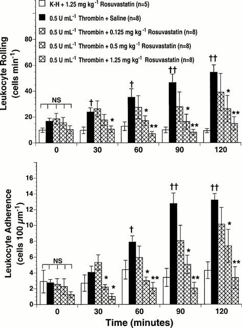
Effect of rosuvastatin on thrombin-stimulated leukocyte rolling (upper panel) and leukocyte adherence (lower panel) in rat mesenteric venules.Br J Pharmacol.2001 Jun;133(3):406-12. |
|---|
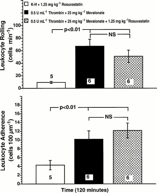
Mevalonic acid blocks the inhibitory effect of rosuvastatin on thrombin-stimulated leukocyte rolling (upper panel) and leukocyte adherence (lower panel).Br J Pharmacol.2001 Jun;133(3):406-12. |
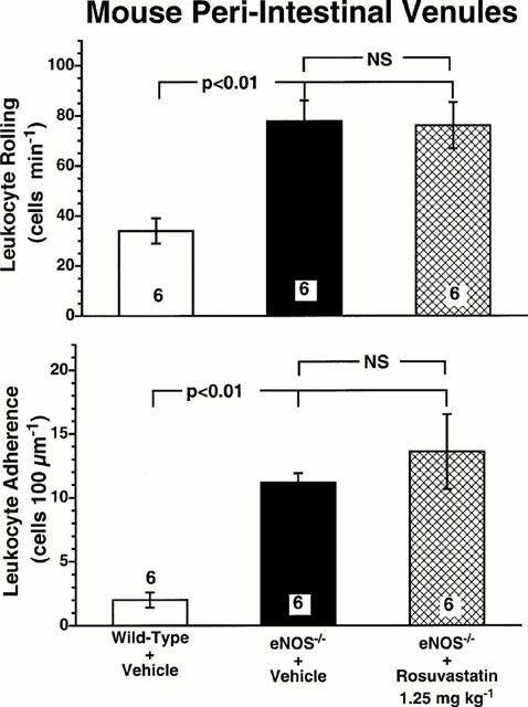
Leukocyte rolling (upper panel) and leukocyte adherence (lower panel) in peri-intestinal venules of wild-type mice, eNOS−/−mice, and eNOS−/−mice given 1.25 mg kg−1rosuvastatin.Br J Pharmacol.2001 Jun;133(3):406-12. |
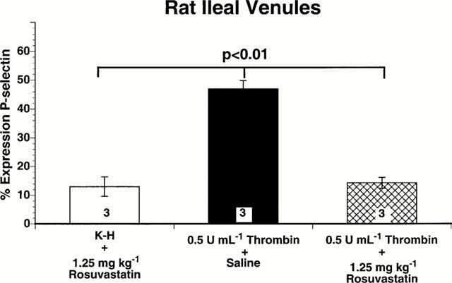
Immunohistochemical analysis of P-selectin expression on rat ileal venules, expressed as percentage of venules staining positive for P-selectin.Br J Pharmacol.2001 Jun;133(3):406-12. |
|---|
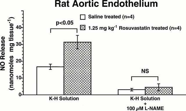
Effect of rosuvastatin on NO release in rat aortic segments. Basal release of nitric oxide is expressed as nanomoles per mg tissue.Br J Pharmacol.2001 Jun;133(3):406-12. |
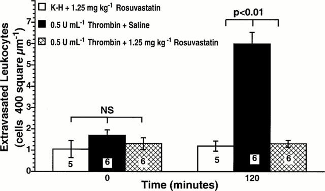
Effect of rosuvastatin on thrombin-stimulated leukocyte extravasation. Rat mesenteries were superfused with either K-H buffer alone or with 0.5 u ml−1thrombin. Rosuvastatin (1.25 mg kg−1) was administered intraperitoneally 18 h prior to the study.Br J Pharmacol.2001 Jun;133(3):406-12. |
Products are for research use only; We do not sell to patients
Copyright 2020 InvivoChem LLC | All Rights Reserved