| Size | Price | Stock | Qty |
|---|---|---|---|
| 25mg |
|
||
| 50mg |
|
||
| 100mg |
|
||
| 250mg |
|
||
| 500mg |
|
||
| Other Sizes |


Purity: ≥98%
| Targets |
p110α (IC50 = 3 nM); p110β (IC50 = 33 nM); p110δ (IC50 = 3 nM); p110γ (IC50 = 75 nM); p110α-H1047R (IC50 = 3 nM); p110α-E545K (IC50 = 3 nM); DNA-PK (IC50 = 1.23 μM); mTOR (Ki = 0.58 μM); Autophagy
Pictilisib (GDC-0941) dimesylate (CAS#: 957054-33-0) targets class I PI3K isoforms: PI3Kα (IC50 = 3 nM), PI3Kβ (IC50 = 33 nM), PI3Kδ (IC50 = 3 nM), PI3Kγ (IC50 = 75 nM). It does not significantly inhibit mTOR or other lipid/protein kinases. [4] In cell growth inhibition studies, GI50 values in colorectal cancer cell lines: HCT116 = 1081 nM, DLD1 = 1070 nM, HT29 = 157 nM (72 h, SRB assay). [3] |
|---|---|
| ln Vitro |
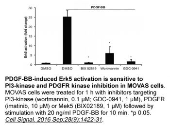
GDC-0941 is equipotent against PI3Kα and PI3Kδ as well as PI3Kα mutants E545-K and H1047-R, displaying modest levels of selectivity against PI3Kβ (10-fold) and PI3Kγ (25-fold), and greater levels of selectivity against members of PI3K class II, III, and IV, including C2β, Vps34, DNA-PK, and mTOR. With IC50 values of 46 nM, 37 nM, and 28 nM, respectively.[1] With an IC50 of 149-944 nM, GDC-0941 treatment effectively reduces the proliferation of HER2-amplified cells that are both trastuzumab-sensitive and -insensitive. With an IC50 of 500 nM or less, GDC-0941 effectively inhibits the proliferation of HER2-amplified cells that have PIK3CA mutations and the viability of HER2-amplified breast cancer cells that lack PTEN and are resistant to trastuzumab.[2] The growth of HCT116, DLD1, and HT29 cells is significantly inhibited by GDC-0941, with GI50 values of 1081 nM, 1070 nM, and 157 nM, respectively. [3] GDC-0941 suppresses the population of centroblasts, induces apoptosis, and inhibits the proliferation of tumor cells.[4]
Pictilisib (GDC-0941) dimesylate (CAS#: 957054-33-0) as a single agent showed growth inhibition in HCT116, DLD1 and HT29 cells with GI50 values of 1081 nM, 1070 nM and 157 nM respectively. [3] In combination with MEK inhibitors (AZD6244 or PD0325901), it exhibited marked synergistic growth inhibition in HCT116 and HT29 cells (combination index values: 0.045 for GDC-0941/PD0325901 at 1×GI50 in HCT116; 0.097 in HT29). [3] Western blot analysis in HCT116 and HT29 cells showed that GDC-0941 (at 1× and 10× GI50) inhibited S6 phosphorylation but had minimal effect on 4EBP1 phosphorylation. It did not consistently inhibit AKT phosphorylation except at 10× GI50 in HCT116. In combination with PD0325901, it enhanced inhibition of S6 phosphorylation. [3] In PTEN⁺/⁻ LKB1⁺/hypo mice with spontaneous B-cell follicular lymphoma, oral administration of Pictilisib (GDC-0941) dimesylate (CAS#: 957054-33-0) (75 mg/kg once daily) for 5 days markedly inhibited Akt activity and phosphorylation (Thr308 and Ser473), suppressed SGK1 activity and phosphorylation (Thr229 and Thr389), inhibited S6K activity (p-S6), increased electrophoretic mobility of 4E-BP1 with dephosphorylation at Thr37/Thr46 and Ser65, and inhibited NDRG1 phosphorylation (SGK1 substrate). [4] After 14 days of treatment, tumours shrank to ~40% of original size; after 42 days, tumours shrank to ~50% of original size. [4] Immunohistochemistry after 14 days showed reduced Ki67 reactivity (<5% vs 10-15% in controls), increased caspase-3 positive apoptotic cells, and diminished p-S6 staining in centroblasts. [4] After 42 days, Ki67 remained <5% and p-S6 reduced. [4] Flow cytometry showed that GDC-0941 treatment dramatically reduced the centroblast population within tumours. [4] Following cessation of drug treatment (after 42 days), tumours re-grew to original size within ~16 days, with increased growth rate (29 ± 6.5 mm³/week vs 11.8 ± 9.0 mm³/week in untreated). [4] |
| ln Vivo |
GDC-0941 has a 78% oral bioavailability due to limited microsomal metabolism [5]. In established human U87MG glioblastoma xenografts in female NCr athymic mice, administration of GDC-0941 at 75 mg/kg/day results in a significant inhibitory effect, with tumor growth inhibition of 83%. [1] In mice with HER2-amplified, trastuzumab-resistant MDA-MB-361.1 xenografts, oral administration of GDC-0941 at 150 mg/kg/day significantly slows tumor progression and induces potent tumor apoptosis. [2] Two weeks of treatment with GDC-0941 (75 mg/kg/day) results in a 40% reduction in the tumor size of spontaneous B-cell follicular lymphomas in PTEN+/-LKB1+/hypo mice. This tumor volume reduction is accompanied by the elimination of Akt, S6K, and SGK (serum and glucocorticoid protein kinase) protein phosphorylation.[4]
Enzyme assays for PI3K isoforms were performed using standard methods. Pictilisib (GDC-0941) dimesylate (CAS#: 957054-33-0) inhibited PI3Kα with IC50 of 3 nM, PI3Kβ 33 nM, PI3Kδ 3 nM, PI3Kγ 75 nM. It did not significantly inhibit any other lipid or protein kinases tested including mTOR. [4] In the colorectal cancer study, growth inhibition was measured using Sulforhodamine B (SRB) assay after 72 h exposure. Cells were treated with serial dilutions of compounds, fixed, stained with SRB, and absorbance measured. GI50 values were calculated. [3] |
| Enzyme Assay |
Scintillation proximity assay; Recombinant human PI3Kα, PI3Kβ, and PI3Kδ are coexpressed in a Sf9 baculovirus system with the p85α regulatory subunit and are purified as GST-fusion proteins using affinity chromatography on glutathione-sepharose. Scintillation proximity assay. Recombinant human PI3Kγ is similarly expressed and purified as monomeric GST-fusions. GDC-0941 is dissolved in DMSO and infused into a 50 μL mixture of 20 mM Tris-HCl (pH 7.5), 4 mM MgCl2, 1 mM DTT, 1 μM ATP, 0.125 μCi [γ-33P]-ATP, and 4% (v/v) DMSO. To start the kinase reaction, the assay mixture is mixed with the recombinant GST-fusion of PI3Kα (5 ng), PI3Kβ (5 ng), PI3Kδ (5 ng), or PI3Kγ (5 ng).
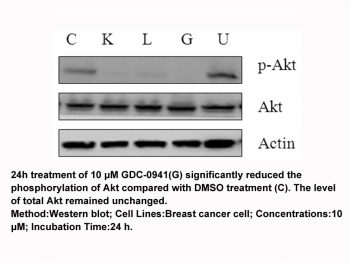
For cell growth inhibition (SRB assay): Cells were seeded in 96-well plates, treated with compounds for 72 h, then fixed with trichloroacetic acid, stained with SRB, and absorbance read at 540 nm. [3] For western blotting in cell lines: Cells were treated with compounds for 24 h, lysed, and proteins separated by SDS-PAGE. Antibodies against p-ERK, p-AKT, p-S6, p-4EBP1, and total proteins were used. [3] For flow cytometry in mouse tumours: Cells extracted from tumours were stained with antibodies against B220, TCRβ, κ and λ light chains, and analysed by flow cytometry. Forward/side scatter was used to identify centroblast population. [4] For immunohistochemistry: Formalin-fixed paraffin-embedded sections were stained with antibodies against B220, CD79a, CD3, Ki67, cleaved caspase-3, p-S6 (Ser235/236), and p-AKT (Ser473). [4] |
| Cell Assay |
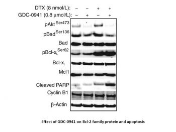
BT474-M1, SKBR-3, AU-565, HCC-1419, ZR75-30, JIMT-1, BT474-EEI, HCC-1954, MCF-7, CALU-3, SKOV-3, and MKN-7 cells are exposed to different concentrations of GDC-0941 for 48 and 72 hours. The CellTiter-Glo Luminescent Cell Viability Assay is used to identify cell viability and proliferation. By using a western blot, the pAkt (Ser473), cleaved caspase-3, and cleaved PARP are all examined. Apoptosis and caspase 3/7 activity are both detected using the Cell Death Detection ELISAplus assay and the Caspase-Glo 3/7 assay, respectively.
For the combination study in colorectal cancer cells (literature 3), no animal experiments were performed with GDC-0941 alone; only in vitro studies. [3] For the spontaneous lymphoma study in PTEN⁺/⁻ LKB1⁺/hypo mice (literature 4): 6-8 month old mice bearing visible cervical lymph node tumours were used. Pictilisib (GDC-0941) dimesylate (CAS#: 957054-33-0) was dissolved in 30% captisol (w/v) and administered by oral gavage once daily at 75 mg/kg for up to 42 days. Vehicle control was 30% captisol. Tumour volume was monitored by MRI (3D RARE-4) at days 0, 14, 28, 42, and after cessation at days 63, 77. After 5 days treatment, mice were culled 2 h after final dose for biochemical analysis. After 14 or 42 days, tumours were collected for histology and immunoblotting. [4] |
| Animal Protocol |
Under the skin, MCF7-neo/HER2 or MX-1 breast cancer cells are injected into female nu/nu mice. Animals are distributed into groups of 10 animals each when tumors reach a mean volume of 200 to 250 mm3. Group sizes are determined by size matching. Once a week, intravenous RP-56976, a formulation of 3% EtOH and 97% saline, is given. Pictilisib (GDC-0941), a daily oral dose of MCT (0.5% methylcellulose, 0.2% Tween-80), is administered. By directly implanting tumors from patients under the skin of NMRI nu/nu mice, the MAXF1162 HER2+/ER+/PR+ patient-derived breast cancer tumor xenograft model was created. Volume of the tumor is calculated. Throughout a study, tumor size measurements are taken twice a week.
Preclinical animal studies: Female BALB/c mice or NCr athymic mice (6‑8 wk old) bearing s.c. U87MG or IGROV‑1 xenografts. Pictilisib (dimesylate salt) was formulated in 10% DMSO, 5% Tween 20, 85% saline and administered orally (p.o.) once daily at doses of 25, 50, 100, 150 mg/kg for up to 19 d. Tumor volumes measured bi‑weekly, and at termination tumors were weighed. Pharmacokinetic studies used i.v. (10 mg/kg) and p.o. (10 mg/kg) dosing in BALB/c mice for bioavailability determination [5]. Clinical trial: First‑in‑human phase I dose‑escalation study in patients with advanced solid tumors. Pictilisib was administered orally once daily on days 1‑21 every 28 days (21/28 schedule) initially, later continuous daily dosing (28/28). Starting dose 15 mg, escalated to 450 mg. Dose‑limiting toxicities (DLTs) and maximum tolerated dose (MTD) determined using modified 3+3 design. RP2D was 330 mg once daily continuous. Pharmacokinetic sampling on days 1 and 15 at multiple time points (pre‑dose, 0.5,1,2,3,4,8,24,48,72 h). Pharmacodynamic assessments included pAKT in platelet‑rich plasma, tumor biopsies for pS6 and pAKT, plasma insulin/glucose, and 18F‑FDG‑PET scans [6]. |
| ADME/Pharmacokinetics |
Pharmacokinetics [6]
Pharmacokinetic parameters of Pitticilib for all dose groups were estimated and summarized in Table 3 and Supplementary Table 1. In the fasting state, Pitticilib was rapidly absorbed after oral administration (median Tmax was 2 hours [range 0.5–8 hours]); this was independent of dose and remained constant after multiple administrations. The terminal plasma elimination half-life (T1/2) on day 1 ranged from 13.1 to 24.1 hours. Within the studied dose range, drug exposure (Cmax and AUC0–24) was observed to increase proportionally with dose (Figure 1). Similar pharmacokinetic characteristics were observed on day 15. The cumulative index (AUCDay15/AUCDay1) ranged from 1.2 to 2.2, indicating minimal drug accumulation after multiple administrations. The absorption, metabolism, and excretion of [14C]Pitticilib (a selective class 1A phosphatidylinositol 3-kinase (PI3K) small molecule inhibitor) were characterized in rats, dogs, and humans following single oral administration of target doses of 30 mg/kg, 5 mg/kg, and 60 mg, respectively. Pitticilib was rapidly absorbed in all species, with a time to peak concentration (Tmax) of less than 2 hours. In systemic circulation, Pitticilib constituted the major component of total radioactivity in all species, exceeding 86.6%. In rats, dogs, and humans, 98%, 80%, and 95% of Pitticilib and its associated radioactivity were recovered in urine and feces, respectively, with less than 2% excreted in urine and the remainder in feces. In rats and dogs, over 40% of drug-related radioactivity was excreted via bile, indicating that bile excretion is the primary route of excretion. Unmetabolized Pitticilib was present in low concentrations in the bile of rats and dogs. In rats, the major metabolite in bile was O-glucuronide of partially oxidized indazole (M20, 21% of the dose), while in dogs it was M7, the ring-opening metabolite of oxidized piperazine (10.8% of the dose). Novel metabolites detected in rat bile were oxidized glutathione (GSH) conjugates (M18, M19), suggesting that Pitticilib may generate active intermediates. Nuclear magnetic resonance (NMR) further confirmed that the structure of M18 was an N-hydroxylated and GSH-conjugated metabolite of the indazole ring. Xenobiotica. 2021 Jul;51(7):796-810. https://pubmed.ncbi.nlm.nih.gov/33938357/ Preclinical mouse PK: Following p.o. administration of Pictilisib (10 mg/kg free base) to female BALB/c mice, oral bioavailability was 78%. After 50 mg/kg p.o. to U87MG tumor‑bearing mice, Cmax in plasma and tumor reached at 0.5 h, with tumor/plasma ratio ~0.8. Tumor concentrations remained above GI50 for >8 h. Plasma half‑life ~4.5 h. Volume of distribution (Vdss) 1.9 L/kg, clearance 33 mL/min/kg [5]. Human clinical PK: Pictilisib was rapidly absorbed with median Tmax of 2 h (range 0.5‑8 h). Terminal half‑life on day 1 ranged from 13.1 to 24.1 h. Dose‑proportional increases in Cmax and AUC0‑24 were observed across 15‑450 mg doses. Accumulation index (AUC day 15/day 1) ranged from 1.2 to 2.2. For the RP2D of 330 mg, geometric mean Cmax = 2.07 μM and AUC0‑24 = 22.8 hr·μM on day 15 [6]. |
| Toxicity/Toxicokinetics |
Safety and Tolerability [6] Pitticilib was well tolerated at a dose of 330 mg (21/28 dosing regimen); most adverse events were mild to moderate, and there were no treatment-related deaths (Table 2). At the dose levels assessed, the toxicity profiles of the 21/28 and 28/28 dosing regimens appeared to be not significantly different. Treatment-related adverse events occurring in ≥10% of patients included nausea, diarrhea, vomiting, fatigue, dysgeusia, decreased appetite, and rash. In addition to two dose-limiting toxicities (DLT) of grade 3 rash in the 450 mg dose group, a third patient in this dose group also developed a grade 2 rash; despite this, the patient received 8 months of Pitticilib treatment concurrently with oral antihistamines and emollients. Of the 10 patients treated with 330 mg once daily (28/28 regimen), 2 developed grade 1 or 2 rashes and 2 developed grade 3 rashes (after the dose-limiting toxicity definition window); all of these rashes subsided after discontinuation of the drug and the use of supportive care, including emollients and corticosteroids. [6] Other clinically significant grade ≥3 drug-related adverse events included grade 4 hyperglycemia (n=1, 130 mg) and grade 3 pneumonia (n=1, 340 mg). Grade 4 hyperglycemia was transient, without clinically significant symptoms, signs or acidosis, and occurred in a patient with cholangiocarcinoma who had previously undergone pancreaticoduodenectomy and had started low-dose prednisolone 2 days prior to the event. A breast cancer patient who had previously received thoracic radiotherapy developed grade 3 pneumonia at the end of the first treatment cycle, accompanied by grade 1 dyspnea, decreased DLCO, and ground-glass opacities on high-resolution CT (HRCT); these symptoms were relieved after 2 weeks of discontinuation of the drug and concurrent use of prednisolone. Dyspnea and HRCT changes recurred when Pitticilib was restarted at 240 mg; these symptoms were relieved again after permanent discontinuation of Pitticilib due to disease progression.
Dose-limiting toxicity (DLT) and maximum tolerated dose (MTD) [6] The MTD of once daily 450 mg (21/28 regimen) was exceeded, with two patients experiencing a grade 3 rash as a DLT. This was a maculopapular rash covering 70-80% of the body surface area, which appeared approximately 2 weeks after the start of daily Pitticilib and resolved spontaneously 2 weeks after discontinuation of the drug. In the once-daily 330 mg (21/28 dosing regimen) treatment regimen, one in seven patients developed a grade 3 maculopapular rash with a similar onset and resolution time pattern; this was also determined to be a dose-limiting toxicity (DLT). No DLT was observed in the 28/28 dosing regimen. Pictilisib (GDC-0941) dimesylate (CAS#: 957054-33-0) is a pan-class I PI3K inhibitor that is more synergistic with MEK inhibitors than the dual PI3K/mTOR inhibitor NVP-BEZ235 in colorectal cancer cells. The reduced synergy of NVP-BEZ235 is attributed to its direct mTOR inhibition, which compromises the synergy. Addition of the mTORC1/2 inhibitor KU0063794 to GDC-0941/PD0325901 combination reduced synergy in a concentration-dependent manner. [3] In the PTEN/LKB1-deficient mouse lymphoma model, GDC-0941 treatment induced tumour shrinkage but not complete regression. After drug withdrawal, tumours regrew at an accelerated rate and progressed to diffuse large B-cell lymphoma. [4] |
| References | |
| Additional Infomation |
Pitticilib dimethylsulfonate is a highly bioavailable oral dimethylsulfonate of pitticilib. Pitticilib is a class I phosphatidylinositol 3-kinase (PI3K) small molecule inhibitor with potential antitumor activity. After administration, pitticilib selectively binds to PI3K in an ATP-competitive manner, inhibiting the production of the second messenger phosphatidylinositol-3,4,5-triphosphate (PIP3) and the activation of the PI3K/Akt signaling pathway. This may lead to suppression of the growth, migration, and survival of susceptible tumor cell populations. Activation of the PI3K/Akt signaling pathway is commonly associated with tumorigenesis. Dysregulation of the PI3K/Akt signaling pathway may lead to resistance to multiple antitumor drugs in tumors. Phosphatidylinositol-3-kinase (PI3K) is an important target for cancer therapy because the PI3K/Akt signaling pathway is dysregulated in various tumors. We synthesized a series of thieno[3,2-d]pyrimidine derivatives and evaluated their activity as inhibitors of PI3 kinase p110α. The synthesis, bioactivity and further characterization of these compounds are described in this paper. This work identified compound 17, GDC-0941, a potent, selective, and orally bioavailable PI3K inhibitor currently undergoing human clinical trials for the treatment of cancer. [1] Herceptin (trastuzumab) is a cornerstone of HER2-targeted breast cancer therapy, benefiting patients with adjuvant and metastatic breast cancer. The mechanism of action of trastuzumab is described in this paper, in which antibody therapy disrupts the ligand-independent HER2/HER3 interaction in HER2-amplified cells. This dissociation kinetic parallels HER3 dephosphorylation and uncoupling from PI3K activity, leading to downregulation of the proximal and distal AKT signaling pathways and is associated with the antiproliferative effect of trastuzumab. The selective and potent PI3K inhibitor GDC-0941, when used in combination with trastuzumab, showed extremely high efficacy in treating trastuzumab-resistant cells and tumors. [2]
Background: Combination targeting of MAPK and PI3K signaling pathways may be a necessary condition for achieving optimal efficacy in cancer treatment. This study evaluated the effects of MEK inhibitors AZD6244 and PD0325901 alone and in combination with the mTOR/PI3K dual inhibitor NVP-BEZ235 or the PI3K inhibitor GDC-0941 on three colorectal cancer cell lines. Methods: The growth inhibition, survival, and signal transduction of HCT116, HT29, and DLD1 cell lines were detected by sulforhodamine B (SRB) assay, colony formation assay, and Western blotting, respectively. Results: All MEK/PI3K inhibitor combinations showed significant synergistic growth inhibition; however, GDC-0941 showed a stronger synergistic effect when used in combination with any MEK inhibitor. Compared to GDC-0941, NVP-BEZ235 exhibited stronger inhibition of 4EBP1 phosphorylation, while its inhibitory effects on S6 and AKT phosphorylation were similar to those of GDC-0941. Both PD0325901 and AZD6244 inhibited ERK phosphorylation, and the inhibitory effect on S6 phosphorylation was enhanced when used in combination with MEK/PI3K inhibitors. Compared to GDC-0941, the synergistic effect of NVP-BEZ235 in combination with MEK inhibitors was reduced, possibly due to mTOR inhibition. Furthermore, the addition of the mTORC1/2 inhibitor KU0063794 weakened the synergistic effect of GDC-0941 in combination with PD0325901. Conclusion: These studies confirm that the dual targeting of PI3K and MEK can induce synergistic growth inhibition; however, compared with mTOR/PI3K dual inhibitors, the combination of specific PI3K inhibitors and MEK inhibitors produces a greater synergistic effect. [3] Pictilisib (GDC‑0941) is a potent, selective, orally bioavailable pan‑class I PI3K inhibitor that entered phase I clinical trials under Genentech. It demonstrated on‑target pharmacodynamic effects including >90% reduction of pAKT in platelet‑rich plasma at the RP2D, increases in plasma insulin and glucose (mean fold‑change 3.58 and 1.18 at 1 h post‑dose at 330 mg), and >25% decrease in 18F‑FDG‑PET SUVmax in 7/32 evaluable patients (22%). The recommended phase II dose is 330 mg once daily continuous. Further clinical development includes combination trials with erlotinib, fulvestrant, trastuzumab emtansine, MEK inhibitors, paclitaxel, and chemotherapy regimens [6]. |
| Molecular Formula |
C25H35N7O9S4
|
|
|---|---|---|
| Molecular Weight |
705.84
|
|
| Exact Mass |
705.137
|
|
| Elemental Analysis |
C, 42.54; H, 5.00; N, 13.89; O, 20.40; S, 18.17
|
|
| CAS # |
957054-33-0
|
|
| Related CAS # |
Pictilisib;957054-30-7
|
|
| PubChem CID |
56972143
|
|
| Appearance |
White to light yellow solid powder
|
|
| Melting Point |
>280°C (dec.)
|
|
| Hydrogen Bond Donor Count |
3
|
|
| Hydrogen Bond Acceptor Count |
16
|
|
| Rotatable Bond Count |
5
|
|
| Heavy Atom Count |
45
|
|
| Complexity |
924
|
|
| Defined Atom Stereocenter Count |
0
|
|
| SMILES |
O=S(C)(O)=O.O=S(C)(N1CCN(CC2=CC3N=C(C4C5=C(NN=C5)C=CC=4)N=C(C=3S2)N2CCOCC2)CC1)=O
|
|
| InChi Key |
RFRIKACSFOTIMU-UHFFFAOYSA-N
|
|
| InChi Code |
InChI=1S/C23H27N7O3S2.2CH4O3S/c1-35(31,32)30-7-5-28(6-8-30)15-16-13-20-21(34-16)23(29-9-11-33-12-10-29)26-22(25-20)17-3-2-4-19-18(17)14-24-27-19;2*1-5(2,3)4/h2-4,13-14H,5-12,15H2,1H3,(H,24,27);2*1H3,(H,2,3,4)
|
|
| Chemical Name |
4-[2-(1H-indazol-4-yl)-6-[(4-methylsulfonylpiperazin-1-yl)methyl]thieno[3,2-d]pyrimidin-4-yl]morpholine;methanesulfonic acid
|
|
| Synonyms |
|
|
| HS Tariff Code |
2934.99.9001
|
|
| Storage |
Powder -20°C 3 years 4°C 2 years In solvent -80°C 6 months -20°C 1 month Note: Please store this product in a sealed and protected environment, avoid exposure to moisture. |
|
| Shipping Condition |
Room temperature (This product is stable at ambient temperature for a few days during ordinary shipping and time spent in Customs)
|
| Solubility (In Vitro) |
|
|||
|---|---|---|---|---|
| Solubility (In Vivo) |
|
| Preparing Stock Solutions | 1 mg | 5 mg | 10 mg | |
| 1 mM | 1.4168 mL | 7.0838 mL | 14.1675 mL | |
| 5 mM | 0.2834 mL | 1.4168 mL | 2.8335 mL | |
| 10 mM | 0.1417 mL | 0.7084 mL | 1.4168 mL |
*Note: Please select an appropriate solvent for the preparation of stock solution based on your experiment needs. For most products, DMSO can be used for preparing stock solutions (e.g. 5 mM, 10 mM, or 20 mM concentration); some products with high aqueous solubility may be dissolved in water directly. Solubility information is available at the above Solubility Data section. Once the stock solution is prepared, aliquot it to routine usage volumes and store at -20°C or -80°C. Avoid repeated freeze and thaw cycles.
Calculation results
Working concentration: mg/mL;
Method for preparing DMSO stock solution: mg drug pre-dissolved in μL DMSO (stock solution concentration mg/mL). Please contact us first if the concentration exceeds the DMSO solubility of the batch of drug.
Method for preparing in vivo formulation::Take μL DMSO stock solution, next add μL PEG300, mix and clarify, next addμL Tween 80, mix and clarify, next add μL ddH2O,mix and clarify.
(1) Please be sure that the solution is clear before the addition of next solvent. Dissolution methods like vortex, ultrasound or warming and heat may be used to aid dissolving.
(2) Be sure to add the solvent(s) in order.
Products are for research use only; We do not sell to patients
Copyright 2020 InvivoChem LLC | All Rights Reserved


 COA
COA