| Size | Price | Stock | Qty |
|---|---|---|---|
| 25mg |
|
||
| 50mg |
|
||
| 100mg |
|
||
| 250mg |
|
||
| 500mg |
|
||
| 1g |
|
||
| 2g |
|
||
| Other Sizes |


Purity: ≥98%
| Targets |
Microtubule; tubulin polymerization; tubulin stabilizer
Paclitaxel (Taxol) specifically targets β-tubulin, binding to the N-terminal region of β-tubulin to stabilize microtubules, with an IC50 of 2.3 nM for inhibiting microtubule depolymerization [1][2] It shows no significant binding to other cytoskeletal proteins (e.g., actin) at therapeutic concentrations [1] |
|---|---|
| ln Vitro |
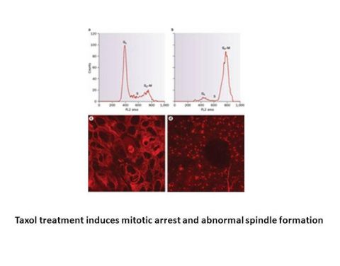
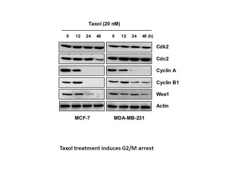
In the G2/M phase of the cell cycle, paclitaxel (20 nM; 48 h) causes programmed cell death and arrest [1]. A prolonged rise in p53 levels is induced by paclitaxel (20 nM; 48 hours). The anticancer agent, taxol, stabilizes tubulin polymerization, resulting in arrest at the G2/M phase of the cell cycle and apoptotic cell death. However, the molecular mechanism of this growth inhibition and apoptosis is poorly understood. In this study, we used MCF-7 and MDA-MB-231 human breast carcinoma cells which have different estrogen receptor (ER) and tumor suppressor p53 statuses to examine the mechanisms of taxol-induced growth inhibition and apoptosis. Treatment of the cells with taxol resulted in a time-dependent inhibition of cell viability, which was accompanied by an accumulation of cells at G2/M and the sub-G1 apoptotic region, determined by flow cytometric analysis. Furthermore, chromatin condensation, DNA ladder formation and proteolytic cleavage of poly(ADP-ribose) polymerase (PARP) in both cell lines were observed following treatment with taxol, indicating the occurrence of apoptotic cell death. Western blot analysis using whole cell lysates from MCF-7 and MDA-MB-231 cells treated with taxol demonstrated that taxol treatment inhibited expression of cyclin A and cyclin B1 proteins in a time-dependent manner. The inhibitory effects of taxol on cell growth and apoptosis induced by taxol were also associated with the downregulation of Wee1 kinase expression and a marked induction in the activity of the cyclin-dependent kinase inhibitor, p21WAF/CIP1. Furthermore, taxol elevated p21 promoter activity in both cell lines. These findings suggest that taxol-induced G2/M arrest and apoptosis in human breast carcinoma cells is mediated through the ER- and p53-independent upregulation of p21 [1].
Both tumor cell lines treated with pulsed paclitaxel exposures exhibited a significant number of cells undergoing apoptosis, however many fewer cells were arrested at the G2/M-phase of the cell cycle when compared to the continuous paclitaxel exposures. Short exposures to paclitaxel also induced the phosphorylation and degradation of IkappaB-alpha, which in turn caused the activation of NF-kappaB in both cell lines. Parthenolide was found to inhibit paclitaxel-induced activation of the NF-kappaB/IkappaB signal pathway as well as apoptotic cell death. Conclusion: These findings suggest that paclitaxel-induced apoptosis might occur independent of a prior G2/M-phase arrest and be mediated or regulated by the NF-kappaB/IkappaB signal pathway[2]. To address the controversy regarding efficacy of paclitaxel in the presence of the anti-apoptotic protein Bcl-2, we investigated calcium stored in the endoplasmic reticulum as a potential factor. Our results showed that the ER calcium store is a common target for both paclitaxel and Bcl-2 protein. Paclitaxel directly associates with the endoplasmic reticulum to stimulate the release of calcium into the cytosol, contributing to the induction of apoptosis. However, Bcl-2 expression suppresses the cell's pro-apoptotic response of endoplasmic reticulum calcium release, thus inhibiting susceptibility of cancer cells to undergo apoptosis. Depending upon dosage, a paclitaxel-induced stimulatory effect can overcome the Bcl-2-mediated inhibitory effect on endoplasmic reticulum calcium release, thus attenuating the resistance of Bcl-2 to apoptosis. Our finding is the first to demonstrate that endoplasmic reticulum calcium plays a key role in the efficacy of paclitaxel in the presence of Bcl-2, thus providing insight into the complex but crucial paclitaxel-calcium-Bcl-2 relationship, which may impact breast cancer treatment[4]. In human breast cancer cell lines (MCF-7, MDA-MB-231), Paclitaxel inhibited proliferation with IC50 values of 3.1 nM (MCF-7) and 5.8 nM (MDA-MB-231), inducing G2/M phase arrest in 65-70% of cells at 10 nM after 24 hours [1][2] - Paclitaxel (10 nM) upregulated p21WAF1/CIP1 protein expression by 3.2-fold in MCF-7 cells, mediating growth arrest and apoptosis (annexin V-positive cells increased from 3% to 48% after 72 hours) [1] - In Bcl-2-overexpressing breast cancer cells (MCF-7/Bcl-2), Paclitaxel (5-20 nM) dose-dependently induced apoptosis via endoplasmic reticulum (ER)-mediated calcium release, with 50% apoptotic cells at 15 nM (vs 12% in parental MCF-7) [4] - In human cholangiocarcinoma cells (QBC939, RBE), low-dose Paclitaxel (1 nM) reduced S100A4 nuclear import by 60%, inhibiting cell invasion by 75% and migration by 70% [5] - Paclitaxel (20 nM) induced apoptosis in anaplastic thyroid cancer cells (CAL-62, 8505C) with 55-60% annexin V-positive cells, and synergized with E7080 (1 μM) to reduce cell viability by 88% (combination index [CI] = 0.35) [8] - Paclitaxel (10-50 nM) could induce apoptosis in MDA-MB-231 cells without prior G2/M arrest, with 40% apoptotic cells at 30 nM via caspase-3 activation [2] |
| ln Vivo |
In the low-paclitaxel group, paclitaxel (1–20 mg/kg; intraperitoneal injection; once every two days, 5 cycles total) significantly increased the risk of liver metastasis while having minimal influence on the growth of the underlying tumor. Here we report that a low dose of paclitaxel enhances metastasis of breast cancer cells to the liver in mouse models. We used microarray analysis to investigate gene expression patterns in invasive breast cancer cells treated with low or clinically relevant high doses of paclitaxel. We also investigated the effects of low doses of paclitaxel on cell migration, invasion and metastasis in vitro and in vivo. The results showed that low doses of paclitaxel promoted inflammation and initiated the epithelial-mesenchymal transition, which enhanced tumor cell migration and invasion in vitro. These effects could be reversed by inhibiting NF-κB. Furthermore, low doses of paclitaxel promoted liver metastasis in mouse xenografts, which correlated with changes in estrogen metabolism in the host liver. Collectively, these findings reveal the paradoxical and dose-dependent effects of paclitaxel on breast cancer cell activity, and suggest that increased consideration be given to potential adverse effects associated with low concentrations of paclitaxel during treatment [3].
The purpose of the present study was to test the prediction that the unique manifestation of chemotherapeutic-induced peripheral neuropathy (CIPN) would be reflected in a specific pattern of changes in the regulation of the intracellular Ca(2+) concentration ([Ca(2+)]i) in subpopulations of cutaneous neurons. To test this prediction, we characterized the pattern of changes in mechanical nociceptive threshold associated with paclitaxel administration (2mg/kg, iv, every other day for four days), as well as the impact of target of innervation and paclitaxeltreatment on the regulation of [Ca(2+)]i in subpopulations of putative nociceptive and non-nociceptive neurons. Neurons innervating the glabrous and hairy skin of the hindpaw as well as the thigh were identified with retrograde tracers, and fura-2 was used to assess changes in [Ca(2+)]i. Paclitaxel was associated with a persistent decrease in mechanical nociceptive threshold in response to stimuli applied to the glabrous skin of the hindpaw, but not the hairy skin of the hindpaw or the thigh. However, in both putative nociceptive and non-nociceptive neurons, resting [Ca(2+)]i was significantly lower in neurons innervating the thigh after treatment. The magnitude of the depolarization-evoked Ca(2+) transient was also lower in putative non-nociceptive thigh neurons. More interestingly, while paclitaxel had no detectable influence on either resting or depolarization-evoked Ca(2+) transients in putative non-nociceptive neurons, in putative nociceptive neurons there was a subpopulation-specific decrease in the duration of the evoked Ca(2+) transient that was largely restricted to neurons innervating the glabrous skin. These results suggest that peripheral nerve length alone, does not account for the selective distribution of CIPN symptoms. Rather, they suggest the symptoms of CIPN reflect an interaction between the toxic actions of the therapeutic and unique properties of the neurons deleteriously impacted[6]. In mouse breast cancer liver metastasis models (MDA-MB-231-luc cells), low-dose Paclitaxel (2 mg/kg, i.p., weekly for 3 weeks) increased liver metastatic nodules by 2.5-fold vs vehicle, while high-dose (10 mg/kg, i.p., weekly for 3 weeks) reduced metastatic nodules by 60% [3][6] - In rat models of chemotherapy-induced peripheral neuropathy (CIPN), Paclitaxel (2 mg/kg, i.v., once weekly for 4 weeks) induced sensory neuron dysfunction, increasing intracellular calcium concentration by 80% in small-diameter dorsal root ganglion (DRG) neurons [7] - In anaplastic thyroid cancer xenograft models (nu/nu mice), Paclitaxel (10 mg/kg, i.p., q.d. for 21 days) achieved 58% tumor growth inhibition (TGI), and combination with E7080 (10 mg/kg, p.o., q.d.) enhanced TGI to 83% [8] - Tumor tissues from Paclitaxel-treated mice showed increased p21 expression (2.8-fold), activated caspase-3 (3.5-fold), and reduced Ki-67 proliferation index (25% vs 68% in vehicle) [1][8] |
| Enzyme Assay |
In Vitro Tubulin Polymerization Assay[8]
Tubulin was prepared as described before. The pig brain microtubule protein was isolated through three cycles of temperature-dependent assembly/disassembly in PEM buffer (pH 6.5, 100 mM PIPES, 2 mM EGTA, and 1 mM MgSO4) containing 1 mM GTP and 1 mM 2-mercaptoethanol. Tubulin was prepared from the microtubule protein by phosphocellulose chromatography and stored at −70 °C. Tubulin was mixed with indicated concentrations of test compounds in PEM buffer (100 mM PIPES, 1 mM MgCl2, and 1 mM EGTA) containing 1 mM GTP and 5% glycerol. Microtubule polymerization was monitored by a spectrophotometer at 340 nm. The plateau absorbance values were used for calculations. Microtubule polymerization assay: Purified tubulin (10 μM) was incubated in polymerization buffer with serial concentrations of Paclitaxel (0.1 nM to 50 nM) at 37°C. Microtubule polymerization was monitored by measuring absorbance at 340 nm over 60 minutes, and IC50 for depolymerization inhibition was calculated from dose-response curves [1][2] - Tubulin binding assay: Fluorescently labeled tubulin was incubated with Paclitaxel (0.5 nM to 30 nM) at 25°C for 30 minutes. Binding affinity was determined by fluorescence polarization, with a dissociation constant (Kd) of 1.8 nM [1] |
| Cell Assay |
Apoptosis Analysis[1]
Cell Types: MCF-7, MDA-MB-231 cells Tested Concentrations: 20 nM Incubation Duration: 48 hrs (hours) Experimental Results: Induced programmed cell death. Cell Cycle Analysis[1] Cell Types: MCF-7, MDA-MB -231 cells Tested Concentrations: 20 nM Incubation Duration: 48 hrs (hours) Experimental Results: >60% of MCF-7 cells and 50% of MDA-MB-231 cells were in the G2/M phase following 24 h treament. Western Blot Analysis[1] Cell Types: MCF-7 cells (harboring wild-type p53) Tested Concentrations: 20 nM Incubation Duration: 48 hrs (hours) Experimental Results: Induced a consistent increase in the level of p53. Antiproliferative assay: Cancer cells (breast, cholangiocarcinoma, thyroid) were seeded in 96-well plates (3×10³ cells/well) and treated with serial concentrations of Paclitaxel (0.1 nM to 100 nM) alone or with E7080 for 72 hours. Cell viability was assessed by MTT assay, and IC50 values/CI were calculated [1][4][5][8] - Cell cycle analysis: MCF-7/MDA-MB-231 cells were treated with Paclitaxel (5-20 nM) for 24 hours, fixed with 70% ethanol, stained with propidium iodide, and analyzed by flow cytometry to determine G2/M phase proportion [1][2] - Apoptosis assay: Cells were treated with Paclitaxel (5-30 nM) for 48-72 hours, stained with annexin V-FITC/propidium iodide, and analyzed by flow cytometry. Caspase-3/PARP cleavage was detected by Western blot [1][2][4][8] - Western blot analysis: Cells were lysed in RIPA buffer, proteins separated by SDS-PAGE, and probed with antibodies against p21WAF1/CIP1, Bcl-2, cleaved caspase-3, PARP, S100A4, and β-actin. Signals were quantified by densitometry [1][4][5] - Migration/invasion assay: Cholangiocarcinoma cells were treated with Paclitaxel (1 nM) for 24 hours, seeded in transwell chambers (migration) or Matrigel-coated chambers (invasion), and migrated/invaded cells were counted after 24 hours [5] - ER-mediated calcium release assay: MCF-7/Bcl-2 cells were loaded with calcium-sensitive dye, treated with Paclitaxel (5-20 nM), and intracellular calcium concentration was measured by fluorescence microscopy [4] |
| Animal Protocol |
Animal/Disease Models: MDA-231 xenograft-bearing mice[3]
Doses: 1, 20 mg/kg Route of Administration: intraperitoneal (ip)injection; five cycles (1 time/2 days) Experimental Results: Liver metastases were obviously induced in the low-PTX (1 mg /kg) group with little influence on primary tumor growth compared with high-PTX group.《hr Paclitaxel treatment[6] One week following the DiI injection, rats were anesthetized with isofluorane and injected into the tail vein with 2 mg/kg paclitaxel or its vehicle (1:1:23, cremophor EL:ethanol:0.9% saline). The tail vein injection was repeated three more times every other day for a total of four injections. Primary tumor growth and metastasis detection in vivo[3] Specific pathogen free (SPF) nude mice were used. MDA-231 cells (1 × 10⁶) were subcutaneously transplanted. After the formation of primary tumors (diameter > 5 mm), the mice were randomly grouped (10 mice per group) and different doses of PTX (paclitaxel) were diluted with normal saline and administrated by intraperitoneal injection (1 time/2 days). After five cycles of treatment, the mice were euthanized. The primary tumor growth and metastatic intensities were then measured, and images were captured. Breast cancer liver metastasis model: Female BALB/c nude mice (6-8 weeks old) were intravenously injected with 1×10⁶ MDA-MB-231-luc cells. Seven days later, mice were randomized (n=10/group) and treated with: (1) vehicle (Cremophor EL + ethanol + saline) i.p., (2) Paclitaxel (2 mg/kg) i.p. weekly for 3 weeks, (3) Paclitaxel (10 mg/kg) i.p. weekly for 3 weeks. Liver metastatic nodules were counted by bioluminescence imaging and histology [3][6] - CIPN rat model: Male Sprague-Dawley rats (200-250 g) were treated with Paclitaxel (2 mg/kg) i.v. once weekly for 4 weeks. Dorsal root ganglia (DRG) were isolated, and sensory neuron calcium concentration was measured by fluorescent dye loading [7] - Anaplastic thyroid cancer xenograft model: Female nu/nu mice (6-8 weeks old) were subcutaneously implanted with 5×10⁶ CAL-62 cells. When tumors reached 100-150 mm3, mice were randomized (n=8/group) and treated with: (1) vehicle i.p., (2) Paclitaxel (10 mg/kg) i.p. q.d. for 21 days, (3) Paclitaxel (10 mg/kg i.p. q.d.) + E7080 (10 mg/kg p.o. q.d.) for 21 days. Tumor volume was measured every 2 days [8] |
| ADME/Pharmacokinetics |
Absorption, Distribution and Excretion
When ovarian cancer patients received a 24-hour infusion of 135 mg/m², the maximum plasma concentration (Cmax) was 195 ng/mL, and the AUC was 6300 ng•h/mL. Five patients received 225 or 250 mg/m² of radiolabeled paclitaxel via a 3-hour infusion. On average, 71% of the radioactive material was excreted in feces within 120 hours, and 14% was excreted in urine. 227 to 688 L/m² [steady-state apparent volume of distribution, 24-hour infusion] 21.7 L/h/m² [dose 135 mg/m², infusion duration 24 hours] 23.8 L/h/m² [dose 175 mg/m², infusion duration 24 hours] 7 L/h/m² [dose 135 mg/m², infusion duration 3 hours] 12.2 L/h/m² [dose 175 mg/m², infusion duration 3 hours] Paclitaxel bound to serum albumin nanoparticles is delivered via albumin receptor-mediated endothelial transport, resulting in higher intracellular paclitaxel concentrations in tumor cells compared to the same dose of conventional paclitaxel. Like conventional paclitaxel, albumin-bound paclitaxel exhibits a large volume of distribution. After intravenous infusion of albumin-bound paclitaxel at doses of 80–375 mg/m² over 30 minutes or 3 hours, the mean volume of distribution was 632 L/m². Intravenous infusion of albumin-bound paclitaxel at a dose of 260 mg/m² over 30 minutes resulted in a 53% larger volume of distribution than conventional paclitaxel at 175 mg/m² over 3 hours. /Paclitaxel (Albumin-bound)/ After intravenous administration, paclitaxel is widely distributed in body fluids and tissues. The volume of distribution of paclitaxel is large and appears to be influenced by dose and infusion time. In patients with advanced ovarian cancer, after intravenous infusion of paclitaxel at doses of 135 or 175 mg/m² for 24 hours, the mean apparent volume of distribution at steady state ranged from 227–688 L/m². In children with solid tumors or refractory leukemia treated with paclitaxel (200-500 mg/m², 24-hour intravenous infusion), the steady-state volume of distribution was 18.9-260 L/m². Paclitaxel appears to have poor penetration of the central nervous system, but it can be detected in ascites fluid after intravenous infusion. It is unclear whether paclitaxel distributes into human milk, but in lactating rats given radiolabeled paclitaxel, the radioactivity concentration in milk was higher than in plasma, decreasing with decreasing plasma drug concentration. Within the dose range of 80-375 mg/m², increases in albumin-bound paclitaxel dose were associated with a proportional increase in AUC. The duration of infusion did not affect the pharmacokinetic distribution of albumin-bound paclitaxel. The mean peak plasma concentration was 18,741 ng/mL 30 minutes or 3 hours after intravenous infusion of 260 mg/m² albumin-bound paclitaxel. /Paclitaxel (Albumin-Bound)/ Significant individual differences exist in peak plasma concentration and area under the plasma concentration-time curve (AUC) after intravenous administration of paclitaxel. Paclitaxel plasma concentrations increase during continuous intravenous administration and decrease immediately after the infusion ends. In patients with advanced ovarian cancer, the mean peak plasma concentrations 24 hours after intravenous infusion of 135 or 175 mg/m² paclitaxel were 195 or 365 ng/mL, respectively; dose increase (30%) led to a disproportionate increase in peak plasma concentration (87%), but the increase in AUC was proportional to the dose increase. In patients with advanced ovarian cancer, the mean peak plasma concentrations 3 hours after continuous intravenous infusion of paclitaxel at a dose of 135 or 175 mg/m² were 2.17 or 3.65 ug/mL, respectively; dose increase (30%) was associated with a disproportionate increase in peak plasma concentration (68%) and AUC (89%). For more complete data on the absorption, distribution, and excretion of TAXOL (8 items), please visit the HSDB records page. Metabolism/Metabolites Hepatitis. In vitro studies using human liver microsomes and tissue sections showed that paclitaxel is primarily metabolized to 6α-hydroxypaclitaxel via the cytochrome P450 isoenzyme CYP2C8; paclitaxel is mainly metabolized in the liver. The metabolism of its major metabolite, 6α-hydroxypaclitaxel, is mediated by the cytochrome P-450 isoenzyme CYP2C8, while the metabolism of its two minor metabolites, 3'-p-hydroxypaclitaxel and 6α,3'-p-dihydroxypaclitaxel, is catalyzed by CYP3A4. In this study, the clearance of non-radioactive paclitaxel in bile and urine was investigated after intravenous injection (10 mg/kg) into rats. Similar to humans, no paclitaxel metabolites were detected in rat urine, and only 10% of the injected paclitaxel was recovered from the urine within 24 hours. In contrast, 11.5% and 29% of the injected paclitaxel were recovered from rat bile as unchanged paclitaxel and metabolites, respectively. Of the nine paclitaxel metabolites detected by HPLC, only one minor metabolite—baccatin III—had its C13 side chain removed, which is essential for the pharmacological activity of paclitaxel. The chemical structures of the two major hydroxylated metabolites were determined by mass spectrometry (fast atomic bombardment and desorption/chemical ionization) and ¹H NMR. One was a paclitaxel derivative with a C3-hydroxylated benzene ring on the C13 side chain, and the other was a paclitaxel derivative with a meta-hydroxylated benzoate ester on the C2 side chain. Although these two major paclitaxel metabolites were as effective as paclitaxel in inhibiting microtubule cold depolymerization, they exhibited 9-fold and 39-fold lower cytotoxicity, respectively, against L1210 leukemia cell growth in vitro. These results demonstrate for the first time the significant hepatic metabolism of paclitaxel. To investigate how the C3' substituent in taxanes affects their metabolism, we compared the metabolism of cefadroxil and paclitaxel (a pair of analogs with slight differences at the C3' position). Two monohydroxylated metabolites (M1 and M2) were detected by liquid chromatography/tandem mass spectrometry after incubation of cefadroxil with human liver microsomes in an NADPH-generating system. We hypothesized that C4'' (M1) and C6α (M2) might be the hydroxylation sites, and confirmed the structure of M1 by ¹H NMR. Chemical inhibition studies and recombinant human cytochrome P450 (P450) assays indicated that 4''-hydroxycephalosporins are primarily generated by CYP3A4, while 6α-hydroxycephalosporins are primarily generated by CYP2C8. The overall biotransformation rates of paclitaxel and cephalosporins differed slightly (184 vs. 145 pmol/min/mg), but in five human liver samples, the average proportions of hydroxylated metabolites at the C13 and C6α side chains of paclitaxel and cephalosporins differed significantly (15:85 vs. 64:36). Compared to paclitaxel, the major hydroxylation site of cephalosporins shifted from C6α to C4'', and the major metabolizing P450 enzyme changed from CYP2C8 to CYP3A4. In incubation systems using rat or miniature pig liver microsomes as substrates, only 4''-hydroxycephalosporins were detected, and their formation could be inhibited by CYP3A inhibitors. AutoDock molecular docking results indicated that cephalosporins tended to undergo 4''-hydroxylation, while paclitaxel tended to undergo 3'-p-hydroxylation. Kinetic studies showed that CYP3A4 catalyzed cephalosporins more efficiently than paclitaxel due to increased V(m). Our results indicate that relatively minor modifications at the C3' position of taxane compounds have a significant impact on their metabolism. Liver. In vitro studies of human liver microsomes and tissue sections showed that paclitaxel is primarily metabolized to 6α-hydroxypaclitaxel via the cytochrome P450 isoenzyme CYP2C8; and further metabolized to two minor metabolites via CYP3A4: 3-p-hydroxypaclitaxel and 6α,3-p-dihydroxypaclitaxel. Elimination pathway: In 5 patients who received radiolabeled paclitaxel at doses of 225 or 250 mg/m² via intravenous infusion over 3 hours, an average of 71% of the radioactive material was excreted in feces within 120 hours, and 14% was excreted in urine. Half-life: In patients with ovarian cancer, the elimination half-life was 52.7 hours after a 24-hour intravenous infusion of 135 mg/m². Biochemical Half-Life Following a 24-hour intravenous infusion of 135 mg/m² in ovarian cancer patients, the elimination half-life was 52.7 hours. At dose levels of 15–275 mg/m² following 1-hour and 6-hour infusions, plasma concentrations decreased within 5.3–17.4 hours. In adult patients with malignant tumors, some studies have shown a biphasic decrease in paclitaxel plasma concentrations 6–24 hours after intravenous infusion, with a mean distribution half-life of 0.34 hours and a mean elimination half-life of 5.8 hours. However, other studies, particularly those with shorter infusion times, have shown that the drug exhibits nonlinear pharmacokinetic behavior. Three hours after an intravenous infusion of 175 mg/m² paclitaxel, the mean distribution half-life was 0.27 hours and the mean elimination half-life was 2.33 hours. After intravenous infusion of 80-375 mg/m² albumin-bound paclitaxel over 30 minutes or 3 hours, ... the terminal half-life of albumin-bound paclitaxel is approximately 27 hours. ... /paclitaxel (albumin-bound) / At therapeutic concentrations, paclitaxel has a human plasma protein binding rate of 99% [1][8] - paclitaxel is primarily metabolized in vitro via hepatic cytochrome P450 3A4 (CYP3A4) and CYP2C8 [8] |
| Toxicity/Toxicokinetics |
Hepatotoxicity
Paclitaxel is associated with elevated serum transaminases in 7% to 26% of patients, but only 2% of patients receiving the highest dose had transaminase levels exceeding the upper limit of normal (ULN) by more than 5 times. Elevated alkaline phosphatase and occasional mild bilirubin elevations occurred at similar rates. These abnormalities are usually asymptomatic, mild, and self-limiting, rarely requiring dose adjustments or discontinuation. Paclitaxel has not been proven to be associated with delayed, specific, clinically significant liver injury (with jaundice). However, hypersensitivity reactions to paclitaxel infusion can be severe and can lead to acute liver necrosis. Liver injury may be relatively mild and without jaundice (Case 1), but it can also be severe, rapidly leading to multiple organ failure and death. At least one case of acute liver failure due to paclitaxel hypersensitivity has been reported in the literature, and the product information for paclitaxel and docetaxel has recently been updated to mention the possibility of toxic death following severe infusion reactions. Because paclitaxel is often used in combination with other anti-tumor drugs, liver injury occurring during treatment cannot always be reliably attributed to paclitaxel and may be related to other specific drugs. Furthermore, the combined use of paclitaxel with other anticancer drugs may lead to hepatitis B virus reactivation, increased risk of opportunistic viral infections, hepatic sinusoidal obstruction syndrome, or sepsis, all of which can cause abnormal liver function or clinically significant liver injury. Probability Score: D (Possibly the cause of acute liver necrosis due to hypersensitivity to the initial infusion). Use during Pregnancy and Lactation ◉ Overview of Use during Lactation Most sources suggest that mothers should avoid breastfeeding while receiving anti-tumor drug treatment. During intermittent treatment, breastfeeding may be safe if the lactation period is appropriately extended. Some studies suggest a 6-10 day suspension of lactation, but recent pharmacokinetic models using worst-case assumptions suggest that 6 days is sufficient to minimize systemic and intestinal toxicity after colostrum. Chemotherapy may adversely affect the normal microbiota and chemical composition of breast milk. Women who receive chemotherapy during pregnancy are more likely to experience breastfeeding difficulties compared to non-pregnant mothers. ◉ Impact on breastfed infants No published information found as of the revision date. ◉ Impact on lactation and breast milk A telephone follow-up study surveyed 74 women who received cancer chemotherapy at the same center during mid- or late-pregnancy to determine their postpartum breastfeeding success. Results showed that only 34% of women were able to exclusively breastfeed their infants, and 66% reported breastfeeding difficulties. In contrast, the breastfeeding success rate was 91% for 22 mothers diagnosed during pregnancy but who did not receive chemotherapy. Other statistically significant correlations included: 1. Mothers with breastfeeding difficulties received an average of 5.5 cycles of chemotherapy, while mothers without breastfeeding difficulties received an average of 3.8 cycles; 2. Mothers with breastfeeding difficulties received their first cycle of chemotherapy an average of 3.4 weeks earlier in pregnancy. Of the nine women who received taxane-containing regimens, seven experienced breastfeeding difficulties. Protein binding 89%-98% is bound to plasma proteins. The presence of cimetidine, ranitidine, dexamethasone, or diphenhydramine does not affect the protein binding of paclitaxel. Paclitaxel induces chemotherapy-induced peripheral neuropathy (CIPN) in rats, characterized by increased intracellular calcium concentrations and impaired sensory function in small-diameter dorsal root ganglion (DRG) neurons [7] -In repeated-dose toxicity studies in mice (21 days, 5-15 mg/kg intraperitoneal injection), paclitaxel caused mild myelosuppression (20% decrease in white blood cell count at 15 mg/kg) and transient weight loss (<5%) [8] -In mice treated with paclitaxel (10 mg/kg intraperitoneal injection for 21 days), no significant histopathological abnormalities (liver/kidney) were observed [8] |
| References |
|
| Additional Infomation |
Depending on state or federal labeling requirements, paclitaxel may cause developmental toxicity, female reproductive toxicity, and male reproductive toxicity. Paclitaxel (Taxol) is available in needle-like (dissolved in an aqueous methanol solution) or as a white fine powder. It is an anticancer drug. Paclitaxel is a tetracyclic diterpenoid compound, originally isolated from the bark of the Pacific yew (Taxus brevifolia). It is a mitotic inhibitor used in cancer chemotherapy. Note that the use of the older generic name \"taxol\" is now restricted because Taxol is a registered trademark. It has effects as a microtubule stabilizer, metabolite, human metabolite, and antitumor drug. It is a tetracyclic diterpenoid compound and a taxane diterpenoid compound. It is functionally related to baccatin III. Paclitaxel is a chemotherapy drug marketed under brand names such as Taxol. Paclitaxel is a mitotic inhibitor used to treat a variety of cancers. It was first isolated in 1971 from the bark of the Pacific yew tree, which contains endophytic fungi capable of synthesizing paclitaxel. Paclitaxel is administered intravenously. A novel formulation containing albumin-bound paclitaxel is marketed under the brand name Abraxane. Paclitaxel is a microtubule inhibitor. Its physiological action is achieved by inhibiting microtubules. Paclitaxel is an antitumor drug whose mechanism of action is through inhibiting cell mitosis. It currently plays a central role in the treatment of ovarian, breast, and lung cancer. Paclitaxel treatment is associated with a low incidence of elevated serum enzymes, but its association with clinically significant acute liver injury has not been definitively established. Paclitaxel has been reported to be present in Aspergillus ochraceopetaliformis, Aspergillus versicolor, and other microorganisms with relevant data. Paclitaxel is a compound with antitumor activity extracted from the Pacific yew (Taxus brevifolia). Paclitaxel binds to tubulin, inhibiting microtubule depolymerization and thus inhibiting cell division. This drug also induces apoptosis by binding to and blocking the function of the apoptosis-inhibiting protein Bcl-2 (B-cell leukemia 2). (NCI04)
Nab-paclitaxel is a nanoparticle formulation of paclitaxel, a natural taxane drug stabilized by albumin and free of polyoxyethylene castor oil (Cremophor EL), and possesses antitumor activity. Paclitaxel binds to and stabilizes microtubules, preventing microtubule depolymerization, thereby inhibiting cell motility, mitosis, and replication. This formulation dissolves paclitaxel without the solvent polyoxyethylene castor oil (Cremophor), thus allowing for the use of larger doses of paclitaxel while avoiding the toxicity associated with polyoxyethylene castor oil. Paclitaxel is a cyclodecane compound isolated from the bark of the Pacific yew (Taxus brevifolia). It stabilizes polymerized microtubules, ultimately leading to cell death. ABI-007 (albumin-bound paclitaxel, Abraxane) is the latest attempt to improve paclitaxel, one of the leading chemotherapy drugs currently available. Both drugs contain the same active ingredient, but albumin-bound paclitaxel is delivered using nanoparticle technology, which binds to the natural protein albumin instead of using the toxic solvent polyoxyethylene castor oil. It is believed that this delivery technology will reduce hypersensitivity reactions and may improve tumor absorption of the drug. Paclitaxel is a mitotic inhibitor used in cancer chemotherapy. It was discovered in 1967 in a study at the National Cancer Institute's Triangle Research Park in the United States, when Monroe E. Wall and Mansuk C. Vani isolated paclitaxel from the bark of the Pacific yew (Taxus brevifolia) and named it paclitaxel. It was later discovered that endophytic fungi in the bark could synthesize paclitaxel. See also: Paclitaxel selibate (active ingredient); Paclitaxel polyether (active ingredient); 7-acetylpaclitaxel (note moved to). Drug Indications For the treatment of Kaposi's sarcoma, as well as lung, ovarian, and breast cancer. Abraxane® is particularly indicated for the treatment of metastatic breast cancer and locally advanced or metastatic non-small cell lung cancer. FDA Label Apealea in combination with carboplatin is indicated for the treatment of adult patients with first-time recurrence of platinum-sensitive epithelial ovarian cancer, primary peritoneal cancer, and fallopian tube cancer. Abraxane monotherapy is indicated for adult patients with metastatic breast cancer who have failed first-line therapy and are not eligible for standard anthracycline therapy. Abraxane in combination with gemcitabine is indicated for the first-line treatment of adult patients with metastatic pancreatic adenocarcinoma. Abraxane in combination with carboplatin is indicated for the first-line treatment of adult patients with non-small cell lung cancer who are not eligible for potential radical surgery and/or radiation therapy. Pazenil monotherapy is indicated for adult patients with metastatic breast cancer who have failed first-line therapy for metastatic disease and are not eligible for standard anthracycline therapy. Pazenil in combination with carboplatin is indicated for the first-line treatment of adult patients with non-small cell lung cancer who are not eligible for potential radical surgery and/or radiation therapy. Pacinib is indicated for the treatment of the following patients: • Advanced AIDS-related Kaposi's sarcoma (AIDS-KS) that has failed prior liposomal anthracycline therapy; • Metastatic breast cancer (MBC) that has failed or is not suitable for standard anthracycline therapy; • Advanced ovarian cancer (AOC) or residual lesions (>1 cm) after initial exploratory laparotomy, as first-line treatment in combination with cisplatin; • Metastatic ovarian cancer (MOC) that has failed platinum-based chemotherapy (excluding taxanes), as second-line treatment; • Non-small cell lung cancer (NSCLC) that is not suitable for potentially radical surgery and/or radiation therapy, as in combination with cisplatin. Limited efficacy data support this indication (see Section 5.1). Treatment of Soft Tissue Sarcomas Treatment of Solid Malignancies Mechanism of Action Paclitaxel interferes with the normal function of microtubule growth. Unlike drugs such as colchicine that induce microtubule depolymerization in vivo, paclitaxel inhibits microtubule function through the opposite effect; it excessively stabilizes microtubule structure. This impairs the cell's ability to flexibly utilize the cytoskeleton. Specifically, paclitaxel binds to the β subunit of tubulin. Tubulin is the \"building block\" of microtubules, and paclitaxel binding locks these building blocks in situ. The resulting microtubule/paclitaxel complex cannot dissociate. This adversely affects cell function because the shortening and elongation of microtubules (a phenomenon known as dynamic instability) is crucial for their function as cellular transport channels. For example, chromosomes rely on this property of microtubules during mitosis. Further research has shown that paclitaxel induces programmed cell death (apoptosis) in cancer cells by binding to an apoptosis-inhibiting protein called Bcl-2 (B-cell leukemia 2), thereby blocking its function. There is evidence that paclitaxel may also induce cell death by triggering apoptosis. Furthermore, paclitaxel and docetaxel may enhance the effects of ionizing radiation by arresting cells in the G2 phase (the period in the cell cycle when cells are most sensitive to radiation). Paclitaxel is an antimicrotubule antitumor drug. Unlike some other common antimicrotubule drugs (such as vinca alkaloids, colchicine, and podophyllotoxin) that inhibit microtubule assembly, paclitaxel and docetaxel (a semi-synthetic taxane) promote microtubule assembly. Microtubules are organelles in a dynamic equilibrium with their constituent components—tubule dimers. They are important components of the spindle apparatus and also participate in maintaining cell shape and movement, as well as the transport of substances between intracellular organelles. Paclitaxel binds reversibly, in a concentration-dependent manner, to the N-terminal domain of the tubulin β subunit, enhancing the polymerization of tubulin (the protein subunit of spindle microtubules) and inducing the formation of stable, nonfunctional microtubules even in the absence of factors typically required for microtubule assembly (such as guanosine triphosphate [GTP]). Paclitaxel promotes microtubule stability even under conditions that would normally lead to microtubule depolymerization in vitro (e.g., low temperature, addition of calcium ions, presence of antimitotic drugs). Although the exact mechanism of action of this drug is not fully elucidated, paclitaxel disrupts the homeostasis within the microtubule system and arrests cells in late G2 and M phases of the cell cycle, thereby inhibiting cell replication. Paclitaxel induces tubulin polymerization to form extremely stable and nonfunctional microtubules. Paclitaxel has shown broad activity in preclinical screening studies and has been observed to have antitumor activity in several classic refractory tumors. These tumors include cisplatin-resistant ovarian cancer undergoing phase II clinical trials and malignant melanoma and non-small cell lung cancer undergoing phase I clinical trials. Paclitaxel is a microtubule stabilizer that inhibits mitosis by preventing microtubule depolymerization and induces G2/M phase arrest and apoptosis in cancer cells [1][2]. Its antitumor mechanism involves multiple pathways: upregulation of p21WAF1/CIP1, endoplasmic reticulum-mediated calcium release to overcome Bcl-2 resistance, and inhibition of S100A4 nuclear importation to inhibit invasion/metastasis[1][4][5]. Paclitaxel has a dose-dependent effect on metastasis: low doses (≤2 mg/kg) may promote liver metastasis of breast cancer, while high doses (≥10 mg/kg) inhibit tumor growth and metastasis[3][6]. It shows synergistic antitumor activity. E7080 is used to treat undifferentiated thyroid cancer, supporting combination therapy strategies[8]. Paclitaxel is clinically used to treat breast cancer, non-small cell lung cancer, ovarian cancer and other solid tumors[1][8]. |
| Molecular Formula |
C47H51NO14
|
|---|---|
| Molecular Weight |
853.91
|
| Exact Mass |
853.33
|
| Elemental Analysis |
C, 66.11; H, 6.02; N, 1.64; O, 26.23
|
| CAS # |
33069-62-4
|
| Related CAS # |
Paclitaxel-d5;1129540-33-5;Paclitaxel-d5 (benzoyloxy);1261254-56-1; 33069-62-4; 186040-50-6 (ceribate); 263351-82-2 (Poliglumex); 117527-50-1 (Paclitaxel-Succinic acid)
|
| PubChem CID |
36314
|
| Appearance |
White to off-white solid powder
|
| Density |
1.4±0.1 g/cm3
|
| Boiling Point |
957.1±65.0 °C at 760 mmHg
|
| Melting Point |
213 °C (dec.)(lit.)
|
| Flash Point |
532.6±34.3 °C
|
| Vapour Pressure |
0.0±0.3 mmHg at 25°C
|
| Index of Refraction |
1.637
|
| Source |
Originated from plants: Taxaceae Taxus chinensis (Pilger) Rehd.
|
| LogP |
7.38
|
| Hydrogen Bond Donor Count |
4
|
| Hydrogen Bond Acceptor Count |
14
|
| Rotatable Bond Count |
14
|
| Heavy Atom Count |
62
|
| Complexity |
1790
|
| Defined Atom Stereocenter Count |
11
|
| SMILES |
O=C(C1=CC=CC=C1)N[C@@H](C2=CC=CC=C2)[C@H](C(O[C@@H]3C(C)=C([C@@H](OC(C)=O)C([C@@]4(C)[C@]([C@@](CO5)(OC(C)=O)[C@@]5([H])C[C@@H]4O)([H])[C@@H]6OC(C7=CC=CC=C7)=O)=O)C(C)(C)[C@@]6(O)C3)=O)O
|
| InChi Key |
RCINICONZNJXQF-MZXODVADSA-N
|
| InChi Code |
InChI=1S/C47H51NO14/c1-25-31(60-43(56)36(52)35(28-16-10-7-11-17-28)48-41(54)29-18-12-8-13-19-29)23-47(57)40(61-42(55)30-20-14-9-15-21-30)38-45(6,32(51)22-33-46(38,24-58-33)62-27(3)50)39(53)37(59-26(2)49)34(25)44(47,4)5/h7-21,31-33,35-38,40,51-52,57H,22-24H2,1-6H3,(H,48,54)/t31-,32-,33+,35-,36+,37+,38-,40-,45+,46-,47+/m0/s1
|
| Chemical Name |
(2aR,4S,4aS,6R,9S,11S,12S,12aR,12bS)-9-(((2R,3S)-3-benzamido-2-hydroxy-3-phenylpropanoyl)oxy)-12-(benzoyloxy)-4,11-dihydroxy-4a,8,13,13-tetramethyl-5-oxo-2a,3,4,4a,5,6,9,10,11,12,12a,12b-dodecahydro-1H-7,11-methanocyclodeca[3,4]benzo[1,2-b]oxete-6,12b-diyl diacetate
|
| Synonyms |
NSC 125973; BMS 181339-01; NSC-125973; BMS181339-01; NSC125973; BMS-181339-01; Trade name: Taxol; Taxol Konzentrat; Anzatax; Asotax; Bristaxol; Praxel; TAX.P88XT4IS4D; Paclitaxel; Taxol A; Yewtaxan; Genaxol; Plaxicel;
|
| HS Tariff Code |
2934.99.9001
|
| Storage |
Powder -20°C 3 years 4°C 2 years In solvent -80°C 6 months -20°C 1 month Note: This product requires protection from light (avoid light exposure) during transportation and storage. |
| Shipping Condition |
Room temperature (This product is stable at ambient temperature for a few days during ordinary shipping and time spent in Customs)
|
| Solubility (In Vitro) |
DMSO: 171 mg/mL (200.3 mM)
Water: 1 mg/mL (Insoluble) Ethanol: 18 mg/mL (21.1 mM) |
|---|---|
| Solubility (In Vivo) |
Solubility in Formulation 1: ≥ 2.08 mg/mL (2.44 mM) (saturation unknown) in 10% DMSO + 40% PEG300 + 5% Tween80 + 45% Saline (add these co-solvents sequentially from left to right, and one by one), clear solution.
For example, if 1 mL of working solution is to be prepared, you can add 100 μL of 20.8 mg/mL clear DMSO stock solution to 400 μL PEG300 and mix evenly; then add 50 μL Tween-80 to the above solution and mix evenly; then add 450 μL normal saline to adjust the volume to 1 mL. Preparation of saline: Dissolve 0.9 g of sodium chloride in 100 mL ddH₂ O to obtain a clear solution. Solubility in Formulation 2: 2.08 mg/mL (2.44 mM) in 10% DMSO + 90% (20% SBE-β-CD in Saline) (add these co-solvents sequentially from left to right, and one by one), suspension solution; with ultrasonication. For example, if 1 mL of working solution is to be prepared, you can add 100 μL of 20.8 mg/mL clear DMSO stock solution to 900 μL of 20% SBE-β-CD physiological saline solution and mix evenly. Preparation of 20% SBE-β-CD in Saline (4°C,1 week): Dissolve 2 g SBE-β-CD in 10 mL saline to obtain a clear solution. View More
Solubility in Formulation 3: ≥ 2.08 mg/mL (2.44 mM) (saturation unknown) in 10% DMSO + 90% Corn Oil (add these co-solvents sequentially from left to right, and one by one), clear solution. Solubility in Formulation 4: 1% DMSO +30% polyethylene glycol+1% Tween 80 : 30 mg/mL Solubility in Formulation 5: 10 mg/mL (11.71 mM) in Corn Oil (add these co-solvents sequentially from left to right, and one by one), suspension solution; with ultrasonication. Solubility in Formulation 6: 10 mg/mL (11.71 mM) in 50% PEG300 50% Saline (add these co-solvents sequentially from left to right, and one by one), suspension solution; with ultrasonication. Preparation of saline: Dissolve 0.9 g of sodium chloride in 100 mL ddH₂ O to obtain a clear solution. |
| Preparing Stock Solutions | 1 mg | 5 mg | 10 mg | |
| 1 mM | 1.1711 mL | 5.8554 mL | 11.7108 mL | |
| 5 mM | 0.2342 mL | 1.1711 mL | 2.3422 mL | |
| 10 mM | 0.1171 mL | 0.5855 mL | 1.1711 mL |
*Note: Please select an appropriate solvent for the preparation of stock solution based on your experiment needs. For most products, DMSO can be used for preparing stock solutions (e.g. 5 mM, 10 mM, or 20 mM concentration); some products with high aqueous solubility may be dissolved in water directly. Solubility information is available at the above Solubility Data section. Once the stock solution is prepared, aliquot it to routine usage volumes and store at -20°C or -80°C. Avoid repeated freeze and thaw cycles.
Calculation results
Working concentration: mg/mL;
Method for preparing DMSO stock solution: mg drug pre-dissolved in μL DMSO (stock solution concentration mg/mL). Please contact us first if the concentration exceeds the DMSO solubility of the batch of drug.
Method for preparing in vivo formulation::Take μL DMSO stock solution, next add μL PEG300, mix and clarify, next addμL Tween 80, mix and clarify, next add μL ddH2O,mix and clarify.
(1) Please be sure that the solution is clear before the addition of next solvent. Dissolution methods like vortex, ultrasound or warming and heat may be used to aid dissolving.
(2) Be sure to add the solvent(s) in order.
 |
|---|
|
Products are for research use only; We do not sell to patients
Copyright 2020 InvivoChem LLC | All Rights Reserved


 COA
COA