| Size | Price | |
|---|---|---|
| 50mg | ||
| Other Sizes |
Purity: ≥98%
| Targets |
PI3Kβ (phosphoinositide 3-kinase beta).
|
|---|---|
| ln Vitro |
Kinase Assay: The inhibition of PI3Kβ, PI3Kα, PI3Kγ, and PI3Kδ is evaluated in an AlphaScreen based enzyme activity assay using human recombinant enzymes. The assay measures PI3K-mediated conversion of PIP2 to PIP3. Biotinylated PIP3, a GST-tagged pleckstrin homology (PH) domain and the two AlphaScreen beads form a complex that elicits a signal upon laser excitation at 680 nm. The PIP3 formed in the enzyme reaction competes with the biotinylated PIP3 for binding to the PH domain thus reducing the signal with increasing enzyme product. The AZD6482 is dissolved in DMSO and added to 384 well plates. PBKβ, PBKα, PBKγ, or PBKδ is added in a Tris buffer (50 mM Tris pH 7.6, 0.05% CHAPS, 5 mM DTT, and 24 mM MgCl2) and allowed to preincubate with AZD6482 for 20 minutes prior to the addition of substrate solution containing PIP2 and ATP. The enzyme reaction is stopped after 20 minutes by addition of stop solution containing EDTA and biotin-PIP3, followed by addition of detection solution containing GST-grpl PH and AlphaScreen beads. Plates are left for a minimum of 5 hours in the dark prior to analysis. The final concentration of DMSO, ATP and PIP2 in the assay are, 0.8%, 4 μM, and 40 μM, respectively. IC50 values are calculated according to the equation, y = {a+[(b-a)/(l+(x/IC50)s)]}, where y = % inhibition; a = 0%; b = 100%; s = the slope of the concentration-response curve; x = AZD6482 concentration.
Cell Assay: BT20, U87MG, and SKOV3 cells are plated at 3,000 cell/well in 96-well culture plates in growth medium with 10% FBS. Cells are incubated overnight and treated with DMSO (0.1% final) or serial diluted compound for 3 days. Resazurin is added to 0.1 mg/mL. Plates are incubated at 37 °C in 5% CO2 for 3 hours. Fluorescence signals are read as emission at 590 nm after excitation at 530 nm. IC50 values are calculated by plotting fluorescence intensity to drug concentration in nonlinear curve. For assay of washed platelet aggregation (WPA), the platelet pellets are isolated from human blood and re-suspended to 2 × 10¹⁵/L in Tyrodes buffer (TB) containing 1 μM hirudin and 0.02 U/mL apyrase. Then, the platelet suspension is left to rest at room temperature for 30 min. Just prior to time for assay, CaCl2 is added to a final concentration of 2 mM. AZD6482, dissolved in DMSO, is added to a 96-well plate prior to the addition of the washed platelet suspension. The platelet suspension is preincubated with AZD6482 for 5 min. Light absorption at 650 nm is recorded before and after a 5 min plate shake and referred to as recording 0 (R0) and Rl. A mouse anti-human CD9 antibody is added (at a donor specific concentration) to each well prior to next 10 min plate shake and light absorption recording; R2. For data analysis, light absorbance in wells with TB are subtracted from all readings before percent aggregation is calculated according the formula: [(R1-R2)/R1] × 100 = % aggregation. Spontaneous aggregation or pro-aggregatory effect of the inhibitor is evaluated by the same formula, [(R0-Rl)/R0] × 100 = % aggregation. IC50 values are calculated according to the equation, y = {a+[(b-a)/(l+(x/IC50)s)]}, where y = % inhibition; a = 0%; b = 100%; s = the slope of the concentration-response curve; x = AZD6482 concentration. A ZD6482 also inhibits PI3Kα, γ, and δ, with IC50 of 80 nM to 1.09 μM, which are significantly lower than its (+)-enantiomer (S-form). AZD6482 is an antiplatelet agent; it blocks platelet activation adhesion/aggregation and promotes platelet disaggregation in assay of washed platelet aggregation (WPA), with an IC50 value of 6 nM. Furthermore, by targeting PI3Kβ, AZD6482 specifically inhibits thrombosis without interfering with normal haemostasis. Therefore, AZD6482 is used as an anti-thrombotic drug for the prophylaxis of thrombotic disorders. AZD6482 S-isomer is an isomer of AZD6482, which is a potent, selective, and ATP-competitive PI3Kβ inhibitor with an IC50 of 10 nM in cell-free assays. It displays 8-fold selectivity for PI3Kβ over PI3Kδ, 87-fold over PI3Kα, and 109-fold over PI3Kγ. The compound's in vitro activity is characterized by its ability to inhibit PI3Kβ kinase activity, thereby blocking the production of phosphatidylinositol 3,4,5-trisphosphate (PIP3) and downstream signaling. |
| ln Vivo |
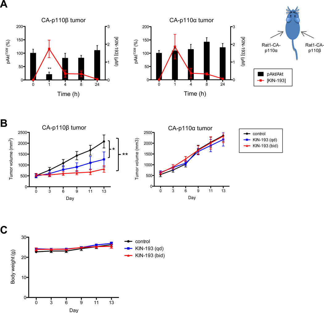
To determine the pharmacodynamics of AZD 6482 (KIN-193) in tumors in vivo, rat fibroblast (Rat1) cells are engineered to express both p53DD, a dominant negative mutant of p53, and a constitutively activated myr-p110β (Rat1-CA-p110β) to enable these cells to form xenograft tumors in mice. For comparison, an isogenic Rat1 cell line expressing p53DD and myr-p110α (Rat1-CA-p110α) is also generated. Rat1-CA-p110α and Rat1-CA-p110β cells are introduced subcutaneously into the contralateral flanks of athymic mice such that tumors driven by activated p110α or p110β would be exposed to identical conditions and that concern about animal-to-animal variability could be eliminated. When tumors reach a volume of ~500 mm3, the tumor-bearing mice receives a single IP injection of AZD 6482 (10 mg/kg). The plasma concentration of AZD 6482 is highest at 1 hour post-injection and declined to undetectable levels by 4h. Concentrations of AZD 6482 in both the CA-p110α- and CA-p110β-driven tumors parallel the plasma concentrations. Analyses of tumor lysates harvested at various time points after AZD 6482 injection reveal that the phosphorylation of AKT is significantly reduced at 1hour after AZD 6482 injection in Rat1-CA-p110β tumors, but remain unchanged in Rat1-CAp110α tumors.
Specific in vivo activity data for AZD6482 S-isomer is not detailed in the available literature. Based on its mechanism of action as a PI3Kβ inhibitor, it is expected to demonstrate effects in preclinical models where PI3Kβ plays a role, such as thrombosis and certain cancers. PI3Kβ is involved in platelet activation and thrombus formation, making it a potential target for antithrombotic therapy. |
| Enzyme Assay |
In vitro kinase inhibition assays are performed using purified PI3Kβ enzyme and a lipid substrate to determine IC50 values for inhibition of PI3Kβ kinase activity. The assay typically involves incubating the enzyme with varying concentrations of the compound and measuring the production of PIP3. Selectivity profiling is conducted against other PI3K isoforms (PI3Kα, PI3Kδ, PI3Kγ) to determine specificity. ATP-competitive inhibition can be assessed by varying ATP concentrations.
|
| Cell Assay |
Cell-based assays are conducted to evaluate PI3Kβ inhibition in cells. Cells are treated with the compound and stimulated with growth factors or other agonists, and the phosphorylation of downstream signaling molecules such as AKT is assessed by Western blot. Platelet aggregation assays are performed to evaluate the compound's effects on platelet function. Cell proliferation and survival assays are conducted to assess the functional effects of PI3Kβ inhibition.
|
| Animal Protocol |
Mice are dosed once by ip injection with AZD 6482 formulated in 7.5% NMP, 40% PEG400, 52.5% dH2O at 0.1 mL/10g body weight and 10 mg/kg. Mice; Approximately 6-8 week-old female nude mice are injected s.c. with Rat1-Myr-HA-p110α(Rat1-CAp110α) cells (1×10⁶ cells in 40% matrigel) in one flank (site 1) and Rat1-Myr-HA-p110β (Rat1-CAp110β) cells (0.5×10⁶ cells in 10% matrigel) in the contralateral flank (site 2). When tumors grow to ~500 mm3, mice are dosed once by ip injection with AZD 6482 formulated in 7.5% NMP, 40% PEG400, 52.5% dH2O at 0.1 mL/10g body weight and 10 mg/kg. Tumors are collected at 0, 1, 4, 8, and 24 h following compound administration and blood samples are obtained by direct heart puncture. Serum is separated and stored at -80°C. The drug concentrations in serum and tumor samples are assessed by LC-MS/MS analysis by the DMPK group.
In vivo efficacy is evaluated in animal models of thrombosis or cancer. The compound is administered via appropriate routes, and endpoints include thrombus formation, tumor growth inhibition, and survival. Platelet function is assessed ex vivo using platelet aggregation assays. The compound's ability to inhibit PI3Kβ-mediated signaling in vivo is evaluated by measuring AKT phosphorylation in target tissues. |
| References |
J Thromb Haemost.2012Oct;10(10):2127-36;Cancer Discov. 2012 May;2(5):425-33.
|
| Molecular Formula |
C22H24N4O4
|
|---|---|
| Molecular Weight |
408.45
|
| Exact Mass |
408.179
|
| CAS # |
1173900-37-2
|
| Related CAS # |
1173900-33-8 (AZD6438);1173900-37-2 (S-isomer);
|
| PubChem CID |
57789333
|
| Appearance |
Typically exists as solid at room temperature
|
| Density |
1.4±0.1 g/cm3
|
| Boiling Point |
635.5±65.0 °C at 760 mmHg
|
| Flash Point |
338.1±34.3 °C
|
| Vapour Pressure |
0.0±2.0 mmHg at 25°C
|
| Index of Refraction |
1.664
|
| LogP |
4.07
|
| Hydrogen Bond Donor Count |
2
|
| Hydrogen Bond Acceptor Count |
7
|
| Rotatable Bond Count |
5
|
| Heavy Atom Count |
30
|
| Complexity |
838
|
| Defined Atom Stereocenter Count |
1
|
| SMILES |
CC1=CN2C(=O)C=C(N=C2C(=C1)[C@H](C)NC3=CC=CC=C3C(=O)O)N4CCOCC4
|
| InChi Key |
IRTDIKMSKMREGO-HNNXBMFYSA-N
|
| InChi Code |
InChI=1S/C22H24N4O4/c1-14-11-17(15(2)23-18-6-4-3-5-16(18)22(28)29)21-24-19(12-20(27)26(21)13-14)25-7-9-30-10-8-25/h3-6,11-13,15,23H,7-10H2,1-2H3,(H,28,29)/t15-/m0/s1
|
| Chemical Name |
2-[[(1S)-1-(7-methyl-2-morpholin-4-yl-4-oxopyrido[1,2-a]pyrimidin-9-yl)ethyl]amino]benzoic acid
|
| HS Tariff Code |
2934.99.9001
|
| Storage |
Powder -20°C 3 years 4°C 2 years In solvent -80°C 6 months -20°C 1 month |
| Shipping Condition |
Room temperature (This product is stable at ambient temperature for a few days during ordinary shipping and time spent in Customs)
|
| Solubility (In Vitro) |
DMSO: 82 mg/mL (200.75 mM)
Water: 1 mg/mL (Insoluble) Ethanol: 10 mg/mL (24.48 mM) |
|
|---|---|---|
| Solubility (In Vivo) |
|
| Preparing Stock Solutions | 1 mg | 5 mg | 10 mg | |
| 1 mM | 2.4483 mL | 12.2414 mL | 24.4828 mL | |
| 5 mM | 0.4897 mL | 2.4483 mL | 4.8966 mL | |
| 10 mM | 0.2448 mL | 1.2241 mL | 2.4483 mL |
*Note: Please select an appropriate solvent for the preparation of stock solution based on your experiment needs. For most products, DMSO can be used for preparing stock solutions (e.g. 5 mM, 10 mM, or 20 mM concentration); some products with high aqueous solubility may be dissolved in water directly. Solubility information is available at the above Solubility Data section. Once the stock solution is prepared, aliquot it to routine usage volumes and store at -20°C or -80°C. Avoid repeated freeze and thaw cycles.
Calculation results
Working concentration: mg/mL;
Method for preparing DMSO stock solution: mg drug pre-dissolved in μL DMSO (stock solution concentration mg/mL). Please contact us first if the concentration exceeds the DMSO solubility of the batch of drug.
Method for preparing in vivo formulation::Take μL DMSO stock solution, next add μL PEG300, mix and clarify, next addμL Tween 80, mix and clarify, next add μL ddH2O,mix and clarify.
(1) Please be sure that the solution is clear before the addition of next solvent. Dissolution methods like vortex, ultrasound or warming and heat may be used to aid dissolving.
(2) Be sure to add the solvent(s) in order.
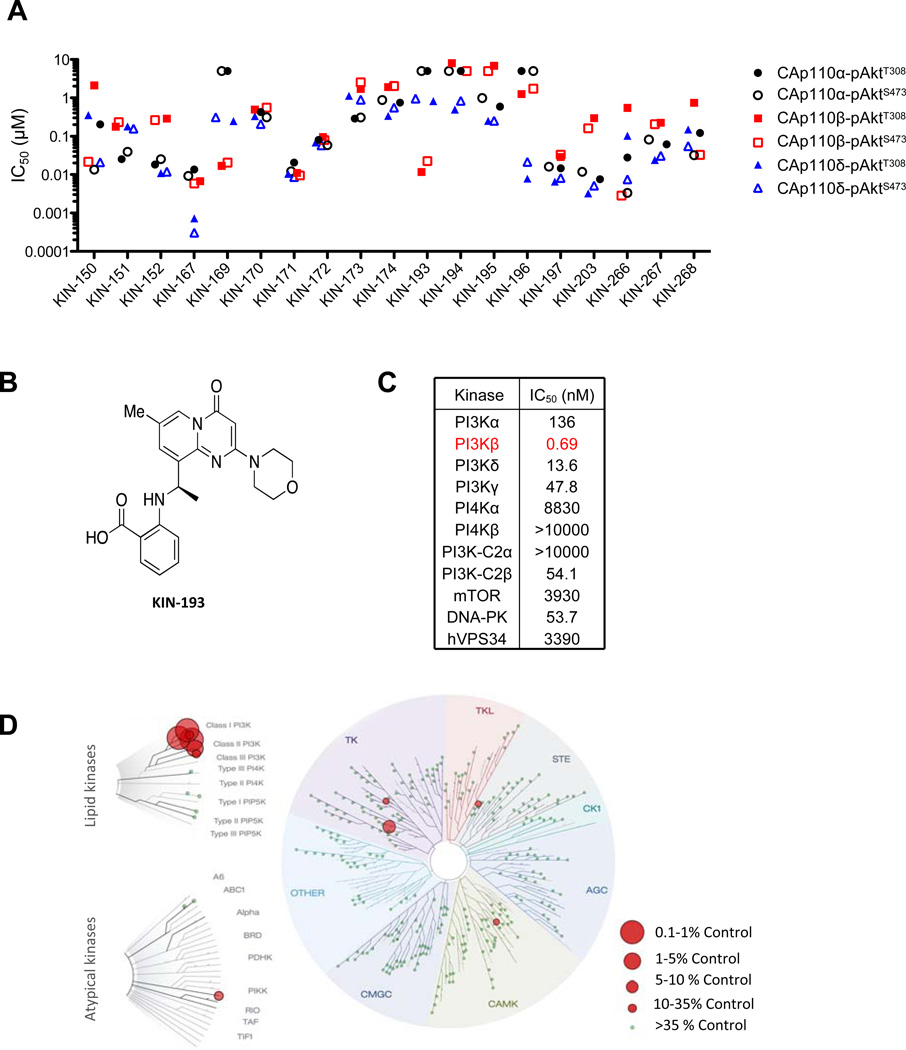
Identification of KIN-193 (AZD6482) as a p110β specific inhibitor.Cancer Discov. 2012 May;2(5):425-33. |
In vivocharacterization of the anti-cancer potential of KIN-193.A,Pharmacokinetics and pharmocodynamics of KIN-193 in mice.Cancer Discov. 2012 May;2(5):425-33. |
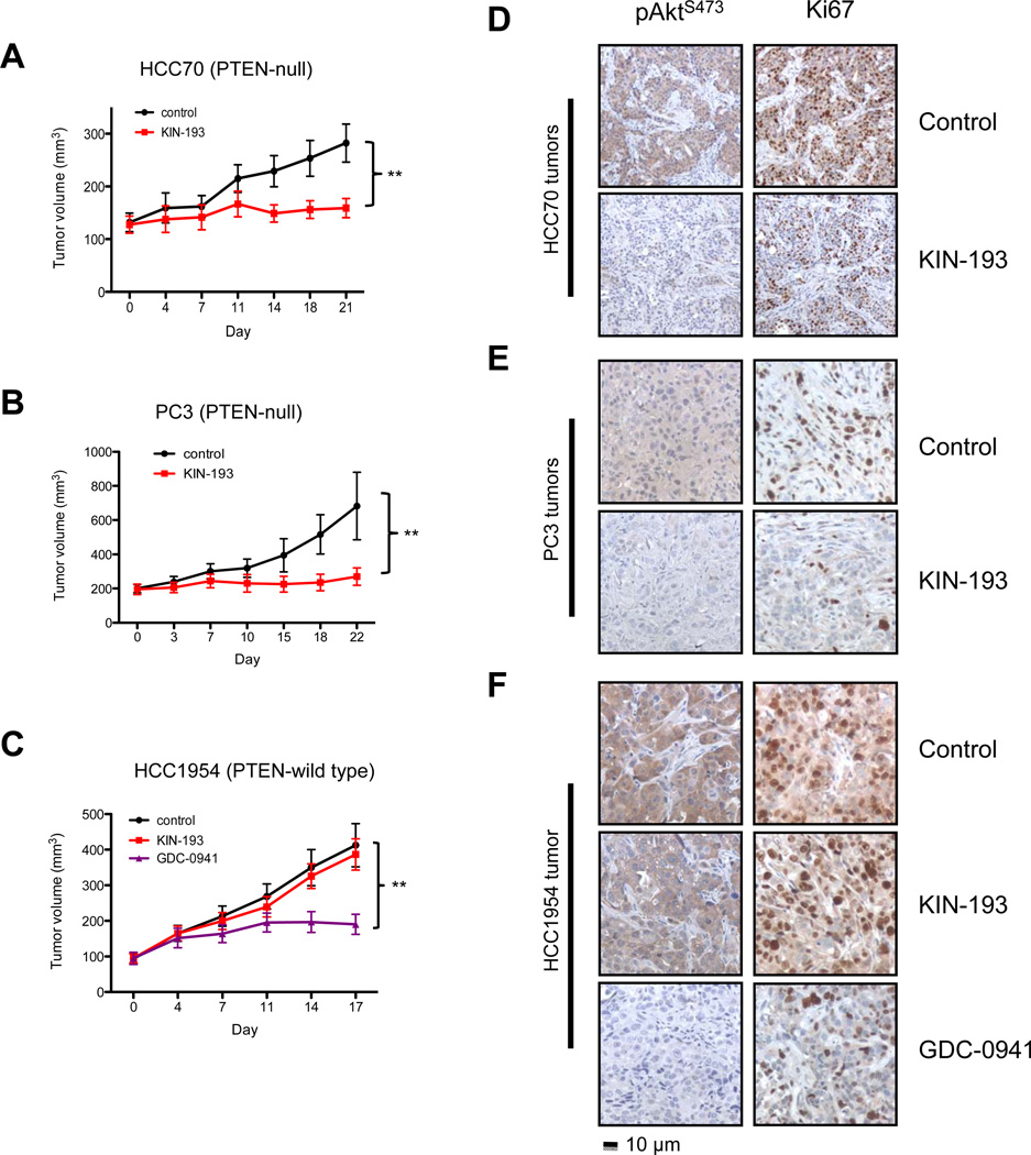
In vivoeffect of KIN-193 on PTEN-deficient tumors.Cancer Discov. 2012 May;2(5):425-33. |
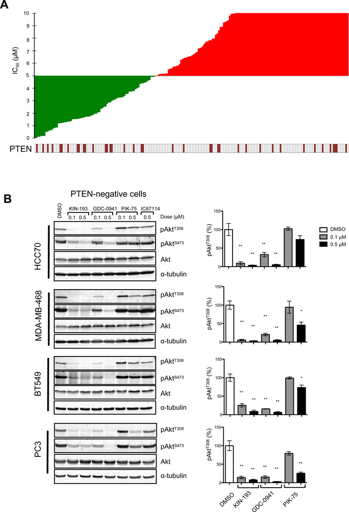
Effects of KIN-193 on PTEN-deficient cancer cells.Cancer Discov. 2012 May;2(5):425-33. |
Products are for research use only; We do not sell to patients
Copyright 2020 InvivoChem LLC | All Rights Reserved


 COA