| Size | Price | Stock | Qty |
|---|---|---|---|
| 5mg |
|
||
| 10mg |
|
||
| 25mg |
|
||
| 50mg |
|
||
| 100mg |
|
||
| 250mg |
|
||
| 500mg |
|
||
| Other Sizes |


Purity: ≥98%
| Targets |
Cdc42 (Kd = 11.4 μM)
Cdc42-Intersectin (ITSN) protein-protein interaction (IC50 = 3.2 μM for disrupting the interaction in vitro) [1] |
|---|---|
| ln Vitro |
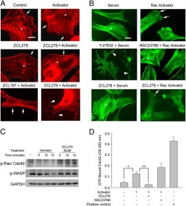
ZCL278 as a potent, cell-permeable inhibitor of Cdc42 that inhibits actin-based cellular processes such as cell motility and Golgi organization.ZCL278 eliminates microspike formation and perturbs GM130-docked Golgi structures in Swiss 3T3 fibroblast cultures, which are two of the most notable Cdc42-mediated subcellular events. Unlike the selective Rac inhibitor NSC23766, ZCL278 lowers the perinuclear accumulation of active Cdc42. In a metastatic prostate cancer cell line (PC-3), ZCL278 inhibits Cdc42-mediated neuronal branching and growth cone dynamics along with actin-based motility and migration without compromising cell viability[1]. While ZCL278 does not inhibit the nonenveloped poliovirus, it does inhibit the function of Cdc42 as an entry inhibitor for the dengue virus, vesicular stomatitis virus, lymphocytic choriomeningitis virus, and Junin virus (JUNV). ZCL278 effectively suppresses chemically induced filopodium formation in cells, a process that depends on Cdc42 activity. In the first round of dose-response studies, ZCL278 is not toxic at 200 μM concentrations, but according to flow cytometry, it inhibits JUNV with an IC50 of about 14 μM[2].
Disruption of Cdc42-ITSN interaction: ZCL278 dose-dependently inhibited the binding between Cdc42 and the SH3 domain of ITSN, with an IC50 of 3.2 μM in GST pull-down assay [1] - Golgi apparatus disorganization: Treatment of HeLa cells and MDA-MB-231 cells with ZCL278 (5-20 μM) for 4 hours resulted in dose-dependent disorganization of the Golgi apparatus, as visualized by immunofluorescence staining of Golgi marker protein (GM130). At 20 μM, >70% of cells showed fragmented or dispersed Golgi structures [1] - Inhibition of cell motility: ZCL278 (5-20 μM) suppressed the migration of MDA-MB-231 breast cancer cells and HeLa cervical cancer cells in Transwell migration assays. At 15 μM, the migration rate was reduced by 58% (MDA-MB-231) and 62% (HeLa) compared to vehicle controls [1] - Reduction of cell invasion: In Matrigel invasion assays, ZCL278 (10-20 μM) dose-dependently inhibited the invasive capacity of MDA-MB-231 cells, with 20 μM resulting in a 75% reduction in invasive cell number [1] - No significant cytotoxicity: ZCL278 at concentrations up to 25 μM did not affect the viability of HeLa or MDA-MB-231 cells after 24 hours of treatment, as determined by cell viability assay [1] |
| ln Vivo |
ZCL278 causes a more than 33-fold reduction in the amount of JUNV RNA in the spleen, with no JUNV RNA present in 5 out of 8 mice. ZCL278 can stop JUNV replication, as evidenced by these outcomes, which are comparable to those observed in mice given gabapentin[2].
|
| Enzyme Assay |
The protein Cdc42 that has been lyophilized is reconstituted to a concentration of 5 mg/mL using a buffer that comprises 50 mM Tris, 0.5 mM MgCl2, 50 mM NaCl, 3% (wt/vol) sucrose, and 0.6% dextran. We next dilute the stock solution to 1 μM in 5 mM phosphate buffer (pH 7.4). Before each fluorescent measurement, aliquots of ZCL278 are added to a quartz cuvettete containing Cdc42 solution and incubated for five minutes. After each addition, tryptophan fluorescence at 350 nm is measured using an excitation wavelength of 275 nm. GraphPad's equimolar specific binding model is used to fit the titration curve, and the Kd is computed[1].
GST pull-down assay for Cdc42-ITSN interaction: Recombinant GST-tagged ITSN SH3 domain and His-tagged Cdc42 were purified and incubated with serial dilutions of ZCL278 in binding buffer at 4°C for 2 hours. GST beads were added to the mixture and incubated for another 1 hour at 4°C. The beads were washed thoroughly, and bound proteins were eluted, separated by SDS-PAGE, and detected by immunoblotting with anti-His antibody. The intensity of the immunoreactive band corresponding to Cdc42 was quantified, and IC50 value was calculated from the dose-response curve [1] |
| Cell Assay |
ZCL278, NSC23766, or the Cdc42 activator are added to PC-3 cells during a 24-hour incubation period in order to measure cell viability. With a Countess Automated Cell Counter, the trypan blue dye exclusion method is used to determine the number of live and dead cells. Any null hypothesis with a probability level less than 95% is rejected, and P values are assigned for every experiment[1].
Immunofluorescence staining for Golgi structure: HeLa or MDA-MB-231 cells were seeded on coverslips and grown to 50-60% confluence. Cells were treated with ZCL278 (5-20 μM) for 4 hours, then fixed with paraformaldehyde and permeabilized with Triton X-100. After blocking, cells were incubated with primary antibody against GM130 (Golgi marker) followed by fluorescent secondary antibody. Nuclei were stained with a nuclear dye, and cells were imaged using a confocal microscope. The percentage of cells with disorganized Golgi was counted from at least 200 cells per treatment group [1] - Transwell migration assay: MDA-MB-231 or HeLa cells were resuspended in serum-free medium containing ZCL278 (5-20 μM) and seeded into the upper chamber of Transwell inserts. The lower chamber was filled with medium containing 10% fetal bovine serum as a chemoattractant. After incubation for 24 hours at 37°C, cells that migrated to the lower surface of the insert were fixed, stained, and counted under a microscope. Migration rate was calculated relative to vehicle-treated controls [1] - Matrigel invasion assay: Transwell inserts were coated with Matrigel and allowed to solidify. MDA-MB-231 cells were resuspended in serum-free medium with ZCL278 (10-20 μM) and seeded into the upper chamber. The lower chamber contained medium with 10% fetal bovine serum. After 48 hours of incubation, invasive cells on the lower surface were fixed, stained, and counted. Invasion inhibition rate was calculated compared to vehicle controls [1] - Cell viability assay: HeLa or MDA-MB-231 cells were seeded in 96-well plates and treated with ZCL278 (0-25 μM) for 24 hours. A cell viability reagent was added, and absorbance was measured at the appropriate wavelength. Cell viability was expressed as a percentage of vehicle-treated controls [1] |
| Animal Protocol |
Mice: C57BL/6 mice that are four weeks old are given intravenous injections of either ZCL278 (100 μg/g) or gabapentin. The mice receive an intraperitoneal injection of JUNV Candid #1 (1×10 6 PFU) in a maximum volume of 1 mL using a 27 1/2-gauge needle, one hour post-treatment. When the trial is over, the mice are killed, their spleens are homogenized using a Dounce homogenizer, they are centrifuged to produce a cell pellet and supernatant, and the levels of RNA expression are assessed.
|
| References | |
| Additional Infomation |
ZCL278 is a small molecule designed to target the protein-protein interaction between Cdc42 (a Rho GTPase) and Intersectin (ITSN). ITSN is a scaffold protein involved in vesicle transport and cytoskeleton regulation [1]. Its mechanism of action is to block the binding of Cdc42-ITSN, thereby disrupting the integrity of the Golgi apparatus, impairing cell motility and invasion, without causing significant cytotoxicity [1]. ZCL278 can serve as an important pharmacological tool for studying the role of Cdc42-ITSN interaction in Golgi apparatus organization, cell migration, and cancer cell invasion [1].
|
| Molecular Formula |
C21H19BRCLN5O4S2
|
|
|---|---|---|
| Molecular Weight |
584.89
|
|
| Exact Mass |
582.975
|
|
| Elemental Analysis |
C, 43.12; H, 3.27; Br, 13.66; Cl, 6.06; N, 11.97; O, 10.94; S, 10.96
|
|
| CAS # |
587841-73-4
|
|
| Related CAS # |
|
|
| PubChem CID |
1791111
|
|
| Appearance |
White to light yellow solid powder
|
|
| Density |
1.6±0.1 g/cm3
|
|
| Index of Refraction |
1.687
|
|
| LogP |
4.45
|
|
| Hydrogen Bond Donor Count |
3
|
|
| Hydrogen Bond Acceptor Count |
8
|
|
| Rotatable Bond Count |
7
|
|
| Heavy Atom Count |
34
|
|
| Complexity |
800
|
|
| Defined Atom Stereocenter Count |
0
|
|
| SMILES |
O=C(NC(NC1=CC=C(S(=O)(NC2=NC(C)=CC(C)=N2)=O)C=C1)=S)COC3=CC=C(Br)C=C3Cl
|
|
| InChi Key |
XKZDWYDHEBCGCG-UHFFFAOYSA-N
|
|
| InChi Code |
InChI=1S/C21H19BrClN5O4S2/c1-12-9-13(2)25-20(24-12)28-34(30,31)16-6-4-15(5-7-16)26-21(33)27-19(29)11-32-18-8-3-14(22)10-17(18)23/h3-10H,11H2,1-2H3,(H,24,25,28)(H2,26,27,29,33)
|
|
| Chemical Name |
2-(4-bromo-2-chlorophenoxy)-N-[[4-[(4,6-dimethylpyrimidin-2-yl)sulfamoyl]phenyl]carbamothioyl]acetamide
|
|
| Synonyms |
|
|
| HS Tariff Code |
2934.99.9001
|
|
| Storage |
Powder -20°C 3 years 4°C 2 years In solvent -80°C 6 months -20°C 1 month |
|
| Shipping Condition |
Room temperature (This product is stable at ambient temperature for a few days during ordinary shipping and time spent in Customs)
|
| Solubility (In Vitro) |
DMSO: ~100 mg/mL (~170.9 mM)
Water: 1 mg/mL (Insoluble) Ethanol: 1 mg/mL |
|---|---|
| Solubility (In Vivo) |
Solubility in Formulation 1: ≥ 2.5 mg/mL (4.27 mM) (saturation unknown) in 10% DMSO + 40% PEG300 + 5% Tween80 + 45% Saline (add these co-solvents sequentially from left to right, and one by one), clear solution.
For example, if 1 mL of working solution is to be prepared, you can add 100 μL of 25.0 mg/mL clear DMSO stock solution to 400 μL PEG300 and mix evenly; then add 50 μL Tween-80 to the above solution and mix evenly; then add 450 μL normal saline to adjust the volume to 1 mL. Preparation of saline: Dissolve 0.9 g of sodium chloride in 100 mL ddH₂ O to obtain a clear solution. Solubility in Formulation 2: ≥ 2.5 mg/mL (4.27 mM) (saturation unknown) in 10% DMSO + 90% Corn Oil (add these co-solvents sequentially from left to right, and one by one), clear solution. For example, if 1 mL of working solution is to be prepared, you can add 100 μL of 25.0 mg/mL clear DMSO stock solution to 900 μL of corn oil and mix evenly. (Please use freshly prepared in vivo formulations for optimal results.) |
| Preparing Stock Solutions | 1 mg | 5 mg | 10 mg | |
| 1 mM | 1.7097 mL | 8.5486 mL | 17.0972 mL | |
| 5 mM | 0.3419 mL | 1.7097 mL | 3.4194 mL | |
| 10 mM | 0.1710 mL | 0.8549 mL | 1.7097 mL |
*Note: Please select an appropriate solvent for the preparation of stock solution based on your experiment needs. For most products, DMSO can be used for preparing stock solutions (e.g. 5 mM, 10 mM, or 20 mM concentration); some products with high aqueous solubility may be dissolved in water directly. Solubility information is available at the above Solubility Data section. Once the stock solution is prepared, aliquot it to routine usage volumes and store at -20°C or -80°C. Avoid repeated freeze and thaw cycles.
Calculation results
Working concentration: mg/mL;
Method for preparing DMSO stock solution: mg drug pre-dissolved in μL DMSO (stock solution concentration mg/mL). Please contact us first if the concentration exceeds the DMSO solubility of the batch of drug.
Method for preparing in vivo formulation::Take μL DMSO stock solution, next add μL PEG300, mix and clarify, next addμL Tween 80, mix and clarify, next add μL ddH2O,mix and clarify.
(1) Please be sure that the solution is clear before the addition of next solvent. Dissolution methods like vortex, ultrasound or warming and heat may be used to aid dissolving.
(2) Be sure to add the solvent(s) in order.
|
|---|
|
Products are for research use only; We do not sell to patients
Copyright 2020 InvivoChem LLC | All Rights Reserved


 COA
COA