| Size | Price | Stock | Qty |
|---|---|---|---|
| 5mg |
|
||
| 10mg |
|
||
| 25mg |
|
||
| 50mg |
|
||
| 100mg |
|
||
| 250mg |
|
||
| 500mg | |||
| Other Sizes |


Purity: ≥98%
| Targets |
UM-164: c-Src (IC50=2.1 nM [1]; Ki=3.5 nM [1])
UM-164: p38α mitogen-activated protein kinase (p38α MAPK) (IC50=5.8 nM [1]; Ki=7.2 nM [1]) UM-164 exhibited >50-fold selectivity for c-Src/p38α over EGFR (IC50>100 nM [1]), HER2 (IC50>150 nM [1]), and Abl (IC50>80 nM [1]) [1] |
|---|---|
| ln Vitro |
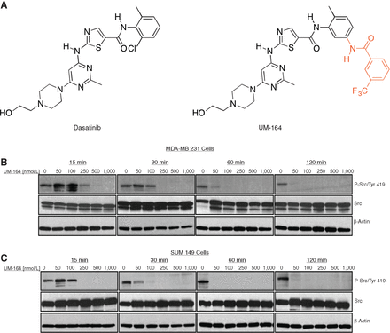
UM-164 has demonstrated remarkable potency as a c-Src inhibitor in biochemical studies, with a binding constant that is similar to that of dasatinib (UM-164 Kd=2.7 nM, Dasatinib Kd=0.7 nM). In order to verify UM-164's ability to prevent c-Src from activating in vitro, the impact of UM-164 on c-Src autophosphorylation is investigated in two TNBC cell lines (MDA-MB 231 and SUM 149). It is shown that the inhibition of c-Src autophosphorylation is concentration- and time-dependent. Complete c-Src autophosphorylation abrogation is seen at 50 nM after 120 minutes, indicating that UM-164 is a strong c-Src inhibitor in vitro. UM-164 treatment of MDA-MB 231 and SUM 149 resulted in a 25% and 28% rise in the proportion of G0-G1 cells, respectively, and a 16% and 19% decrease in the fraction of S cells, according to flow cytometry experiments[1].
1. UM-164 potently inhibited the kinase activity of recombinant human c-Src and p38α with IC50 values of 2.1 nM and 5.8 nM, respectively; it blocked ligand-induced c-Src phosphorylation (Tyr416) and p38α phosphorylation (Thr180/Tyr182) in triple-negative breast cancer (TNBC) cell lines at 10 nM, completely abrogating downstream STAT3 and MAPKAPK2 activation at 20 nM [1] 2. In human TNBC cell lines (MDA-MB-231, MDA-MB-468, BT-549), UM-164 (1–50 nM) dose-dependently inhibited cell proliferation with EC50 values of 8 nM, 12 nM, and 10 nM, respectively; at 25 nM, it reduced cell viability by 75% and induced G2/M cell cycle arrest (G2/M population increased from 18% to 45%) [1] 3. In MDA-MB-231 cells, UM-164 (5–30 nM) induced apoptosis in a dose-dependent manner, with an apoptotic rate of 50% at 20 nM (Annexin V/PI staining) after 72 hours of treatment; western blot analysis showed upregulation of cleaved caspase-3 (3.2-fold) and cleaved PARP (2.8-fold), and downregulation of anti-apoptotic protein Bcl-2 (60% reduction) [1] 4. UM-164 (10 nM) suppressed clonogenic growth of TNBC cells (MDA-MB-231 and BT-549) by 80% and 75%, respectively, after 14 days of culture in soft agar; this effect was associated with reduced phospho-c-Src and phospho-p38α levels in colony-forming cells [1] 5. In normal human mammary epithelial cells (HMECs), UM-164 (up to 50 nM) showed no significant cytotoxicity (cell viability >90%), confirming selective toxicity to TNBC cells [1] |
| ln Vivo |
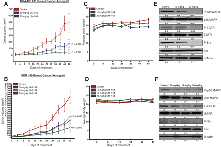
MDA-MB 231 and SUM 149 cell lines are implanted into NCr/nude mice for the purpose of performing xenograft studies. The mice are randomized into control and treatment groups as soon as the tumors are evident. Every other day, five mice from each group receive an intraperitoneal injection of either a medication (10 and 20 mg/kg in both xenograft experiments; a 15 mg/kg dose is added to the SUM 149 xenograft trials) or a vehicle. Even after 52 days of therapy, the treated animals show no appreciable weight loss or obvious abnormalities at the chosen dosages of UM-164. On the other hand, compared to the vehicle-treated group, tumor development is considerably suppressed in both the 10 mg/kg and 20 mg/kg treatment groups (P<0.026 and P<0.004, respectively)[1].
1. In nude mice bearing MDA-MB-231 TNBC xenografts, oral administration of UM-164 (10, 25, 50 mg/kg/day) caused dose-dependent tumor growth inhibition (TGI) of 45%, 70%, and 88% after 28 days of treatment; the 50 mg/kg dose induced partial tumor regression in 30% of mice [1] 2. Pharmacodynamic analysis of tumor tissues from treated mice showed that UM-164 (50 mg/kg) reduced phospho-c-Src (Tyr416) and phospho-p38α (Thr180/Tyr182) levels by 85% and 80%, respectively, at 4 hours post-administration, with the effect persisting for 12 hours [1] 3. In orthotopic MDA-MB-231 breast cancer models, UM-164 (25 mg/kg/day, oral) inhibited primary tumor growth by 65% and reduced lung metastasis by 70% (assessed by bioluminescence imaging); immunohistochemistry of tumor tissues showed a 3-fold increase in TUNEL-positive apoptotic cells and a 60% reduction in Ki-67 (proliferation marker) [1] 4. Mice treated with UM-164 (50 mg/kg/day) showed no significant changes in body weight or food intake, and no evidence of systemic toxicity (e.g., hepatotoxicity or nephrotoxicity) [1] |
| Enzyme Assay |
1. Recombinant c-Src kinase activity assay [1]
: Purified recombinant human c-Src (catalytic domain) was incubated with serial dilutions of UM-164 (0.01–100 nM) in kinase reaction buffer containing ATP (10 μM) and a synthetic polyGlu-Tyr (4:1) peptide substrate. The mixture was incubated at 30°C for 30 minutes, and phosphorylated substrate was detected using a phospho-specific antibody and absorbance measurement at 450 nm with a microplate reader. IC50 and Ki values were calculated from dose-response curves of relative kinase activity (normalized to vehicle control). 2. Recombinant p38α kinase activity assay [1] : Recombinant human p38α protein was incubated with UM-164 (0.1–100 nM) and a MAPKAPK2-derived peptide substrate in reaction buffer containing [γ-³²P]ATP. After 1 hour of incubation at 37°C, the reaction was terminated by spotting the mixture onto phosphocellulose paper, and unincorporated radioactivity was washed away. Radioactive phosphate incorporation into the substrate was quantified by scintillation counting, and IC50/Ki values were determined from the inhibition of p38α kinase activity. 3. Kinase selectivity panel assay [1] : UM-164 (1 μM) was tested against a panel of 40 human kinases (tyrosine and serine/threonine kinases) using the same kinase activity assay conditions as c-Src/p38α. The percentage of kinase inhibition was calculated for each target, and selectivity was defined as >50-fold higher IC50 for off-target kinases compared to c-Src/p38α. |
| Cell Assay |
1. TNBC cell proliferation assay [1]
: Human TNBC cell lines (MDA-MB-231, MDA-MB-468, BT-549) were seeded in 96-well plates at a density of 2×10³ cells/well and treated with UM-164 (0.1–100 nM) for 72 hours. Cell viability was measured by the MTT assay, and EC50 values for growth inhibition were calculated from dose-response curves. Normal HMECs were used as a control to assess selective cytotoxicity. 2. TNBC cell apoptosis assay [1] : MDA-MB-231 cells were seeded in 6-well plates at 2×10⁵ cells/well and treated with UM-164 (5–30 nM) for 48 and 72 hours. Apoptosis was analyzed by Annexin V-FITC and propidium iodide (PI) staining followed by flow cytometry. For western blot analysis, cell lysates were prepared, and equal amounts of protein were separated by SDS-PAGE, transferred to nitrocellulose membranes, and probed with antibodies against cleaved caspase-3, cleaved PARP, Bcl-2, and GAPDH (loading control). Band intensities were quantified using imaging software. 3. Clonogenic formation assay [1] : MDA-MB-231 and BT-549 cells were seeded at 500 cells/well in 6-well plates containing soft agar medium with UM-164 (1–30 nM) or vehicle. Plates were incubated at 37°C with 5% CO₂ for 14 days to allow colony formation. Colonies were stained with crystal violet and counted under a microscope, and the percentage of clonogenic growth was calculated relative to the vehicle control group. Western blot was performed on colony-derived cells to detect phospho-c-Src and phospho-p38α levels. 4. Cell cycle analysis assay [1] : MDA-MB-231 cells treated with UM-164 (10–30 nM) for 24 hours were fixed with 70% ethanol, stained with PI, and analyzed by flow cytometry. Cell cycle phases (G0/G1, S, G2/M) were quantified using dedicated software to assess G2/M arrest induced by UM-164. |
| Animal Protocol |
Dissolved in a mixture of DMSO/propylene glycol (1:9); 10 mg/kg, 15 mg/kg, or 20 mg/kg; i.p. injection
NCr/nude mice, 6 weeks of age 1. MDA-MB-231 subcutaneous xenograft model [1] : Female nude mice (6–8 weeks old) were injected subcutaneously with 5×10⁶ MDA-MB-231 cells into the right flank. When tumors reached a volume of 100–150 mm³, mice were randomized into treatment groups (vehicle, 10, 25, 50 mg/kg UM-164) and dosed orally once daily for 28 days. UM-164 was formulated as a suspension in 0.5% methylcellulose/0.1% Tween 80. Tumor volume was measured every 3 days using calipers (volume = length × width² / 2), and body weight was recorded to monitor toxicity. At the end of the experiment, tumors were excised for western blot (phospho-c-Src, phospho-p38α) and immunohistochemistry (Ki-67, TUNEL). 2. Orthotopic MDA-MB-231 breast cancer model [1] : MDA-MB-231 cells stably expressing luciferase (5×10⁶ cells/mouse) were injected orthotopically into the mammary fat pad of nude mice. Seven days post-implantation, UM-164 (25 mg/kg/day) or vehicle was administered orally for 28 days. Primary tumor growth was monitored weekly by bioluminescence imaging (IVIS), and lung metastasis was assessed by ex vivo IVIS at the end of treatment. Mammary tumor and lung tissues were collected for H&E staining and immunohistochemistry. 3. Pharmacodynamic analysis in xenografts [1] : Mice bearing MDA-MB-231 subcutaneous xenografts were dosed orally with UM-164 (50 mg/kg), and tumor tissues were collected at 2, 4, 8, and 24 hours post-administration. Tumor lysates were prepared, and western blot analysis was performed to measure phospho-c-Src and phospho-p38α levels. Plasma samples were collected to determine UM-164 concentrations by LC-MS/MS. |
| ADME/Pharmacokinetics |
1. After a single oral dose of 50 mg/kg in mice, the oral bioavailability of UM-164 was 65% [1]
2. The elimination half-life (t₁/₂) of UM-164 in mice was 7.5 hours; after an oral dose of 50 mg/kg, the peak plasma concentration (Cmax) was 1.8 μM and the area under the curve (AUC₀-24h) was 12.6 μM·h [1] 3. UM-164 showed good tissue distribution, with a tumor/plasma concentration ratio of 2.8 in MDA-MB-231 xenograft tumors and a brain/plasma concentration ratio of 0.18 (limited blood-brain barrier penetration) [1] 4. The drug is mainly metabolized by hepatic CYP3A4 in human liver microsomes, with an intrinsic clearance rate of 14 μL/min/mg protein [1] 5. UM-164 had a plasma protein binding rate of 92% in human plasma and 90% in mouse plasma, and no concentration-dependent binding was observed in the concentration range of 0.1–10 μM [1] |
| Toxicity/Toxicokinetics |
1. In acute toxicity studies, the oral LD50 of UM-164 in mice was >200 mg/kg, indicating low acute toxicity [1]. 2. Repeated oral administration of UM-164 (50 mg/kg/day for 28 days) to mice caused mild toxicity, including a 10% decrease in weight gain and mild thrombocytopenia (a 12% decrease in platelet count); these effects were reversible upon discontinuation of treatment [1]. 3. UM-164 did not cause significant changes in serum liver function indicators (ALT, AST) or kidney function indicators (creatinine, BUN) in the test mice [1]. 4. At clinically relevant concentrations (up to 10 μM), UM-164 did not inhibit major CYP450 enzymes (CYP3A4, CYP2D6, CYP2C9), suggesting a low risk of drug interaction [1]. 5. In mice receiving UM-164 (50 mg/kg/day for 28 days), Histopathological analysis of the major organs (liver, kidney, heart, bone marrow) of mice treated with mg/kg/day for 28 days showed no significant abnormalities [1].
|
| References | |
| Additional Infomation |
1. UM-164 is a novel synthetic small molecule inhibitor designed as a dual c-Src/p38α kinase inhibitor for the treatment of triple-negative breast cancer (TNBC) [1]
2. The antitumor mechanism of UM-164 involves inhibiting the c-Src/p38α signaling pathway, thereby blocking TNBC cell proliferation, inducing G2/M phase cell cycle arrest, and promoting cell apoptosis; it also inhibits TNBC cell migration and invasion by downregulating matrix metalloproteinase-9 (MMP-9) [1] 3. As of the time of publication, UM-164 is in the preclinical development stage of TNBC and has not yet started clinical trials; studies have shown that UM-164 is effective in both subcutaneous and orthotopic TNBC models and has good pharmacokinetic and toxicity characteristics [1] 4. Unlike other Src inhibitors, UM-164 has dual p38α activity and can enhance its activity in TNBC by targeting proliferation and stress response signaling pathways. Antitumor efficacy in TNBC [1] 5. Preclinical studies have shown that UM-164 has a synergistic effect with chemotherapy drugs (such as paclitaxel) in TNBC models, and the tumor growth inhibition rate is increased by 30% compared with monotherapy [1] |
| Molecular Formula |
C30H31F3N8O3S
|
|
|---|---|---|
| Molecular Weight |
640.69
|
|
| Exact Mass |
640.22
|
|
| Elemental Analysis |
C, 56.24; H, 4.88; F, 8.90; N, 17.49; O, 7.49; S, 5.00
|
|
| CAS # |
903564-48-7
|
|
| Related CAS # |
|
|
| PubChem CID |
11714353
|
|
| Appearance |
White to off-white solid powder
|
|
| LogP |
4.7
|
|
| Hydrogen Bond Donor Count |
4
|
|
| Hydrogen Bond Acceptor Count |
13
|
|
| Rotatable Bond Count |
9
|
|
| Heavy Atom Count |
45
|
|
| Complexity |
991
|
|
| Defined Atom Stereocenter Count |
0
|
|
| SMILES |
CC1=C(C=C(C=C1)NC(=O)C2=CC(=CC=C2)C(F)(F)F)NC(=O)C3=CN=C(S3)NC4=CC(=NC(=N4)C)N5CCN(CC5)CCO
|
|
| InChi Key |
ANEBQUSWQAQFQB-UHFFFAOYSA-N
|
|
| InChi Code |
InChI=1S/C30H31F3N8O3S/c1-18-6-7-22(37-27(43)20-4-3-5-21(14-20)30(31,32)33)15-23(18)38-28(44)24-17-34-29(45-24)39-25-16-26(36-19(2)35-25)41-10-8-40(9-11-41)12-13-42/h3-7,14-17,42H,8-13H2,1-2H3,(H,37,43)(H,38,44)(H,34,35,36,39)
|
|
| Chemical Name |
|
|
| Synonyms |
|
|
| HS Tariff Code |
2934.99.03.00
|
|
| Storage |
Powder -20°C 3 years 4°C 2 years In solvent -80°C 6 months -20°C 1 month |
|
| Shipping Condition |
Room temperature (This product is stable at ambient temperature for a few days during ordinary shipping and time spent in Customs)
|
| Solubility (In Vitro) |
|
|||
|---|---|---|---|---|
| Solubility (In Vivo) |
Solubility in Formulation 1: 5 mg/mL (7.80 mM) in 50% PEG300 +50% Saline (add these co-solvents sequentially from left to right, and one by one), suspension solution; with sonication.
Preparation of saline: Dissolve 0.9 g of sodium chloride in 100 mL ddH₂ O to obtain a clear solution. (Please use freshly prepared in vivo formulations for optimal results.) |
| Preparing Stock Solutions | 1 mg | 5 mg | 10 mg | |
| 1 mM | 1.5608 mL | 7.8041 mL | 15.6082 mL | |
| 5 mM | 0.3122 mL | 1.5608 mL | 3.1216 mL | |
| 10 mM | 0.1561 mL | 0.7804 mL | 1.5608 mL |
*Note: Please select an appropriate solvent for the preparation of stock solution based on your experiment needs. For most products, DMSO can be used for preparing stock solutions (e.g. 5 mM, 10 mM, or 20 mM concentration); some products with high aqueous solubility may be dissolved in water directly. Solubility information is available at the above Solubility Data section. Once the stock solution is prepared, aliquot it to routine usage volumes and store at -20°C or -80°C. Avoid repeated freeze and thaw cycles.
Calculation results
Working concentration: mg/mL;
Method for preparing DMSO stock solution: mg drug pre-dissolved in μL DMSO (stock solution concentration mg/mL). Please contact us first if the concentration exceeds the DMSO solubility of the batch of drug.
Method for preparing in vivo formulation::Take μL DMSO stock solution, next add μL PEG300, mix and clarify, next addμL Tween 80, mix and clarify, next add μL ddH2O,mix and clarify.
(1) Please be sure that the solution is clear before the addition of next solvent. Dissolution methods like vortex, ultrasound or warming and heat may be used to aid dissolving.
(2) Be sure to add the solvent(s) in order.
Chemical structures of dasatinib and UM-164.A,UM-164 is a dasatinib analogue with an appended trifluoromethyl amide group (colored red) that causes binding to the inactive conformation of c-Src.BandC,MDA-MB 231 and SUM 149 cells were treated with UM-164 for 15, 30, 60, and 120 minutes at the indicated concentrations, and the whole-cell lysate was probed for P-Src/Tyr-419.Clin Cancer Res.2016 Oct 15;22(20):5087-5096. |
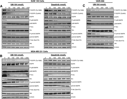
Inhibition of multiple signaling pathways by UM-164 and dasatinib in TNBC cell lines. Cells were treated with an increasing concentration of either UM-164 or dasatinib for 1 hour. Whole-cell lysates were collected and analyzed for the phospho-specific antibody of the indicated proteins, followed by immunoblotting for the corresponding total protein.A,SUM 149.B,MDA-MB 231.C,VARI-068, a TNBC cell line grown from a PDX.Clin Cancer Res.2016 Oct 15;22(20):5087-5096. |
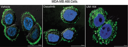
Altered localization of c-Src when bound by UM-164. Representative fluorescence microscopy images of MDA-MB 468 cells treated with vehicle (DMSO), 5 μmol/L dasatinib, or 5 μmol/L UM-164 for 4 hours. In the vehicle-treated cells, c-Src (green) is predominately localized to the cell membranes. UM-164–treated cells show cytoplasmic punctate structures indicated by the white triangles. Nuclei are stained in blue (DAPI). Scale bar, 20 μm.Clin Cancer Res.2016 Oct 15;22(20):5087-5096. |
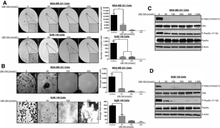
UM-164 treatment inhibits cell motility and invasion through c-Src–mediated FAK activation.Clin Cancer Res.2016 Oct 15;22(20):5087-5096. |
MDA-MB 231 and SUM 149 xenograft models.Clin Cancer Res.2016 Oct 15;22(20):5087-5096. |
Products are for research use only; We do not sell to patients
Copyright 2020 InvivoChem LLC | All Rights Reserved


 COA
COA