| Size | Price | Stock | Qty |
|---|---|---|---|
| 5mg |
|
||
| 10mg |
|
||
| 25mg |
|
||
| 50mg |
|
||
| 100mg |
|
||
| 250mg |
|
||
| 500mg | |||
| Other Sizes |


Purity: ≥98%
| Targets |
Mcl-1 (Ki=260 nM); Bcl-2 (Ki=290 nM); Bcl-xL (Ki=1110 nM)
Bcl-2 (Ki = 290 nM, fluorescence polarization assay for Bcl-2-BH3 peptide interaction) [1] Bcl-2 (Ki = 210 nM, fluorescence polarization assay), Mcl-1 (Ki = 1.1 μM, same assay); no significant binding to Bcl-xL (Ki > 10 μM) [2] |
|---|---|
| ln Vitro |
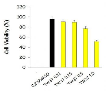
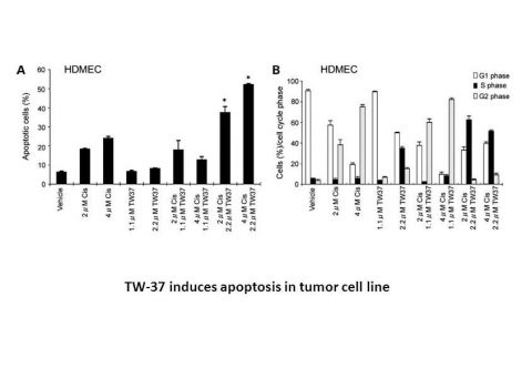
TW-37 (TW37) is a novel nonpeptide small-molecule inhibitor designed using a structure-based design strategy. TW-37 targets the proapoptotic Bcl-2 proteins Bak, Bax, and Bid in the BH3-binding groove of Bcl-2. TW-37 binds to recombinant Bcl-2 and Bcl-xL proteins with Ki values of 290 and 1110 nM, respectively, in fluorescence polarization-based binding assays. TW-37 does not have any cytotoxic effects on fibroblasts at concentrations up to 50 M, but it does have an IC50 of 1.8 M for endothelial cells. Apoptosis, a process mediated by mitochondrial depolarization and activation of caspase-9 and caspase-3, is the mechanism of TW-37-induced endothelial cell death. Coexposure to the growth factor milieu secreted by tumor cells does not counteract the effect of TW-37 on the apoptosis of endothelial cells. At subapoptotic TW-37 concentrations (0.005–0.05 M), the angiogenic chemokines CXCL1 and CXCL8 are expressed and the angiogenic potential of endothelial cells is inhibited (i.e., migration and capillary sprouting assays)[1].
TW-37 is a potent Bcl-2 and Mcl-1 inhibitor. TW-37 binds to recombinant Bcl-2, Bcl-xL, and Mcl-1 proteins with Ki values of 290, 1,110, and 260 nM, respectively, in fluorescence polarization-based binding assays[2].
Antiangiogenic activity (HUVECs): TW-37 inhibited HUVEC proliferation with an IC50 of 0.8 μM (72-hour MTT assay), reduced cell migration by 65% at 1 μM (Boyden chamber assay), and suppressed tube formation by 70% at 1 μM (Matrigel assay). Western blot showed 40% lower VEGF expression and 2.5-fold higher cleaved caspase-3 in HUVECs treated with 1 μM TW-37 [1] Antiproliferative activity (DLBCL cells): TW-37 inhibited SU-DHL-4 (IC50 = 0.3 μM), OCI-Ly3 (IC50 = 0.5 μM), and SU-DHL-6 (IC50 = 0.7 μM) cells (72-hour MTT assay). Annexin V/PI staining revealed 55% apoptosis in SU-DHL-4 cells treated with 0.5 μM TW-37 for 48 hours (vs. 8% in control) [2] Bcl-2/Mcl-1 regulation (DLBCL cells): TW-37 (0.3-1 μM) reduced Bcl-2 (40%) and Mcl-1 (50%) protein levels in SU-DHL-4 cells (24-hour treatment, Western blot); no change in Bcl-2/Mcl-1 mRNA (qPCR), indicating post-translational regulation [2] Antitumor activity (breast cancer cells): TW-37 (1 μM) inhibited MDA-MB-231 cell proliferation by 50% (72-hour MTT) and reduced VEGF secretion by 35% (ELISA) [1] |
| ln Vivo |
We examine the biological impact of TW-37 (TW37) on human microvascular endothelial cells in vivo using a murine model of humanized vasculature. According to this model, both 3 and 30 mg/kg TW-37 are significantly less effective than the vehicle control in terms of the total number of blood vessels (P0.05). Along with a decrease in the overall number of blood vessels, the treated groups are also experiencing an unusually high number of occluded vessels. By counting completely blocked vessels and calculating their percentage of all vessels, the levels of vessel occlusion are evaluated. When compared to control, TW-37 concentration significantly increases the number of occluded vessels[1].
CAM angiogenesis model: TW-37 (5 μg/egg, local application) reduced blood vessel density by 55% vs. vehicle (DMSO/saline); no CAM tissue toxicity [1] Mouse corneal angiogenesis model: BALB/c mice received VEGF pellets; TW-37 (10 μg/eye, topical, daily for 7 days) reduced neovascularization area by 60% vs. vehicle (0.1% DMSO/PBS) [1] MDA-MB-231 xenograft (nude mice): TW-37 (20 mg/kg, ip, twice weekly for 3 weeks) reduced tumor volume by 50% and weight by 45%. Tumors showed 40% lower CD31 (endothelial marker) and 2.5-fold higher cleaved caspase-3 (IHC) [1] SU-DHL-4 xenograft (SCID mice): TW-37 (15 mg/kg, ip, 5 days/week for 4 weeks) reduced tumor volume by 65% and weight by 60%. Tumors had lower Bcl-2/Mcl-1 (Western blot) and Ki-67 (from 60% to 20%, IHC) [2] |
| Enzyme Assay |
For this assay, the 21-residue BH3 peptide QEDIIRNIARHLAQVGDSMDR derived from Bid labeled with 6-carboxyfluorescein succinimidyl ester (FAM-Bid) and recombinant proteins derived from human Bcl-2,Bcl-X L,and Mcl-1 are employed. It is determined that FAM-Bid has a Ki of 11 nM to Bcl-2 protein,25 nM to Bcl-XL protein,and 5.7 nM to Mcl-1 protein. The competitive binding assay for Bcl-XL is same as that for Bcl-2 with the following exceptions: 30 nM Bcl-XL protein and 2.5 nM FAM-Bid peptide in the following assay buffer [50 mM Tris-Bis (pH 7.4) and 0.01% bovine gamma-globulin].
Fluorescence polarization assay (Bcl-2/Mcl-1-BH3 binding): Recombinant Bcl-2/Mcl-1 (100 nM) was incubated with fluorescein-labeled BH3 peptide (50 nM) in buffer (20 mM Tris-HCl pH 7.5, 150 mM NaCl) for 30 minutes. Serially diluted TW-37 (10 nM-10 μM) was added, and polarization was measured at 485 nm/535 nm. Ki values were calculated via competition binding curves [2] Co-IP assay (Bcl-2-Bax interaction): SU-DHL-4 lysates were pre-incubated with TW-37 (0.1-1 μM) for 1 hour, then mixed with anti-Bcl-2 antibody (overnight, 4°C) and Protein A/G beads (2 hours). Bound Bax was detected by Western blot; 0.5 μM TW-37 reduced Bcl-2-Bax complexes by 50% [2] |
| Cell Assay |
The sulforhodamine B (SRB) cytotoxicity assay is used as described. Briefly, optimal cell density for cytotoxicity assay is determined by growth curve analysis. HDMECs are seeded in a 96-well plate and allowed to adhere overnight. Drug or control is diluted in EGM2-MV and layered onto cells, which are allowed to incubate for times as indicated in the figures. Alternatively, HDMECs are coincubated with TW37 and 0 to 100 ng/mL recombinant human VEGF (rhVEGF)165 or 0 to 100 ng/mL recombinant human CXCL8. Cells are fixed on the plates by addition of cold trichloroacetic acid (10% final concentration) and incubation for 1 hour at 4 °C. Cellular protein is stained by addition of 0.4% SRB in 1% acetic acid and incubation at room temperature for 30 minutes. Unbound SRB is removed by washing with 1% acetic acid and the plates are air dried. Bound SRB is resolubilized in 10 mM unbuffered Tris-base and absorbance is determined on a microplate reader at 560 nm. Test results are normalized against initial plating density and drug-free controls. Data are obtained from triplicate wells per condition and are representative of at least three independent experiments
HUVEC proliferation (MTT): HUVECs (5×10³/well, 96-well plate) were treated with TW-37 (0.01-5 μM) for 72 hours. MTT reagent was added, formazan dissolved in DMSO, and absorbance measured at 570 nm to calculate IC50 [1] HUVEC migration (Boyden chamber): HUVECs in serum-free medium + TW-37 (0.1-1 μM) were added to upper chambers; lower chambers had 10% FBS. After 24 hours, fixed/stained cells were counted to calculate migration inhibition [1] HUVEC tube formation: Matrigel-coated 96-well plates were seeded with HUVECs (1×10⁴/well) + TW-37 (0.1-1 μM). After 6 hours, tube length was measured; 1 μM TW-37 reduced length by 70% [1] DLBCL viability (MTT): SU-DHL-4/OCI-Ly3 (1×10⁴/well) were treated with TW-37 (0.05-2 μM) for 72 hours; IC50 calculated via absorbance at 570 nm [2] DLBCL apoptosis (Annexin V/PI): SU-DHL-4 cells treated with TW-37 (0.2-0.8 μM) for 48 hours were stained with Annexin V-FITC/PI and analyzed by flow cytometry [2] Western blot (Bcl-2/VEGF): Cells (HUVECs/SU-DHL-4) treated with TW-37 (0.1-1 μM) for 24 hours were lysed in RIPA buffer. Proteins were separated by SDS-PAGE, transferred to PVDF membranes, and probed with anti-Bcl-2, Mcl-1, VEGF, cleaved caspase-3, and β-actin antibodies [1,2] |
| Animal Protocol |
~40 mg/kg
TW-37 is resuspended in Tween 80/ethanol (1:1 ratio, diluted 10-fold with 0.9% saline before use). Athymic NCr-nu/nu mice bearing SK-Mel-147 melanoma xenografts CAM assay: Fertilized eggs (3-day incubation) had a window opened; TW-37 (5 μg in 50 μL DMSO/saline) or vehicle was applied to CAM. After 48 hours, CAM was fixed and vessel density counted [1] Corneal angiogenesis assay: BALB/c mice (6-8 weeks) were anesthetized; VEGF pellets (50 ng) were implanted into corneas. TW-37 (10 μg in 5 μL 0.1% DMSO/PBS) or vehicle was topically administered daily for 7 days. Corneas were excised and neovascularization area measured [1] MDA-MB-231 xenograft: Female nude mice (6-8 weeks) received 2×10⁶ MDA-MB-231 cells (100 μL Matrigel/PBS 1:1, sc). When tumors reached 100 mm³, mice were dosed with vehicle (5% DMSO + 95% saline) or TW-37 (20 mg/kg, ip, twice weekly for 3 weeks). Tumor volume (length×width²/2) and weight were measured every 3 days [1] SU-DHL-4 xenograft: Male SCID mice (6-8 weeks) received 5×10⁶ SU-DHL-4 cells (100 μL Matrigel/PBS 1:1, sc). When tumors reached 150 mm³, mice were dosed with vehicle (10% DMSO + 40% cremophor EL + 50% saline) or TW-37 (15 mg/kg, ip, 5 days/week for 4 weeks). Tumor volume and weight were measured every 2 days [2] |
| Toxicity/Toxicokinetics |
Acute toxicity (mice): A single intraperitoneal injection of TW-37 (50 mg/kg) did not result in death; transient weight loss (<5%) recovered within 4 days. Serum ALT (≤45 U/L), AST (≤90 U/L), and creatinine (≤0.8 mg/dL) were all within the normal range [1]. Chronic toxicity (xenograft): Mice treated with TW-37 (20 mg/kg intraperitoneal injection, 3 weeks [1]; 15 mg/kg intraperitoneal injection, 4 weeks [2]) showed no significant change in weight (mean: -2% to +3%, control group -1% to +4%). No liver/kidney/heart/lung lesions were observed (histopathology) [1,2]. Plasma protein binding: TW-37 was 85% bound in mouse plasma (ultrafiltration method) [2].
|
| References |
|
| Additional Infomation |
N-[4-(2-tert-butylphenyl)sulfonylphenyl]-2,3,4-trihydroxy-5-[(2-propyl-2-ylphenyl)methyl]benzamide belongs to the benzamide class of compounds. TW-37 is a small molecule Bcl-2 inhibitor that has been studied for its anticancer properties. TW-37 is a non-peptide Bcl-2 small molecule inhibitor that also has Mcl-1 activity, which distinguishes it from Bcl-2 specific inhibitors (e.g., ABT-737)[2]. Mechanism: (1) It induces mitochondrial apoptosis by displacing the pro-apoptotic BH3 protein (Bax/Bad) from Bcl-2/Mcl-1[1,2]. (2) It inhibits angiogenesis by reducing VEGF and endothelial cell dysfunction[1]. TW-37 can overcome Mcl-1-mediated resistance. DLBCL cells (SU-DHL-4, high Mcl-1) IC50 = 0.3 μM [2] No FDA warnings or clinical indications reported (2006-2007, preclinical development) [1,2]
|
| Molecular Formula |
C33H35NO6S
|
|
|---|---|---|
| Molecular Weight |
573.7
|
|
| Exact Mass |
573.218
|
|
| Elemental Analysis |
C, 69.09; H, 6.15; N, 2.44; O, 16.73; S, 5.59
|
|
| CAS # |
877877-35-5
|
|
| Related CAS # |
|
|
| PubChem CID |
11455910
|
|
| Appearance |
White to off-white solid powder
|
|
| Density |
1.3±0.1 g/cm3
|
|
| Boiling Point |
723.7±60.0 °C at 760 mmHg
|
|
| Flash Point |
391.5±32.9 °C
|
|
| Vapour Pressure |
0.0±2.4 mmHg at 25°C
|
|
| Index of Refraction |
1.631
|
|
| LogP |
8.74
|
|
| Hydrogen Bond Donor Count |
4
|
|
| Hydrogen Bond Acceptor Count |
6
|
|
| Rotatable Bond Count |
8
|
|
| Heavy Atom Count |
41
|
|
| Complexity |
963
|
|
| Defined Atom Stereocenter Count |
0
|
|
| SMILES |
O=C(C1C(O)=C(O)C(O)=C(CC2C(C(C)C)=CC=CC=2)C=1)NC1C=CC(S(C2C(C(C)(C)C)=CC=CC=2)(=O)=O)=CC=1
|
|
| InChi Key |
PQAPVTKIEGUPRN-UHFFFAOYSA-N
|
|
| InChi Code |
InChI=1S/C33H35NO6S/c1-20(2)25-11-7-6-10-21(25)18-22-19-26(30(36)31(37)29(22)35)32(38)34-23-14-16-24(17-15-23)41(39,40)28-13-9-8-12-27(28)33(3,4)5/h6-17,19-20,35-37H,18H2,1-5H3,(H,34,38)
|
|
| Chemical Name |
5-(2-isopropylbenzyl)-N-(4-(2-tert-butylphenylsulfonyl)phenyl)-2,3,4-trihydroxybenzamide
|
|
| Synonyms |
TW-37; TW37; TW 37
|
|
| HS Tariff Code |
2934.99.9001
|
|
| Storage |
Powder -20°C 3 years 4°C 2 years In solvent -80°C 6 months -20°C 1 month |
|
| Shipping Condition |
Room temperature (This product is stable at ambient temperature for a few days during ordinary shipping and time spent in Customs)
|
| Solubility (In Vitro) |
DMSO: ~115mg/mL (~200.5 mM)
Water: 1 mg/mL (Insoluble) Ethanol: ~4 mg/mL (~7.0 mM) |
|---|---|
| Solubility (In Vivo) |
Solubility in Formulation 1: ≥ 2.5 mg/mL (4.36 mM) (saturation unknown) in 10% DMSO + 40% PEG300 + 5% Tween80 + 45% Saline (add these co-solvents sequentially from left to right, and one by one), clear solution.
For example, if 1 mL of working solution is to be prepared, you can add 100 μL of 25.0 mg/mL clear DMSO stock solution to 400 μL PEG300 and mix evenly; then add 50 μL Tween-80 to the above solution and mix evenly; then add 450 μL normal saline to adjust the volume to 1 mL. Preparation of saline: Dissolve 0.9 g of sodium chloride in 100 mL ddH₂ O to obtain a clear solution. Solubility in Formulation 2: 2.08 mg/mL (3.63 mM) in 10% DMSO + 90% (20% SBE-β-CD in Saline) (add these co-solvents sequentially from left to right, and one by one), suspension solution; with ultrasonication. For example, if 1 mL of working solution is to be prepared, you can add 100 μL of 20.8 mg/mL clear DMSO stock solution to 900 μL of 20% SBE-β-CD physiological saline solution and mix evenly. Preparation of 20% SBE-β-CD in Saline (4°C,1 week): Dissolve 2 g SBE-β-CD in 10 mL saline to obtain a clear solution. View More
Solubility in Formulation 3: ≥ 2.08 mg/mL (3.63 mM) (saturation unknown) in 10% DMSO + 90% Corn Oil (add these co-solvents sequentially from left to right, and one by one), clear solution. Solubility in Formulation 4: 30% Propylene glycol , 5% Tween 80 , 65% D5W: 30 mg/mL |
| Preparing Stock Solutions | 1 mg | 5 mg | 10 mg | |
| 1 mM | 1.7431 mL | 8.7154 mL | 17.4307 mL | |
| 5 mM | 0.3486 mL | 1.7431 mL | 3.4861 mL | |
| 10 mM | 0.1743 mL | 0.8715 mL | 1.7431 mL |
*Note: Please select an appropriate solvent for the preparation of stock solution based on your experiment needs. For most products, DMSO can be used for preparing stock solutions (e.g. 5 mM, 10 mM, or 20 mM concentration); some products with high aqueous solubility may be dissolved in water directly. Solubility information is available at the above Solubility Data section. Once the stock solution is prepared, aliquot it to routine usage volumes and store at -20°C or -80°C. Avoid repeated freeze and thaw cycles.
Calculation results
Working concentration: mg/mL;
Method for preparing DMSO stock solution: mg drug pre-dissolved in μL DMSO (stock solution concentration mg/mL). Please contact us first if the concentration exceeds the DMSO solubility of the batch of drug.
Method for preparing in vivo formulation::Take μL DMSO stock solution, next add μL PEG300, mix and clarify, next addμL Tween 80, mix and clarify, next add μL ddH2O,mix and clarify.
(1) Please be sure that the solution is clear before the addition of next solvent. Dissolution methods like vortex, ultrasound or warming and heat may be used to aid dissolving.
(2) Be sure to add the solvent(s) in order.
|
|---|
|
|
Products are for research use only; We do not sell to patients
Copyright 2020 InvivoChem LLC | All Rights Reserved


 COA
COA