| Size | Price | Stock | Qty |
|---|---|---|---|
| 10mg |
|
||
| 25mg |
|
||
| 50mg |
|
||
| 100mg |
|
||
| 250mg |
|
||
| 500mg |
|
||
| 1g | |||
| Other Sizes |


Purity: ≥98%
| Targets |
ACCA/acetyl-CoA carboxylase-α; PPARalpha; apoptosis
Acetyl-CoA carboxylase-alpha (ACCA)[1] Acetyl-CoA carboxylase (ACC) [2] Acetyl-CoA carboxylase-α (ACCA) [3] |
|---|---|
| ln Vitro |
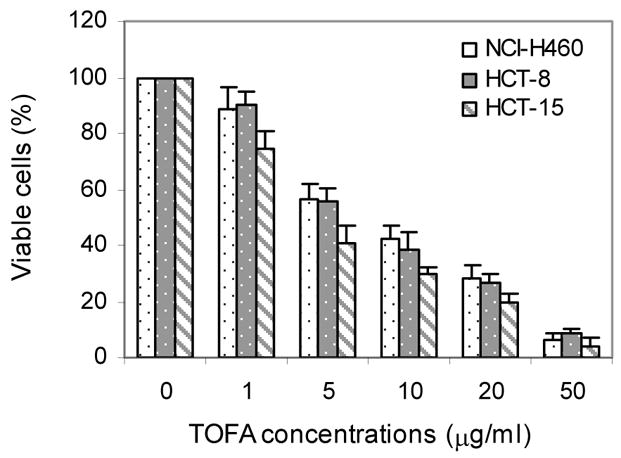
TOFA (5-tetradecyloxy-2-furoic acid) exhibits cytotoxicity against NCI-H460 lung cancer cells as well as HCT-8 and HCT-15 colon cancer cells, with IC50 values of around 5.0, 5.0, and 4.5 μg/mL, in that order. In a dose-dependent manner, TOFA at 1.0–20.0 μg/mL efficiently inhibits the synthesis of fatty acids and causes cell death [1]. The IC50 values of ~26.1 and 11.6 μg/mL for TOFA indicated that it was cytotoxic to COC1 and COC1/DDP cells, respectively. TOFA stops the cell cycle in the G0/G1 phase, triggers apoptosis, and inhibits the growth of cancer cells in a time- and dose-dependent way [2]. Acetyl-CoA-carboxylase-α, or ACCA, is an essential enzyme that controls the production of fatty acids. In most PCa cell lines, TOFA-induced ACCA inhibition results in decreased fatty acid production, caspase activation, and cell death [3].
TOFA (5-tetradecyloxy-2-furoic acid), an allosteric inhibitor of ACCA, is cytotoxic to lung cancer cells NCI-H460 and colon carcinoma cells HCT-8 and HCT-15, with an IC(50) at approximately 5.0, 5.0, and 4.5 microg/ml, respectively. TOFA at 1.0-20.0 microg/ml effectively blocked fatty acid synthesis and induced cell death in a dose-dependent manner. The cell death was characterized with PARP cleavage, DNA fragmentation, and annexin-V staining, all of which are the features of the apoptosis. Supplementing simultaneously the cells with palmitic acids (100 microM), the end-products of the fatty acid synthesis pathway, prevented the apoptosis induced by TOFA. Taken together, these data suggest that TOFA is a potent cytotoxic agent to lung and colon cancer cells, inducing apoptosis through disturbing their fatty acid synthesis.[1] The aim of the present study was to investigate the effects of 5‑tetradecyloxy‑2‑furoic acid (TOFA), an allosteric inhibitor of ACC, on the proliferation and cell cycle progression of the ovarian cancer cell lines COC1 and COC1/DDP. TOFA was found to be cytotoxic to COC1 and COC1/DDP cells with a 50% inhibitory concentration (IC50) of ~26.1 and 11.6 µg/ml, respectively. TOFA inhibited the proliferation of the cancer cells examined in a time‑ and dose‑dependent manner, arrested the cells in the G0/G1 cell cycle phase and induced apoptosis. The expression of the cell cycle regulating proteins cyclin D1 and cyclin-dependent kinase (CDK) 4, as well as the expression of the apoptosis‑related proteins caspase‑3 and Bcl‑2, were detected by western blot analysis. Cyclin D1, CDK4 and Bcl‑2 protein expression was inhibited by TOFA, while caspase‑3 was cleaved and activated. [2] Inhibition of ACCA by TOFA (5-tetradecyl-oxy-2-furoic acid) decreases fatty acid synthesis and induces caspase activation and cell death in most PCa cell lines. Our data suggest that TOFA can kill cells via the mitochondrial pathway since we found cytochrome c release after TOFA treatment in androgen sensitive cell lines. The results also imply that the pro-apoptotic effect of TOFA may be mediated via a decrease of neuropilin-1(NRP1) and Mcl-1expression. We have previously reported that Mcl-1 is under AR regulation and plays an important role in resistance to drug-induced apoptosis in prostate cancer cells, and NRP1 is known to regulate Mcl-1 expression. [3] 1. TOFA exhibited cytotoxicity to human lung cancer NCI-H460 cells, colon carcinoma HCT-8 and HCT-15 cells with IC50 values of approximately 5.0 μg/ml, 5.0 μg/ml, and 4.5 μg/ml, respectively; TOFA (1.0-20.0 μg/ml) blocked fatty acid synthesis and induced cell death in a dose-dependent manner [1] 2. TOFA induced apoptosis in NCI-H460, HCT-8 and HCT-15 cells, characterized by PARP cleavage, DNA fragmentation, and increased annexin-V positive cells (early + late apoptotic cells); supplementation with 100 μM palmitic acid reversed TOFA-induced apoptosis [1] 3. Silencing ACCA with siRNA (50 nM) induced apoptosis in cancer cells, and co-treatment with TOFA (10 μg/ml) further enhanced the apoptotic effect [1] 4. TOFA was cytotoxic to ovarian cancer COC1 and COC1/DDP cells with IC50 values of ~26.1 μg/ml and 11.6 μg/ml, respectively; it inhibited cell proliferation in a time- and dose-dependent manner, arrested cells in G0/G1 phase, and induced apoptosis [2] 5. TOFA downregulated the expression of cyclin D1, CDK4 and Bcl-2 proteins, and cleaved/activated caspase-3 in ovarian cancer COC1 and COC1/DDP cells [2] 6. TOFA reduced fatty acid synthesis in prostate cancer (PCa) cells, induced caspase activation and cell death in most PCa cell lines (independent of p53 status); it triggered cytochrome c release in androgen-sensitive PCa cell lines (mediating apoptosis via mitochondrial pathway) [3] 7. TOFA decreased the expression of neuropilin-1 (NRP1) and Mcl-1 in PCa cells; NRP1 expression was under androgen receptor (AR) control, and Mcl-1 was regulated by AR and involved in drug resistance [3] 8. Dihydrotestosterone (DHT) treatment increased ACCA expression and fatty acid synthesis in LNCaP cells, while TOFA reversed the elevated fatty acid synthesis induced by DHT [3] |
| ln Vivo |
COC1/DDP cell growth in ovarian tumor mouse xenografts is inhibited by TOFA. The tumor growth rate was significantly inhibited by TOFA in comparison to control mice treated with DMSO (1649±356.3 vs. 5128±390.4 mm)3. The tissues of the lung, kidney, spleen, heart, and intestines did not exhibit any toxicity. TOFA may develop into a promising small molecule medication for the treatment of ovarian cancer by blocking ACC [2].
TOFA inhibits growth of ovarian cancer xenografts in mice[2] To investigate whether TOFA suppresses tumor growth in vivo, the effects of TOFA were investigated using a human ovarian cancer mouse xenograft model. COC1/DDP cells were injected into female nude mice, and tumor sizes were measured following TOFA treatment. The tumor growth rate was significantly inhibited by TOFA compared with the DMSO-treated control mice (1649±356.3 vs. 5128±390.4 mm3; Fig. 4A). To test for TOFA toxicity, the effect of TOFA was examined on multiple mouse organs with H&E staining (Fig. 4B). No toxicity was observed in the heart, liver, spleen, lung, kidney and intestinal tissues (data not shown).[2] 1. TOFA inhibited the growth of COC1/DDP ovarian cancer cell xenografts in mice [2] |
| Enzyme Assay |
Apoptosis[1]
Apoptosis was evaluated by PARP cleavage, DNA fragmentation, and annexin-V staining and FACScan analysis. PARP cleavage was detected by Western blot as described above; DNA fragmentation was examined as addressed previously; and annexin-V staining and FACScan was performed as previously described. Fatty acid synthesis[1] Cells were pulsed with 1 μCi 2-14C-labeled acetate (53 mCi/mmol) per well of 12-well plates for 4 hours at 37°C, 5% CO2, followed by lipid synthesis analysis as previously described. 1. Fatty acid synthesis assay: Cancer cells (NCI-H460, HCT-8, HCT-15) were exposed to TOFA at different concentrations for 24 hours; 4 hours before harvesting, cells were pulsed with 1 μCi 2-¹⁴C-labeled acetate per well of 12-well plates, then fatty acid synthesis was analyzed to evaluate the inhibitory effect of TOFA on fatty acid synthesis (reflecting ACCA activity indirectly) [1] |
| Cell Assay |
Cell proliferation assay[2]
\TOFA was dissolved in dimethylsulphoxide (DMSO) to make a 50 mg/ml stock solution. The cells (1×10⁴ cells/well) were seeded in 96-well plates and incubated with TOFA at various concentrations (0, 1, 5, 10, 20 and 50 μg/ml). Cell proliferation was assessed 24, 48 and 72 h following TOFA treatment by measuring the reduction of the tetrazolium salt WST-8 to formazan using the Cell Counting kit-8 (CCK-8) purchased from Dojindo according to the manufacturer’s instructions. At each time point, 10 μl of the CCK-8 solution was added to each well and cultured at 37°C for 2 h. The supernatant from each plate was collected and the absorbance was measured at 450 nm. The inhibition rate of cell proliferation and the 50% inhibitory concentration (IC50; calculated by nonlinear regression) was determined according to the following equation: Inhibition rate (%) = [(Ac−Ae)/(Ac−Ab)] × 100%, where Ae, absorbance of the culture media in experimental wells; Ac, absorbance of the culture medium in control wells and Ab, absorbance of the culture medium in blank wells. Cell cycle analysis[2] \COC1 and COC1/DDP cells were seeded in 6-well plates at a density of 2×10⁵ cells/well and treated with or without TOFA (5 or 10 μg/ml) for 24, 48 and 72 h. For cell cycle analysis, the cells were centrifuged at 466 × g for 5 min and washed twice with cold phosphate-buffered saline (PBS). The cells were fixed with 70% ice-cold ethanol and stored at −20°C. The cells were washed with PBS 24 h later, treated with RNase A and incubated at 37°C for 30 min according to the manufacturer’s protocol. Propidium iodide (PI) was added (400 μl of a 20-μg/ml solution), the cells were incubated in the dark for 30 min and the absorbance was measured at 488 nm. Apoptosis assay[2] \Ovarian cancer cells COC1 and COC1/DDP (2×10⁵ cells/well) were treated with TOFA (0, 1, 5, 10, 20 and 50 μg/ml) for 24, 48 and 72 h. Subsequently, an apoptosis detection kit was used according to the manufacturer’s instructions. The cells were collected by centrifugation at 466 × g for 5 min, washed twice with cold PBS and suspended in 100 μl binding buffer. Annexin V-FITC (5 μl) and PI (5 μl) were added to the culture media of cells and the cells were incubated for 15 min at room temperature in the dark. Binding buffer (400 μl) was added and fluorescent intensities were determined by flow cytometry at 488 nm TOFA suppresses ovarian cancer cell proliferation[2] \To investigate the effect of TOFA on ovarian cancer cells, COC1 and COC1/DDP cell lines were treated with various concentrations of TOFA (1–50 μg/ml) for 24–72 h, and cell proliferation was assessed. The various concentrations of TOFA used were found to inhibit COC1 and COC1/DDP cell growth in a concentration- and time-dependent manner (Fig. 1). TOFA was also shown to be highly cytotoxic to ovarian cancer cells. The IC50 values of COC1 and COC1/DDP cells for TOFA were ~26.1 and ~11.2 μg/ml, respectively, after 48 h of treatment. These data indicate that fatty acid synthesis plays an important role in the proliferation of the ovarian cancer cell lines COC1 and COC1/DDP. TOFA induces cell cycle arrest[2] \To investigate whether TOFA induced cell cycle arrest in ovarian cancer cells, COC1 and COC1/DDP cells were cultured with TOFA for 24, 48 and 72 h, followed by cell counting using FACScan analysis. COC1 cells treated with TOFA were arrested in the G0/G1 phase. Particularly, treatment with 5 μg/ml TOFA for 48 and 72 h increased the percentage of COC1 cells in this phase to 50.2 and 51.1%, compared with the percentage of the untreated control groups, 28.1 and 34.4% (P<0.01), respectively (Fig. 2B and C). The treatment of COC1/DDP cells with TOFA (10 μg/ml) for 72 h increased the percentage of cells in the G0/G1 phase from 38.3 (control) to 43.0% (P<0.05; Fig. 2G). These results indicate that cell cycle progression from the G1 to the S phase is associated with fatty acid synthesis. To investigate the mechanism underlying cell cycle arrest, the protein levels of cyclin D1 and cyclin-dependent kinase (CDK) 4, two proteins that regulate progression from the G1 to the S phase, were examined using western blot analysis (Fig. 2D and H). TOFA treatment of COC1 and COC1/DDP cells decreased cyclin D1 protein levels in a dose-dependent manner. However, CDK4 protein levels were increased in TOFA-treated COC1 cells (1–20 μg/ml), followed by a subsequent decrease when 50 μg/ml TOFA was used (Fig. 2D). CI-H460, human lung cancer cells, and HCT-8 and HCT-15 cells (5,000/well) are seeded in 96-well plates overnight and then exposed to TOFA at indicated concentrations (0, 1, 5, 10, 20, 50 µg/mL) for 72 hours. Viable cells are detected using MTT assay. 1. MTT cytotoxicity/proliferation assay (NCI-H460, HCT-8, HCT-15 cells): Cells were seeded into 96-well plates at 5,000 cells/well and incubated overnight; fresh medium containing TOFA at different concentrations was added, followed by 72-hour incubation; viable cells were assessed by MTT cell proliferation kit [1] 2. Western blot for ACCA expression: Soluble proteins (100 μg per sample) from NCI-H460, HCT-8 and HCT-15 cells were extracted, and ACCA protein levels were detected by Western blot with beta-actin as loading control [1] 3. PARP cleavage detection (apoptosis): NCI-H460, HCT-8 and HCT-15 cells (2×10⁶) were incubated overnight, then treated with TOFA at different concentrations for 24 hours; soluble proteins (100 μg per sample) were extracted, and PARP cleavage was detected by Western blot with beta-actin as loading control [1] 4. DNA fragmentation assay: Medium and PBS wash solutions of TOFA-treated cells were pooled with harvested cells; DNA ladders were prepared and visualized to detect apoptosis [1] 5. Annexin-V staining and FACScan analysis: PBS wash solutions and trypsinized cell suspensions of TOFA-treated cells were pooled; annexin-V staining was performed, and FACScan was used to quantify total apoptotic cells (early + late apoptotic cells) [1] 6. siRNA transfection and apoptosis assay: Chemically synthesized ACCA siRNA (50 nM) was delivered into cancer cells (1×10⁵); cells were treated with/without TOFA (10 μg/ml), then apoptotic cells were assessed by annexin staining and FACScan (scrambled siRNA as control) [1] 7. Palmitic acid rescue assay: NCI-H460, HCT-8, HCT-15 cells (2×10⁶) were incubated overnight, then treated with TOFA (10 μg/ml) and 100 μM palmitic acid simultaneously for 24 hours; apoptosis was detected by annexin-V staining and FACScan [1] 8. MTT assay for ovarian cancer cells (COC1, COC1/DDP): Cells were treated with TOFA at different concentrations for specific time periods; cell proliferation/viability was measured by MTT assay to determine IC50 and time/dose-dependent inhibitory effect [2] 9. Cell cycle analysis: COC1 and COC1/DDP cells were treated with TOFA, then cell cycle distribution (G0/G1 phase arrest) was analyzed [2] 10. Western blot for ovarian cancer cells: COC1 and COC1/DDP cells were treated with TOFA; protein expression of cyclin D1, CDK4, Bcl-2 and cleaved caspase-3 was detected by Western blot [2] 11. Fatty acid synthesis assay for PCa cells: LNCaP cells were treated with DHT and/or TOFA; fatty acid synthesis level was measured to evaluate the effect of TOFA [3] 12. Caspase activation assay for PCa cells: PCa cells were treated with TOFA; caspase activation was detected to evaluate apoptotic effect [3] 13. Cytochrome c release assay: Androgen-sensitive PCa cells were treated with TOFA; cytochrome c release from mitochondria was detected to assess mitochondrial apoptotic pathway [3] 14. Western blot for PCa cells: LNCaP cells were treated with DHT and/or TOFA; ACCA, AR, NRP1 and Mcl-1 protein expression levels were detected by Western blot [3] |
| Animal Protocol |
50 mg/kg; i.p.
Mice COC1/DDP tumor xenografts in mice[2] Female athymic BALB/c nude mice, 4–5 weeks old, weighing 17–20 g were used. COC1/DDP cells were collected by centrifugation at 300 × g for 5 min and suspended in RPMI-1640 medium at a density of 2×10⁶ cells/100 μl. The cells were subcutaneously injected into both right and left flanks of each mouse. Twenty days later, mice were randomly allocated into one of the 2 groups (n=5 mice/group): i) mice treated with 50 μl DMSO (control group) or ii) mice treated with TOFA (50 mg/kg). The drugs were injected intraperitoneally daily for two weeks. Tumor volumes were recorded every two days by measuring dimensions (length and width) with Vernier calipers. The mice were sacrificed four weeks after the final treatment. Tumor weights were measured by a scale. The formula used to determine tumor sizes was A × B2 × 0.5236 (A, length; B, width; all measured in mm). For histopathological examination, tumors and multiple organs were fixed in 10% neutral-buffered formalin and wax embedded. The tissues were cut into 4-mm sections and stained with hematoxylin and eosin (H&E). 1. Ovarian cancer xenograft model: COC1/DDP ovarian cancer cells were implanted into mice to establish xenograft tumors; TOFA was administered (specific dose, route, frequency and formulation not described), and tumor growth was monitored to evaluate the inhibitory effect of TOFA [2] |
| References |
|
| Additional Infomation |
5-(tetradecyloxy)-2-furanic acid is a furan compound with a structure similar to 2-furanic acid, except that the hydrogen at the 5-position is replaced by a tetradecyloxy group. It possesses multiple functions, including as an EC 6.4.1.2 (acetyl-CoA carboxylase) inhibitor, a PPARα agonist, an antitumor agent, and an apoptosis inducer. It is a furanic acid and also an aromatic ether. Its functions are related to those of 2-furanic acid.
1. TOFA (5-tetradecyloxy-2-furanoic acid) is an allosteric inhibitor of ACCA; ACCA is the rate-limiting enzyme in long-chain fatty acid synthesis and is upregulated in various human cancers[1] 2. TOFA induces apoptosis in cancer cells by interfering with fatty acid synthesis; palmitic acid (the end product of fatty acid synthesis) can rescue TOFA-induced apoptosis[1] 3. TOFA is an allosteric inhibitor of ACC; ACC is a key enzyme in fatty acid synthesis and is highly expressed in various human cancers, which is crucial for cancer cell growth[2] 4. TOFA is a promising small molecule drug that can treat ovarian cancer by inhibiting ACC[2] 5. AR binds to the intron region of the human ACCA gene in vivo; the level of ACCA protein in LNCaP cells depends on the expression of AR[3] 6. Tumor-associated adipogenesis can protect cancer cells from damage by carcinogens and therapeutic agents; in androgen-responsive cancer cells, increased fatty acid/cholesterol synthesis is regulated by androgens [3]. 7. TOFA is an effective cell death inducer in prostate cancer cells, and its pro-apoptotic effect may be achieved by reducing the expression of NRP1 and Mcl-1 [3]. |
| Molecular Formula |
C19H32O4
|
|
|---|---|---|
| Molecular Weight |
324.45
|
|
| Exact Mass |
324.23
|
|
| Elemental Analysis |
C, 70.33; H, 9.94; O, 19.72
|
|
| CAS # |
54857-86-2
|
|
| Related CAS # |
|
|
| PubChem CID |
115175
|
|
| Appearance |
White to off-white solid powder
|
|
| Density |
1.0±0.1 g/cm3
|
|
| Boiling Point |
441.7±25.0 °C at 760 mmHg
|
|
| Melting Point |
112-115ºC
|
|
| Flash Point |
220.9±23.2 °C
|
|
| Vapour Pressure |
0.0±1.1 mmHg at 25°C
|
|
| Index of Refraction |
1.483
|
|
| LogP |
7.82
|
|
| Hydrogen Bond Donor Count |
1
|
|
| Hydrogen Bond Acceptor Count |
4
|
|
| Rotatable Bond Count |
15
|
|
| Heavy Atom Count |
23
|
|
| Complexity |
293
|
|
| Defined Atom Stereocenter Count |
0
|
|
| SMILES |
O=C(C1=CC=C(OCCCCCCCCCCCCCC)O1)O
|
|
| InChi Key |
CZRCFAOMWRAFIC-UHFFFAOYSA-N
|
|
| InChi Code |
InChI=1S/C19H32O4/c1-2-3-4-5-6-7-8-9-10-11-12-13-16-22-18-15-14-17(23-18)19(20)21/h14-15H,2-13,16H2,1H3,(H,20,21)
|
|
| Chemical Name |
5-tetradecoxyfuran-2-carboxylic acid
|
|
| Synonyms |
|
|
| HS Tariff Code |
2934.99.9001
|
|
| Storage |
Powder -20°C 3 years 4°C 2 years In solvent -80°C 6 months -20°C 1 month |
|
| Shipping Condition |
Room temperature (This product is stable at ambient temperature for a few days during ordinary shipping and time spent in Customs)
|
| Solubility (In Vitro) |
DMSO: ~4 mg/mL ( 12.32 mM)
Water: Insoluble Ethanol: Insoluble |
|---|---|
| Solubility (In Vivo) |
Solubility in Formulation 1: ≥ 1 mg/mL (3.08 mM) (saturation unknown) in 10% DMSO + 90% Corn Oil (add these co-solvents sequentially from left to right, and one by one), clear solution.
For example, if 1 mL of working solution is to be prepared, you can add 100 μL of 10.0 mg/mL clear DMSO stock solution to 900 μL of corn oil and mix evenly. Solubility in Formulation 2: 5%DMSO+ 40%PEG300+ 5%Tween 80+ 50%ddH2O : 3mg/ml (9.25mM) (Please use freshly prepared in vivo formulations for optimal results.) |
| Preparing Stock Solutions | 1 mg | 5 mg | 10 mg | |
| 1 mM | 3.0821 mL | 15.4107 mL | 30.8214 mL | |
| 5 mM | 0.6164 mL | 3.0821 mL | 6.1643 mL | |
| 10 mM | 0.3082 mL | 1.5411 mL | 3.0821 mL |
*Note: Please select an appropriate solvent for the preparation of stock solution based on your experiment needs. For most products, DMSO can be used for preparing stock solutions (e.g. 5 mM, 10 mM, or 20 mM concentration); some products with high aqueous solubility may be dissolved in water directly. Solubility information is available at the above Solubility Data section. Once the stock solution is prepared, aliquot it to routine usage volumes and store at -20°C or -80°C. Avoid repeated freeze and thaw cycles.
Calculation results
Working concentration: mg/mL;
Method for preparing DMSO stock solution: mg drug pre-dissolved in μL DMSO (stock solution concentration mg/mL). Please contact us first if the concentration exceeds the DMSO solubility of the batch of drug.
Method for preparing in vivo formulation::Take μL DMSO stock solution, next add μL PEG300, mix and clarify, next addμL Tween 80, mix and clarify, next add μL ddH2O,mix and clarify.
(1) Please be sure that the solution is clear before the addition of next solvent. Dissolution methods like vortex, ultrasound or warming and heat may be used to aid dissolving.
(2) Be sure to add the solvent(s) in order.
Cytotoxicity of TOFA.Biochem Biophys Res Commun.2009 Jul 31;385(3):302-6. |
|---|
 Apoptosis induced by TOFA.Biochem Biophys Res Commun.2009 Jul 31;385(3):302-6. |
 Inhibition of fatty acid synthesis by TOFA. Palmitate rescues of apoptosis induced by TOFA.Biochem Biophys Res Commun.2009 Jul 31;385(3):302-6. |
Products are for research use only; We do not sell to patients
Copyright 2020 InvivoChem LLC | All Rights Reserved


 COA
COA