| Size | Price | Stock | Qty |
|---|---|---|---|
| 5mg |
|
||
| 10mg |
|
||
| 25mg |
|
||
| 50mg |
|
||
| Other Sizes |


Purity: ≥98%
| Targets |
Src HuH7 (GI50 = 9 nM); Src PLC/PRF/5 (IC50 = 13 nM); Src Hep3B (IC50 = 26 nM); Src HepG2 (IC50 = 60 nM)
The primary target of Tirbanibulin is Src kinase, a non-receptor tyrosine kinase involved in cell proliferation, survival, motility, and angiogenesis. Unlike conventional kinase inhibitors that target the ATP-binding site, Tirbanibulin targets the peptide substrate site of Src. This unique binding mechanism may contribute to its selectivity and reduced off-target effects. By inhibiting Src activity, Tirbanibulin disrupts downstream signaling pathways involved in cancer cell growth and proliferation. |
|---|---|
| ln Vitro |
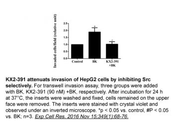
Tirbanibulin (KX2-391) is a Src inhibitor that targets the substrate pocket of Src. Four hepatic cell cancer (HCC) cell lines, Huh7 (GI50=9 nM), PLC/PRF/5 (GI50=13 nM), Hep3B (GI50=26 nM), and HepG2 (GI50=60 nM), exhibit steep dose-response curves when compared to KX2-391[1]. Certain leukemia cells, such as those derived from chronic leukemia cells with the T3151 mutation, that are resistant to currently available commercial drugs are found to be inhibited by tirbanibulin (KX2-391). Tirbanibulin (KX2-391) demonstrates GI50 values of 23 nM and 39 nM, respectively, when assessed in Src-driven cell growth assays using NHK3/c-Src527F and SYF/c-Src527F cells that have been engineered.
Tirbanibulin demonstrates potent in vitro antiproliferative activity against cancer cell lines with GI50 values ranging from 9 to 60 nM. The compound's unique mechanism of targeting the peptide substrate site of Src provides selectivity and distinguishes it from ATP-competitive kinase inhibitors. In cellular assays, Tirbanibulin inhibits Src-mediated signaling and induces growth inhibition in various cancer cell types. Its activity has been characterized in preclinical studies supporting its development for cancer and dermatological applications. |
| ln Vivo |
Tirbanibulin (KX2-391) taken orally has been demonstrated in pre-clinical animal models of cancer to inhibit primary tumor growth and to suppress metastasis[2].
Tirbanibulin has been evaluated in clinical studies for the topical treatment of actinic keratosis, demonstrating efficacy, rapid lesion clearance, and a favorable safety profile with mostly mild local skin reactions. It is approved for this indication, indicating successful translation from preclinical to clinical development. The compound's in vivo activity in animal models of cancer and skin conditions has supported its clinical development, though specific animal model data are not detailed in the available sources. |
| Enzyme Assay |
The Src inhibitor tirbanibulin (KX2-391) targets the Src substrate pocket. The hepatic cell cancer (HCC) cell lines Huh7 (GI50=9 nM), PLC/PRF/5 (GI50=13 nM), Hep3B (GI50=26 nM), and HepG2 (GI50=60 nM) exhibit steep dose-response curves when treated with tirbanibulin (KX2-391).
The Src kinase inhibition assay for Tirbanibulin involves incubating the compound with recombinant Src kinase, ATP, and a peptide substrate. The reaction is carried out in a kinase buffer optimized for Src activity. After incubation, the amount of phosphorylated substrate is quantified using a luminescence-based kinase assay or a radiometric assay. IC50 values are calculated from dose-response curves. The compound's unique mechanism of targeting the peptide substrate site can be assessed using peptide substrate competition assays. |
| Cell Assay |
Hep3B, HepG2, PLC/PRF/5, Huh7, and other liver cell lines are frequently grown and kept in basal medium with 2% fetal bovine serum (FBS) at 37°C and 5% CO2. In each well of a 96-well plate, cells are seeded at 4.0×10³/190 μL and 8.0×10³/190 μL in basal medium containing 1.5% FBS. Before adding Tirbanibulin (KX2-391) at concentrations ranging from 6,564 to 0.012 nM in triplicates, these are cultured for an additional night at 37°C and 5% CO2. Three days are spent incubating treated cells. On day three, 10 μL of a 5-mg/mL 3-(4,5-dimethylthiazol-2-yl)-2,5-diphenyltetrazolium bromide (MTT) solution is added to each well, and the cells are incubated for four hours. 10% SDS is added to diluted HCl to dissolve the formazan product. The optical density is measured at 570 nm. Parallel experiments are conducted with Tirbanibulin (KX2-391) to compare its potency and activity. With GraphPad Prism 5 statistical software, growth inhibition curves, 50% inhibition concentration (GI50), and 80% inhibition concentration (GI80) are calculated. Both the optical density at wavelength of 570 nm (OD570) signal format and the normalized data representing the percentage of maximum response are reported.
To evaluate the antiproliferative activity of Tirbanibulin, cancer cells are plated in 96-well plates and treated with varying concentrations of the compound. After 72 hours of incubation, cell viability is measured using an MTT or SRB assay. The GI50 (concentration causing 50% growth inhibition) is calculated from the dose-response curve. The compound's effect on Src signaling can be assessed by measuring Src phosphorylation and downstream signaling targets using Western blot analysis. Apoptosis induction can be evaluated using annexin V-FITC staining and flow cytometry. |
| Animal Protocol |
Mouse bearing MDA-MB-231 tumors; Oral gavage; 1, 5mg/kg dose
Xenograft procedures and KX-01 oral dosing were as described (11). Briefly, mammary fat pad tumors were established by injecting 5×10⁶ MDA-MB-231 cells in 150μl of PBS-Matrigel mixture (1:2) orthotopically and bilaterally into the mammary fat pads of female NUDE mice (two tumors/mouse). Treatments were started when tumors reached ∼80-100mm3. The first study used MDA-MB-231 xenografts and was performed using vehicle (ultra-pure water) and two doses of KX-01 (1, 5mg/kg) administered twice/day (BID) by oral gavage (using metal 22g feeding needle) for 28 days. A similar experiment was performed with MDA-MB-157 xenografts (another ER/PR/HER2 negative model) to assess KX-01 response. A second study was performed to test combination of KX-01 with paclitaxel on tumor growth. MDA-MD-231 tumor xenograft bearing mice were treated with vehicle or KX-01 (5mg/kg) BID, paclitaxel by intraperitoneal injection (IP) once/week, or combination of KX-0+paclitaxel. Treatments were for 40 days for all groups. A third study used MDA-MB-157 xenografts with the same combination treatment. A fourth study tested the effect of KX-01 or combination with paclitaxel for 24 days on larger MDA-MB-231 tumors (∼300mm3). Tumors were allowed to reach ∼300mm3 before beginning treatments. In this experiment mice were treated with KX-01 at a higher dose of 15mg/kg, and mice were treated once/day instead of twice/day. Paclitaxel was used at a dose of 20mg/kg IP once/week. In all experiments, tumor caliper measurements were taken twice/week and tumor volume was by calculated by the formula: 0.523×LM2 (where L-large diameter, M-small diameter). At the end the experiments animals were sacrificed and tumors and mouse organs removed. Tissues were either stored in 10% neutral buffered formalin for paraffin embedding, or snap frozen for measurement of chromosome-17 by real-time PCR, and embedded for frozen sectioning for CD-31 staining. Immunohistochemistry (IHC) was performed as described on paraffin-embedded tumor tissues [3]. The in vivo efficacy of Tirbanibulin for actinic keratosis is evaluated in clinical studies rather than animal models. For topical application, the compound is formulated as an ointment and applied to affected skin areas once daily for several days. The primary efficacy endpoint is the complete clearance of actinic keratosis lesions. In preclinical development, Tirbanibulin has likely been evaluated in xenograft models of cancer and in models of skin conditions, though specific protocols are not detailed in the available sources. |
| ADME/Pharmacokinetics |
Specific pharmacokinetic data for Tirbanibulin are not provided in the available sources. The compound has a molecular weight of 504.45 and a molecular formula of C26H31Cl2N3O3. As an FDA-approved topical drug for actinic keratosis, its pharmacokinetic profile has been characterized in clinical studies. Topical administration results in minimal systemic absorption, limiting systemic exposure and contributing to its favorable safety profile. The compound is stored at -20°C for long-term stability.
|
| Toxicity/Toxicokinetics |
Tirbanibulin has been evaluated in clinical studies and is approved for the topical treatment of actinic keratosis. Clinical studies demonstrate its efficacy, rapid lesion clearance, and favorable safety profile with mostly mild local skin reactions. As a topical agent, systemic toxicity is minimal due to low systemic absorption. The compound is intended for topical use only and is not approved for systemic administration. Standard safety precautions should be followed when handling this compound.
|
| References |
|
| Additional Infomation |
Tirbanibulin dihydrochloride is also known as KX2-391 dihydrochloride and KX-01 dihydrochloride. It is an inhibitor of Src that targets the peptide substrate site of Src, with GI50 of 9-60 nM in cancer cell lines. It is approved for the topical treatment of actinic keratosis, a precancerous skin condition, offering a non-invasive therapeutic option. Clinical studies demonstrate its efficacy, rapid lesion clearance, and favorable safety profile. The compound has a CAS number of 1038395-65-1.
|
| Molecular Formula |
C₂₆H₃₁CL₂N₃O₃
|
|
|---|---|---|
| Molecular Weight |
504.45
|
|
| Exact Mass |
503.174
|
|
| Elemental Analysis |
C, 61.23; H, 5.96; Cl, 14.46; N, 8.57; O, 9.79
|
|
| CAS # |
1038395-65-1
|
|
| Related CAS # |
Tirbanibulin;897016-82-9;Tirbanibulin Mesylate;1080645-95-9
|
|
| PubChem CID |
24989633
|
|
| Appearance |
Light yellow to yellow solid powder
|
|
| LogP |
5.7
|
|
| Hydrogen Bond Donor Count |
3
|
|
| Hydrogen Bond Acceptor Count |
5
|
|
| Rotatable Bond Count |
9
|
|
| Heavy Atom Count |
34
|
|
| Complexity |
540
|
|
| Defined Atom Stereocenter Count |
0
|
|
| InChi Key |
CPTPOZGQCQXHJO-UHFFFAOYSA-N
|
|
| InChi Code |
InChI=1S/C26H29N3O3.2ClH/c30-26(28-19-21-4-2-1-3-5-21)18-24-9-6-23(20-27-24)22-7-10-25(11-8-22)32-17-14-29-12-15-31-16-13-29;;/h1-11,20H,12-19H2,(H,28,30);2*1H
|
|
| Chemical Name |
N-benzyl-2-[5-[4-(2-morpholin-4-ylethoxy)phenyl]pyridin-2-yl]acetamide;dihydrochloride
|
|
| Synonyms |
|
|
| HS Tariff Code |
2934.99.9001
|
|
| Storage |
Powder -20°C 3 years 4°C 2 years In solvent -80°C 6 months -20°C 1 month Note: Please store this product in a sealed and protected environment (e.g. under nitrogen), avoid exposure to moisture. |
|
| Shipping Condition |
Room temperature (This product is stable at ambient temperature for a few days during ordinary shipping and time spent in Customs)
|
| Solubility (In Vitro) |
|
|||
|---|---|---|---|---|
| Solubility (In Vivo) |
Solubility in Formulation 1: ≥ 2.5 mg/mL (4.96 mM) (saturation unknown) in 10% DMSO + 40% PEG300 + 5% Tween80 + 45% Saline (add these co-solvents sequentially from left to right, and one by one), clear solution.
For example, if 1 mL of working solution is to be prepared, you can add 100 μL of 25.0 mg/mL clear DMSO stock solution to 400 μL PEG300 and mix evenly; then add 50 μL Tween-80 to the above solution and mix evenly; then add 450 μL normal saline to adjust the volume to 1 mL. Preparation of saline: Dissolve 0.9 g of sodium chloride in 100 mL ddH₂ O to obtain a clear solution. Solubility in Formulation 2: ≥ 2.5 mg/mL (4.96 mM) (saturation unknown) in 10% DMSO + 90% (20% SBE-β-CD in Saline) (add these co-solvents sequentially from left to right, and one by one), clear solution. For example, if 1 mL of working solution is to be prepared, you can add 100 μL of 25.0 mg/mL clear DMSO stock solution to 900 μL of 20% SBE-β-CD physiological saline solution and mix evenly. Preparation of 20% SBE-β-CD in Saline (4°C,1 week): Dissolve 2 g SBE-β-CD in 10 mL saline to obtain a clear solution. View More
Solubility in Formulation 3: ≥ 2.5 mg/mL (4.96 mM) (saturation unknown) in 10% DMSO + 90% Corn Oil (add these co-solvents sequentially from left to right, and one by one), clear solution. Solubility in Formulation 4: 4% DMSO+30% PEG 300+ddH2O: 5 mg/mL |
| Preparing Stock Solutions | 1 mg | 5 mg | 10 mg | |
| 1 mM | 1.9824 mL | 9.9118 mL | 19.8236 mL | |
| 5 mM | 0.3965 mL | 1.9824 mL | 3.9647 mL | |
| 10 mM | 0.1982 mL | 0.9912 mL | 1.9824 mL |
*Note: Please select an appropriate solvent for the preparation of stock solution based on your experiment needs. For most products, DMSO can be used for preparing stock solutions (e.g. 5 mM, 10 mM, or 20 mM concentration); some products with high aqueous solubility may be dissolved in water directly. Solubility information is available at the above Solubility Data section. Once the stock solution is prepared, aliquot it to routine usage volumes and store at -20°C or -80°C. Avoid repeated freeze and thaw cycles.
Calculation results
Working concentration: mg/mL;
Method for preparing DMSO stock solution: mg drug pre-dissolved in μL DMSO (stock solution concentration mg/mL). Please contact us first if the concentration exceeds the DMSO solubility of the batch of drug.
Method for preparing in vivo formulation::Take μL DMSO stock solution, next add μL PEG300, mix and clarify, next addμL Tween 80, mix and clarify, next add μL ddH2O,mix and clarify.
(1) Please be sure that the solution is clear before the addition of next solvent. Dissolution methods like vortex, ultrasound or warming and heat may be used to aid dissolving.
(2) Be sure to add the solvent(s) in order.
|
Products are for research use only; We do not sell to patients
Copyright 2020 InvivoChem LLC | All Rights Reserved


 COA
COA