| Size | Price | Stock | Qty |
|---|---|---|---|
| 5mg |
|
||
| 10mg |
|
||
| 25mg |
|
||
| 50mg |
|
||
| 100mg |
|
||
| 250mg |
|
||
| 500mg |
|
||
| 1g | |||
| Other Sizes |


Purity: ≥98%
| Targets |
PARP2 ( Ki = 0.87 nM ); PARP1 ( Ki = 1.2 nM )
Talazoparib (BMN 673; MDV3800) is a potent and selective inhibitor of poly(ADP-ribose) polymerases (PARP), with high affinity for PARP1 (IC50 = 0.87 nM) and PARP2 (IC50 = 2.5 nM) in recombinant enzyme assays. It exhibits minimal inhibition of other PARP family members (e.g., PARP3, PARP6) with IC50 >1000 nM. Notably, it is a potent PARP "trapper," forming stable drug-PARP-DNA complexes with ~10-fold higher trapping efficiency than olaparib [1] - Talazoparib (BMN 673; MDV3800) does not inhibit other DNA repair enzymes (e.g., ATM, ATR, DNA-PK) or kinases (e.g., PI3K, AKT) even at concentrations up to 10 μM, confirming PARP-specific activity [3] |
|---|---|
| ln Vitro |
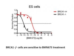
BMN-673 selectively binds to PARP and inhibits the base-excision repair pathway, which is PARP-mediated DNA repair of single strand breaks. This increases the rate at which DNA strand breaks accumulate, encourages genomic instability, and ultimately results in apoptosis. BRCA-1 or BRCA-2 mutated cancer cells are specifically killed by BMN 673. In BRCA-1 mutant (MX-1, IC50 = 0.3 nM) and BRCA-2 mutant cells (Capan-1, IC50 = 5 nM) cells, BMN 673 exhibits single-agent cytotoxicity. In contrast, the IC50 of BMN 673 varies between 90 nM and 1.9 μM in MRC-5 normal human fibroblast and other tumor cell lines with wild-type BRCA-1 and BRCA-2 genes.[1]
Additionally, BMN 673 considerably increases the cytotoxic efficacy of SN-38 and temozolomide in cultured human cancer cells. For this class of PARP inhibitors, off-target molecular screening did not reveal any appreciable non-specific activity.[2] Antiproliferative activity in HR-deficient cells: Talazoparib (BMN 673; MDV3800) shows strong selectivity for homologous recombination (HR)-deficient cells. IC50 values (72 h, MTT assay): BRCA1-mutant MDA-MB-436 (breast cancer, 0.45 μM), BRCA2-mutant Capan-1 (pancreatic cancer, 0.32 μM), BRCA1-mutant OVCAR-8 (ovarian cancer, 0.51 μM); vs. HR-proficient MCF-7 (breast cancer, IC50 = 18 μM), HCT116 (colorectal cancer, IC50 = 22 μM) [1] - PARP inhibition and PARP trapping: In MDA-MB-436 cells, Talazoparib (BMN 673; MDV3800) (0.1–1 μM) dose-dependently reduced PAR polymer levels (by 80–95% at 0.5 μM, western blot) and increased PARP-DNA trapping (detected by chromatin immunoprecipitation, 4.2-fold higher than olaparib at 0.3 μM). It also induced G2/M cell cycle arrest (45% G2/M cells vs. 17% control) and increased γ-H2AX foci (5.8-fold vs. control) at 0.5 μM [1] - Synergy with DNA-damaging agents: Combination of Talazoparib (BMN 673; MDV3800) (0.1 μM) with carboplatin (0.5 μg/mL) in BRCA2-mutant Capan-1 cells reduced viability to 12% (combination) vs. 65% (Talazoparib alone) and 70% (carboplatin alone), with a combination index (CI) of 0.35. Similar synergy was observed with olaparib, but Talazoparib required 10-fold lower concentrations [3] - Activity in PARP inhibitor-resistant cells: Talazoparib (BMN 673; MDV3800) (1 μM) inhibited growth of olaparib-resistant BRCA2-mutant cells (IC50 = 1.2 μM) by restoring PARP trapping and γ-H2AX accumulation, whereas olaparib (10 μM) had no effect [3] |
| ln Vivo |
BMN 673 exhibits >50% oralbioavailability and pharmacokinetic characteristics that permit single-daily dosing in rat pharmacokinetic studies. Daily oral dosing of BMN 673 significantly and dose-dependently increases the antitumor effects of cytotoxic therapies in MX-1 xenograft tumor model studies.[2]
BMN 673 is readily orally bioavailable, with more than 40% absolute oral bioavailability in rats when dosed in carboxylmethyl cellulose. Oral administration of BMN 673 elicited remarkable antitumor activity in vivo; xenografted tumors that carry defects in DNA repair due to BRCA mutations or PTEN deficiency were profoundly sensitive to oral BMN 673 treatment at well-tolerated doses in mice. Synergistic or additive antitumor effects were also found when BMN 673 was combined with temozolomide, SN38, or platinum drugs.
Conclusion: BMN 673 is currently in early-phase clinical development and represents a promising PARP1/2 inhibitor with potentially advantageous features in its drug class.[1]
Combinatorial clinical trials of PARP inhibitors with immunotherapies are ongoing, yet the immunomodulatory effects of PARP inhibition have been incompletely studied. Here, we sought to dissect the mechanisms underlying PARP inhibitor-induced changes in the tumor microenvironment of BRCA1-deficient triple-negative breast cancer (TNBC). We demonstrate that the PARP inhibitor olaparib induces CD8+ T-cell infiltration and activation in vivo, and that CD8+ T-cell depletion severely compromises antitumor efficacy. Olaparib-induced T-cell recruitment is mediated through activation of the cGAS/STING pathway in tumor cells with paracrine activation of dendritic cells and is more pronounced in HR-deficient compared with HR-proficient TNBC cells and in vivo models. CRISPR-mediated knockout of STING in cancer cells prevents proinflammatory signaling and is sufficient to abolish olaparib-induced T-cell infiltration in vivo. These findings elucidate an additional mechanism of action of PARP inhibitors and provide a rationale for combining PARP inhibition with immunotherapies for the treatment of TNBC. SIGNIFICANCE: This work demonstrates cross-talk between PARP inhibition and the tumor microenvironment related to STING/TBK1/IRF3 pathway activation in cancer cells that governs CD8+ T-cell recruitment and antitumor efficacy. The data provide insight into the mechanism of action of PARP inhibitors in BRCA-associated breast cancer.[3] BRCA-mutant breast cancer xenografts: Female nude mice (6–8 weeks old) bearing subcutaneous BRCA1-mutant MDA-MB-436 tumors were treated with Talazoparib (BMN 673; MDV3800) (1 mg/kg, oral, daily) for 21 days. Tumor growth inhibition (TGI) was 85% (treated volume: 220 mm³ vs. vehicle: 1470 mm³, P<0.001). Combination with carboplatin (5 mg/kg, intraperitoneal, weekly) increased TGI to 94% [1] - Ovarian cancer PDX model: Female NOD/SCID mice (8 weeks old) implanted with BRCA2-mutant patient-derived ovarian cancer tissue were treated with Talazoparib (BMN 673; MDV3800) (0.5 mg/kg, oral, daily) for 35 days. Tumor weight was reduced by 82% vs. vehicle, and median survival was prolonged from 45 days (vehicle) to 78 days (P<0.01). No tumor regrowth was observed in 40% of treated mice for 60 days post-treatment [3] - PARP trapping in vivo: In MDA-MB-436 xenografts, Talazoparib (BMN 673; MDV3800) (1 mg/kg, oral) increased PARP-DNA complexes in tumor tissue by 5.1-fold (chromatin immunoprecipitation) and reduced PAR levels by 80% vs. vehicle, confirming in vivo PARP inhibition and trapping [1] |
| Enzyme Assay |
In order to determine the PARP inhibitor Ki, enzyme assays were carried out in 96-well FlashPlate using 0.5 U PARP1 enzyme, 0.25x activated DNA, 0.2 mCi [3H] NAD, and 5 mmol/L cold NAD (Sigma) in a final volume of 50 mL reaction buffer that contained 10% glycerol (v/v), 25 mmol/L HEPES, 12.5 mmol/L MgCl2, 50 mmol/L KCl, 1 mmol/L dithiothreitol (DTT), and 0.01% NP-40 (v/v), and pH 7.6. NAD was added to the PARP reaction mixture, either with or without inhibitors, to start the reaction, and it was then incubated for one minute at room temperature. The reaction was then stopped by adding 50 microliter of ice-cold 20% trichloroacetic acid (TCA) to each well. After the plate was sealed and shaken for an additional 120 minutes at room temperature, centrifugation was performed. Top-Count was used to determine the radioactive signal bound to the FlashPlate. The Michaelis-Menten equation was used to calculate PARP1 Km at different substrate concentrations (ranging from 1 to 100 mmol/L NAD). Using the formula Ki ¼ IC50/[1þ (substrate)/Km], compound Ki was computed from the enzyme inhibition curve. Using the same assay protocol, Km for the PARP2 enzyme and compound Ki were found. However, instead of using 30 ng of PARP2, 0.25x activated DNA, 0.2 mCi [3H] NAD, and 20 mmol/L cold NAD, the reaction was run for 30 minutes at room temperature.
PARP enzyme assays[1] \The ability of a test compound to inhibit PARP-1 enzyme activity was assessed using Trevigen’s PARP Assay Kit following manufacturer’s instruction. IC50 values were calculated using GraphPad Prism5 software. For PARP inhibitor Ki determination, enzyme assays were carried out in 96-well FlashPlate with 0.5 unit PARP1 enzyme, 0.25x activated DNA (Trevigen), 0.2 μCi [3H] NAD and 5 μM cold NAD in a final volume of 50 μL reaction buffer containing 10%glycerol(v/v), 25 mM Hepes, 12.5 mM MgCl2, 50 mM KCl, 1 mM DTT and 0.01% NP-40(v/v), pH 7.6. Reactions were initiated by adding NAD to the PARP reaction mixture with or without inhibitors and incubated for 1 min at room temperature. 50 μL of ice-cold 20% TCA was then added to each well to stop the reaction. The plate was sealed and shaken for a further 120 min at RT, followed by centrifugation. Radioactive signal bound to the FlashPlate was determined using TopCount. PARP1 Km was determined using Michaelis–Menten equation from various substrate concentrations (1-100 μM NAD). Compound Ki was calculated from enzyme inhibition curve according to the formula: Ki = IC50/(1+[substrate]/Km). Km for PARP2 enzyme and compound Ki were determined with the same assay protocol except 30 ng PARP2, 0.25x activated DNA, 0.2 μCi [3H] NAD and 20 μM cold NAD were used in the reaction for 30min at room temperature.\ \Biacore binding assay[1] \Recombinant human PARP1 (rhPARP1) catalytic domain (residues 662 – 1011) with N-terminal 6XHis-tag was generated in house and used in binding assay for PARP inhibitor interaction using Biacore T200 (GE Healthcare). rhPARP1 was immobilized on a CM5 sensor chip by amine coupling method. Briefly, one flow cell of a CM5 chip was first activated by a 7-min injection at 10 μL/min of freshly prepared 50 mM NHS: 200 mM EDC (1:1) at rate of 10 μL/min. Then rhPARP1 (100 μg/mL, in 10 mM MES pH 6.5) was injected onto the flow cell for 60-sec at 10 μL/min. The remaining active coupling sites were blocked with a 7-min injection of 1M ethanolamine at 10 μL/min. The immobilization buffer contains 10 mM Hepes pH 7.4, 150 mM NaCl, 0.05% Surfectant P20, 5 mM MgCl2, and 0.5 mM TCEP (tris(2-carboxyethyl)phosphine). The immobilization level was ~7600 RU. For binding kinetics measurement, PARP inhibitors at increasing concentrations (12.5, 25, 50, 100, 200 nM) were injected over the chip surface for 60 sec per injection. The exposure was followed by a dissociation phase of 3600 sec in running buffer (immobilization buffer + 1% DMSO) after the last injection. The flow rate was 50 μL/min. After sensorgrams were corrected for signals from a reference flow, kinetics was calculated with Biacore T200 evaluation software ver.1.0.\ \Intracellular PAR formation assay[1] \Cellular PAR synthesis assay assesses the ability of a test compound to inhibit polymerization of PAR. LoVo human colorectal tumor cells grown in 96-well microtiter plates overnight were pre-treated with increasing concentrations of PARP inhibitors for 30 min before H2O2 was added at a final concentration of 50 mM. After a 5-min treatment at room temperature, cells were fixed for 10 minutes with pre-chilled methanol/acetone(7:3) at −20 °C. Fixed cells were incubated with anti-PAR monoclonal antibody for 60 min, followed by incubation with FITC coupled goat anti-mouse IgG (diluted 1:100) and 1 μg/mL DAPI for 60 min. FITC signal was normalized with DAPI signal, and EC50 values were calculated using GraphPad Prism. Recombinant PARP1/2 activity assay: Purified recombinant human PARP1 or PARP2 was incubated with a biotinylated double-stranded DNA (dsDNA) activator and NAD⁺ (substrate) in assay buffer (50 mM Tris-HCl pH 8.0, 10 mM MgCl₂, 1 mM DTT) at 37°C for 15 min. Serial concentrations of Talazoparib (BMN 673; MDV3800) (0.01–10 nM) were added, and incubation continued for 30 min. The reaction was terminated by adding 10% trichloroacetic acid (TCA). PAR polymer formation was detected via streptavidin-HRP and chemiluminescence. IC50 values were calculated by fitting remaining PARP activity to a four-parameter logistic model [1] - PARP trapping assay: HeLa cells were treated with Talazoparib (BMN 673; MDV3800) (0.1–1 μM) for 2 h, then lysed in chromatin extraction buffer. Chromatin fractions were immunoprecipitated with anti-PARP1 antibody, and bound DNA was quantified by qPCR (targeting a genomic locus with induced DNA breaks). Trapping efficiency was calculated as the fold increase in PARP1-DNA binding vs. vehicle, with Talazoparib showing ~10-fold higher efficiency than olaparib at equimolar concentrations [3] |
| Cell Assay |
A panel of 11 SCLC cell lines (IC50=1.7 to 15 nmol/L), all of which fall within clinically feasible ranges, demonstrate Talazoparib (BMN 673; MDV3800) 's strong inhibitory action. Furthermore, PI3K pathway activity and DNA repair protein expression are correlated with Talazoparib (BMN 673; MDV3800) sensitivity.
Confocal microscopy[1] Cells were seeded on coverslips placed in 6-well plates and after 24 hours treated with several concentrations of olaparib or Talazoparib (BMN 673; MDV3800). 24 hours after treatment the cells were fixed in 10% formalin (3.7% PFA) for 1 hour. Cells were permeabilized with 0.2% Triton X-100 in PBS for 20 minutes, treated with 50 μL DNase I (diluted 1/10 in PBS) for 1 hour at 37°C and then blocked with IFF (PBS + 1% BSA and 2% FBS followed by filter sterilization) for 1 hour. The coverslips were then incubated with rabbit anti-γH2Ax primary (Millipore) and mouse anti-RAD51 primary (both 1:1000 in 50μL IFF) overnight at 4°C. The next day cells were incubated with anti-mouse Alexafluor 546 secondary and anti-rabbit Alexafluor 488 secondary (both 1:1000 in 50μL IFF) for one hour. Cells were then washed in PBS containing DAPI 1:10.000 for 10 minutes and attached on glass plates using Vectashield and nail polish. A minimum of four pictures were made of each coverslip using the Leica confocal microscope, and cells were subsequently counted. At least 100 cells were assessed per coverslip, being positive for γH2Ax if they had more than 5 foci per nucleus. The percentage of positive cells was plotted. MTT antiproliferation assay: HR-deficient (MDA-MB-436, Capan-1, OVCAR-8) or HR-proficient (MCF-7, HCT116) cells were seeded in 96-well plates (5×10³ cells/well) and incubated overnight (37°C, 5% CO₂). Talazoparib (BMN 673; MDV3800) (0.01–50 μM) was added, and cells were cultured for 72 h. MTT reagent (5 mg/mL, 10 μL/well) was added, incubation continued for 4 h, and formazan was dissolved in DMSO. Absorbance at 570 nm was measured, and IC50 was calculated via GraphPad Prism [1] - γ-H2AX immunofluorescence assay: MDA-MB-436 cells were treated with Talazoparib (BMN 673; MDV3800) (0.1–1 μM) for 24 h, fixed with 4% paraformaldehyde, and permeabilized with 0.2% Triton X-100. Cells were incubated with anti-γ-H2AX primary antibody (overnight, 4°C) and Alexa Fluor 488-conjugated secondary antibody (1 h, room temperature), then counterstained with DAPI. γ-H2AX foci per cell were counted (≥100 cells/group) [1] - Clonogenic survival assay: Capan-1 cells were seeded in 6-well plates (200–1000 cells/well) and incubated overnight. Talazoparib (BMN 673; MDV3800) (0.05–0.5 μM) was added, and cells were cultured for 14 days. Colonies (>50 cells) were fixed with methanol and stained with crystal violet. Surviving fraction (SF) = (colony number × plating efficiency)/number of cells seeded [3] |
| Animal Protocol |
0.33 and 0.1 mg/kg; Oral gavage and twice daily for 28 consecutive days. Nude mice bearing established subcutaneous MX-1 tumor xenografts.
Xenograft experiments[1]
Female athymic nu/nu mice (8-10 week old) were used for all in vivo xenograft studies. Mice were quarantined for at least 1 week before experimental manipulation. Exponentially growing cells (LNcap, MDA-MB-468) or in vivo passaged tumor fragments (MX-1) were implanted subcutaneously at the right flank of nude mice. When tumors reached an average volume of ~150 mm3, mice were randomized into various treatment groups (6-8 mice/group) in each study. Mice were visually observed daily and tumors were measured twice weekly by calliper to determine tumor volume using the formula [length/2] × [width2]. Group median tumor volume (mm3) was graphed over time to monitor tumor growth. In single agent studies, olaparib (100mg/kg), Talazoparib (BMN 673; MDV3800) (various doses as indicated), or vehicle (10% DMAc, 6% Solutol and 84% PBS) was administered by oral gavage (p.o.), once daily or Talazoparib (BMN 673; MDV3800) (0.165 mg/kg) twice daily for 28 consecutive days. Mice were continuously monitored for 10 more days after last day of dosing. In cisplatin combination study, Talazoparib (BMN 673; MDV3800) , olaparib, or vehicle was administered p.o. once daily for 8 days starting on day 1. Cisplatin at a dosage of 6 mg/kg or its vehicle (saline) was administered intra-peritoneally (i.p) as a single injection on day 3, 30 minutes after PARP inhibitor was administered. Combination with carboplatin was conducted in a similar way in MX-1 model in which Talazoparib (BMN 673; MDV3800) was administered p.o. once daily for either 8 days or 5 days and carboplatin was injected i.p. at single dose of 35 mg/kg, 30 min after Talazoparib (BMN 673; MDV3800) on day 3.[1] PAR assay in vivo[1] MX-1 tumor xenografts were prepared as described in methods. When tumors reached an average volume of ~150 mm3, olaparib (100 mg/kg), Talazoparib (BMN 673; MDV3800) (1 mg/kg) or vehicle was administered in a single p.o. dosing. Tumors were harvested at 2, 8 and 24 hours after drug dosing, snap frozen in liquid N2. Tumor tissue was then homogenized in PBS on ice and extracted with lysis buffer (25mM Tris pH 8.0, 150mM NaCl, 5mM EDTA, 2mM EGTA, 25mM NaF, 2mM Na3VO4, 1mM Pefabloc, 1% Triton X-100, and protease inhibitor cocktail) containing 1% SDS. Levels of PAR in the tumor lysates were determined by ELISA using PARP in vivo PD Assay II kit. Breast cancer xenograft protocol: Female nude mice (6–8 weeks old) were subcutaneously injected with 5×10⁶ MDA-MB-436 cells (100 μL PBS/matrigel, 1:1) into the right flank. When tumors reached ~100 mm³, mice were grouped (n=6/group): vehicle (0.5% methylcellulose, oral, daily), Talazoparib (BMN 673; MDV3800) (1 mg/kg, dissolved in 0.5% methylcellulose, oral, daily), carboplatin (5 mg/kg, intraperitoneal, weekly), combination. Treatment lasted 21 days. Tumor volume (length × width² / 2) was measured every 3 days [1] - Ovarian cancer PDX protocol: Female NOD/SCID mice (8 weeks old) were implanted subcutaneously with 5 mm³ BRCA2-mutant patient-derived ovarian cancer tissue. When tumors reached ~150 mm³, mice were grouped (n=5/group): vehicle (0.5% methylcellulose, oral, daily) and Talazoparib (BMN 673; MDV3800) (0.5 mg/kg, oral, daily). Treatment lasted 35 days. Survival was monitored, and tumor weight was measured at euthanasia [3] |
| ADME/Pharmacokinetics |
Absorption, Distribution and Excretion
Following a once-daily oral administration of 1 mg taprazole, the mean [coefficient of variation (CV%)] AUC and peak plasma concentration (Cmax) at steady state were 208 (37%) ng·hr/mL and 16.4 (32%) ng/mL, respectively. The mean (CV%) steady-state trough concentration (Ctrough) was 3.53 (61%) ng/mL. Steady state was reached within 2 to 3 weeks of treatment. The time to peak concentration (Tmax) was 1 to 2 hours. High-fat, high-calorie diets increased the mean Cmax by 46% and the median Tmax by 4 hours from 1 hour, but did not affect the AUC. The primary route of excretion is renal. Approximately 68.7% of the total dose of radiolabeled taprazole was recovered in the urine, of which 54.6% was unchanged. Approximately 19.7% was recovered in the feces, of which 13.6% was unchanged. The mean apparent volume of distribution of tprazole is 420 L. The mean apparent oral clearance is 6.45 L/h. Inter-individual variability is 31%. Metabolism/MetabolitesTprazole is minimally metabolized in the liver. Metabolic pathways include monooxidation, dehydrogenation, monodefluorination, cysteine conjugation, and glucuronide conjugation of tprazole. Biological Half-LifeThe mean terminal plasma half-life (± standard deviation) in cancer patients is 90 (±58) hours. Oral bioavailability in rodents: Male Sprague-Dawley rats (250–300 g) were administered tprazole (BMN 673; MDV3800) via gavage (1 mg/kg) or intravenous injection (0.2 mg/kg). Oral bioavailability was 84%. Oral administration: Cmax = 0.9 μg/mL (Tmax = 1.0 h), terminal t1/2 = 5.2 h, AUC0-24h = 6.8 μg·h/mL. Intravenous administration: Cmax = 2.3 μg/mL, t1/2 = 4.8 h, AUC0-∞ = 7.9 μg·h/mL [1] - Human pharmacokinetics (Phase I): In patients with BRCA-mutated cancer, oral administration of Talazoparib (BMN 673; MDV3800) (1 mg daily) resulted in Cmax = 1.2 μg/mL (Tmax = 1.5 h), t1/2 = 11.8 h, and AUC0-24h = 15.6 μg·h/mL. Steady-state concentrations were reached on day 7 with no accumulation (accumulation ratio = 1.05 ± 0.12) [3] - Plasma protein binding: In human plasma, tprazole (BMN 673; MDV3800) had a protein binding rate of 94%, mainly bound to albumin (as determined by 37°C equilibrium dialysis) [1] - Tissue distribution: In MDA-MB-436 xenograft mice, 2 hours after oral administration of tprazole (BMN 673; MDV3800) (1 mg/kg), the tumor tissue concentration was 1.1 μM, approximately twice the plasma concentration (0.5 μM) [1] |
| Toxicity/Toxicokinetics |
Hepatotoxicity
Elevated serum transaminase levels are common during taporabiz treatment, occurring in 33% of patients, but only 1% had transaminase levels exceeding 5 times the upper limit of normal. These elevations are usually transient and without symptoms or jaundice. Similar rates of transaminase elevation were also reported in the control and comparison groups. The clinical use of taporabiz is limited, and no cases of acute liver injury with symptoms or jaundice have been identified. Due to limited clinical experience with taporabiz and other PARP inhibitors, the likelihood of liver injury is unclear. Probability score: E (Unproven, but suspected cause of clinically significant liver injury). Pregnancy and Lactation Effects ◉ Overview of Use During Lactation There is currently no information regarding the clinical use of taporabiz during lactation. Because taporabiz binds to plasma proteins at a rate of 74%, its concentration in milk is likely very low. The manufacturer recommends discontinuing breastfeeding during treatment with tprazole and for one month after the last dose. ◉ Effects on breastfed infants No published information found as of the revision date. ◉ Effects on lactation and breast milk No published information found as of the revision date. Protein binding In vitro studies showed that tprazole has a protein binding rate of 74%, independent of tprazole concentration. Repeat-dose toxicity in rodents: Male/female Sprague-Dawley rats (n=4 per sex per group) were treated with tprazole (BMN 673; MDV3800) (0.5, 2, 10 mg/kg, orally, once daily) for 28 days. No deaths were observed. The No Observed Adverse Effect Level (NOAEL) was 2 mg/kg. At a dose of 10 mg/kg: mild anemia (15% decrease in hemoglobin compared to the control group) and thrombocytopenia (20% decrease in platelet count compared to the control group) occurred, with no histopathological changes observed in the bone marrow or kidneys [1] - Clinical toxicity (Phase I): In 48 patients with BRCA-mutant cancer treated with Talazoparib (BMN 673; MDV3800), common adverse events (AEs) included anemia (65%), fatigue (58%), and nausea (42%). Grade 3/4 adverse events: anemia (21%), thrombocytopenia (15%), and neutropenia (8%). Dose-limiting toxicity (DLT) was grade 4 thrombocytopenia at a daily dose of 2 mg [3] - In vitro normocytotoxicity: Talazoparib (BMN 673; MDV3800) (≤5 μM) showed very low cytotoxicity after 72 hours in normal human peripheral blood mononuclear cells (PBMCs) and dermal fibroblasts (cell viability >85% vs. control group) [1] |
| References | |
| Additional Infomation |
Pharmacodynamics
Tprazole is a cytotoxic antitumor drug. In vitro experiments have shown that tprazole is cytotoxic to cancer cell lines carrying DNA repair gene defects (including BRCA1 and BRCA2). Tprazole has antitumor activity in patient-derived xenograft breast cancer models carrying mutant BRCA1 or mutant BRCA2 or wild-type BRCA1 and BRCA2. Mechanism of Action: Tprazole (BMN 673; MDV3800) exerts its antitumor effect through two mechanisms: (1) inhibiting PARP1/2 enzyme activity, blocking base excision repair (BER) of DNA single-strand breaks; (2) as a potent PARP scavenger, it forms a stable drug-PARP-DNA complex, blocking DNA replication and inducing double-strand breaks, especially in HR-deficient cells (synthetic lethality). Its efficient capture ability explains its potency relative to other PARP inhibitors [1,3] - Clinical Approval and Indications: Talazoparib (BMN 673; MDV3800) has been approved by the FDA for the treatment of BRCA1/2-mutant metastatic breast cancer and advanced ovarian cancer. It is also currently undergoing Phase III clinical trials for HR-deficient pancreatic cancer and prostate cancer [3] - Naming Note: "MDV3800" is the correct code for enzalutamide (a prostate cancer drug), not Talazoparib. This may be a naming error; the official code for Talazoparib is BMN 673 [1,3] |
| Molecular Formula |
C19H14F2N6O
|
|---|---|
| Molecular Weight |
380.35
|
| Exact Mass |
380.119
|
| Elemental Analysis |
C, 60.00; H, 3.71; F, 9.99; N, 22.10; O, 4.21
|
| CAS # |
1207456-01-6
|
| Related CAS # |
1207456-00-5; 1207456-01-6; 1207454-56-5 (racemic); 1373431-65-2
|
| PubChem CID |
135565082
|
| Appearance |
White solid powder
|
| Density |
1.6±0.1 g/cm3
|
| Index of Refraction |
1.775
|
| LogP |
1.91
|
| Hydrogen Bond Donor Count |
2
|
| Hydrogen Bond Acceptor Count |
7
|
| Rotatable Bond Count |
2
|
| Heavy Atom Count |
28
|
| Complexity |
654
|
| Defined Atom Stereocenter Count |
2
|
| SMILES |
FC1=C([H])C2C(N([H])N=C3C=2C(=C1[H])N([H])[C@]([H])(C1C([H])=C([H])C(=C([H])C=1[H])F)[C@@]3([H])C1=NC([H])=NN1C([H])([H])[H])=O
|
| InChi Key |
HWGQMRYQVZSGDQ-HZPDHXFCSA-N
|
| InChi Code |
InChI=1S/C19H14F2N6O/c1-27-18(22-8-23-27)15-16(9-2-4-10(20)5-3-9)24-13-7-11(21)6-12-14(13)17(15)25-26-19(12)28/h2-8,15-16,24H,1H3,(H,26,28)/t15-,16-/m1/s1
|
| Chemical Name |
(11S,12R)-7-fluoro-11-(4-fluorophenyl)-12-(2-methyl-1,2,4-triazol-3-yl)-2,3,10-triazatricyclo[7.3.1.05,13]trideca-1,5(13),6,8-tetraen-4-one
|
| Synonyms |
BMN 673; BMN673; MDV-3800; MDV 3800; 1207456-01-6; Talazoparib (BMN 673); Talzenna; (8S,9R)-5-fluoro-8-(4-fluorophenyl)-9-(1-methyl-1H-1,2,4-triazol-5-yl)-8,9-dihydro-2H-pyrido[4,3,2-de]phthalazin-3(7H)-one; MDV3800; BMN-673; LT673; LT 673; LT-673; Talazoparib; trade name: Talzenna
|
| HS Tariff Code |
2934.99.9001
|
| Storage |
Powder -20°C 3 years 4°C 2 years In solvent -80°C 6 months -20°C 1 month |
| Shipping Condition |
Room temperature (This product is stable at ambient temperature for a few days during ordinary shipping and time spent in Customs)
|
| Solubility (In Vitro) |
DMSO: 25~38 mg/mL (65.7~99.9 mM)
Water: 1 mg/mL (Insoluble) Ethanol: 1 mg/mL |
|---|---|
| Solubility (In Vivo) |
Solubility in Formulation 1: 5 mg/mL (13.15 mM) in 10% DMAC 6% Solutol HS-15 84% PBS (add these co-solvents sequentially from left to right, and one by one), suspension solution; with sonication.
Solubility in Formulation 2: ≥ 2.5 mg/mL (6.57 mM) (saturation unknown) in 10% DMSO + 40% PEG300 + 5% Tween80 + 45% Saline (add these co-solvents sequentially from left to right, and one by one), clear solution. For example, if 1 mL of working solution is to be prepared, you can add 100 μL of 25.0 mg/mL clear DMSO stock solution to 400 μL PEG300 and mix evenly; then add 50 μL Tween-80 to the above solution and mix evenly; then add 450 μL normal saline to adjust the volume to 1 mL. Preparation of saline: Dissolve 0.9 g of sodium chloride in 100 mL ddH₂ O to obtain a clear solution. View More
Solubility in Formulation 3: 1.25 mg/mL (3.29 mM) in 5% DMSO + 95% (20% SBE-β-CD in Saline) (add these co-solvents sequentially from left to right, and one by one), suspension solution; with ultrasonication. Solubility in Formulation 4: ≥ 0.5 mg/mL (1.31 mM) (saturation unknown) in 2% DMSO + 40% PEG300 + 5% Tween80 + 53% Saline (add these co-solvents sequentially from left to right, and one by one), clear solution. Preparation of saline: Dissolve 0.9 g of sodium chloride in 100 mL ddH₂ O to obtain a clear solution. Solubility in Formulation 5: ≥ 0.25 mg/mL (0.66 mM) (saturation unknown) in 1% DMSO + 99% Saline (add these co-solvents sequentially from left to right, and one by one),clear solution. |
| Preparing Stock Solutions | 1 mg | 5 mg | 10 mg | |
| 1 mM | 2.6292 mL | 13.1458 mL | 26.2916 mL | |
| 5 mM | 0.5258 mL | 2.6292 mL | 5.2583 mL | |
| 10 mM | 0.2629 mL | 1.3146 mL | 2.6292 mL |
*Note: Please select an appropriate solvent for the preparation of stock solution based on your experiment needs. For most products, DMSO can be used for preparing stock solutions (e.g. 5 mM, 10 mM, or 20 mM concentration); some products with high aqueous solubility may be dissolved in water directly. Solubility information is available at the above Solubility Data section. Once the stock solution is prepared, aliquot it to routine usage volumes and store at -20°C or -80°C. Avoid repeated freeze and thaw cycles.
Calculation results
Working concentration: mg/mL;
Method for preparing DMSO stock solution: mg drug pre-dissolved in μL DMSO (stock solution concentration mg/mL). Please contact us first if the concentration exceeds the DMSO solubility of the batch of drug.
Method for preparing in vivo formulation::Take μL DMSO stock solution, next add μL PEG300, mix and clarify, next addμL Tween 80, mix and clarify, next add μL ddH2O,mix and clarify.
(1) Please be sure that the solution is clear before the addition of next solvent. Dissolution methods like vortex, ultrasound or warming and heat may be used to aid dissolving.
(2) Be sure to add the solvent(s) in order.
Talazoparib in Combination With Belinostat for Metastatic Breast Cancer, Metastatic Castration Resistant Prostate Cancer, and Metastatic Ovarian Cancer
CTID: NCT04703920
Phase: Phase 1 Status: Active, not recruiting
Date: 2024-11-05
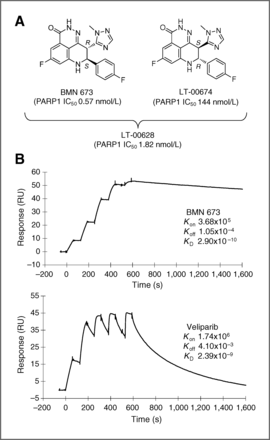
BMN 673 is a potent PARP inhibitor.Clin Cancer Res.2013 Sep 15;19(18):5003-15. |
|---|
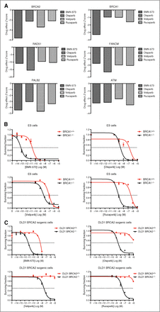
A, siRNAs targeting homologous recombination genes sensitize to PARP1/2 inhibitors.Clin Cancer Res.2013 Sep 15;19(18):5003-15. |
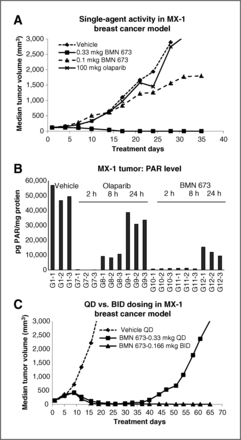
BMN 673 exhibits antitumor activity against a BRCA-mutant tumor model in mice.Clin Cancer Res.2013 Sep 15;19(18):5003-15. |
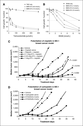
BMN 673 potentiates the effects of DNA-damaging cytotoxic agents.Clin Cancer Res.2013 Sep 15;19(18):5003-15. |
|---|
Products are for research use only; We do not sell to patients
Copyright 2020 InvivoChem LLC | All Rights Reserved


 COA
COA