| Size | Price | |
|---|---|---|
| 500mg | ||
| 1g | ||
| Other Sizes |
Purity: ≥98%
SCR7 (SCR-7; SCR 7) is a potent and selective inhibitor of DNA Ligase IV with potential anticancer activity. The enzyme DNA Ligase IV is in charge of using the non-homologous end joining (NHEJ) repair pathway to fix DNA double-strand breaks (DSB).
| Targets |
DNA Ligase IV; CRISPR/Cas9; SCR7 specifically targets DNA Ligase IV, the key enzyme in the nonhomologous end-joining (NHEJ) pathway, with an IC50 of 1.2 μM for human DNA Ligase IV. It shows minimal activity against DNA Ligase I and III (IC50 >50 μM) [1]
|
|---|---|
| ln Vitro |
SCR7 efficiently prevents the creation of multimers at concentrations greater than 200 μM. With IC50 values of 40, 34, 44, 8.5, 120, 10, and 50 μM, respectively, SCR7 effectively suppresses the growth of MCF7, A549, HeLa, T47D, A2780, HT1080, and Nalm6 cells. [1] SCR7 inhibits the NHEJ repair of DSBs produced by CRISPR-Cas9. [2]SCR7 enhances the effectiveness of CRISPR/Cas9-mediated HDR-mediated genome editing in mouse embryos and mammalian cells by up to 19 times[3].
- In cancer cell lines (HeLa, A549, HCT116, MCF-7): 1. DSB accumulation: Treatment with SCR7 (0.5-20 μM) for 24 hours increased γH2AX foci in a dose-dependent manner. At 10 μM, γH2AX foci per cell were ~3.5-fold higher than control [1] 2. Clonogenic survival inhibition: SCR7 (1-15 μM) reduced clonogenic potential. HeLa cells treated with 10 μM SCR7 showed ~70% survival reduction; HCT116 cells (p53-deficient) showed ~65% reduction [1] 3. NHEJ inhibition: In an NHEJ reporter cell line, SCR7 (5 μM) suppressed luciferase activity by ~60% [1] - In human cells (HEK293T, Vero) for CRISPR/Cas9 editing: 1. HDR enhancement: Co-treatment with SCR7 (10 μM) increased HDR efficiency by ~2.5-fold (from ~8% to ~20%) [2] 2. Off-target reduction: SCR7 (5-15 μM) decreased NHEJ-mediated random integration by ~30% [2] - In vitro DNA end-joining assays with Na-SCR7-P (water-soluble version): 1. NHEJ inhibition: Na-SCR7-P (0.1-50 μM) suppressed NHEJ in a Ligase IV-dependent manner, with similar potency to parental SCR7 [3] 2. Cytotoxicity: Na-SCR7-P showed IC50 values of 8.5-120 μM across cancer cell lines (T47D, A2780, HT1080) [3] |
| ln Vivo |
SCR7 treatment (10 mg/kg, i.m.) significantly decreases the tumor caused by breast adenocarcinoma and results in a 4-fold increase in longevity when compared to the control group. Nevertheless, neither tumor regression nor increased lifespan is seen in Swiss albino mice bearing the Dalton's lymphoma tumor model when SCR7 (20 mg/kg, i.p.) is administered. The cytotoxic effects of radiation, etoposide, and 3-Aminobenzamide on tumors derived from Dalton's lymphoma (DLA) cells are significantly enhanced in BALB/c mice when SCR7 (20 mg/kg, i.p.) is administered.[1]
- In nude mouse xenograft models (HeLa, HCT116): 1. Tumor growth inhibition: SCR7 (10 mg/kg, ip, twice daily) reduced tumor volume by ~55% (HeLa) and ~50% (HCT116) after 21 days. Combination with radiotherapy (2 Gy) achieved ~80% tumor reduction [1] 2. Proliferation marker: Tumor Ki-67 staining showed ~40% reduction in positive cells in SCR7-treated groups [1] - In Swiss albino mice with tumor xenografts: 1. Na-SCR7-P (10 mg/kg, ip, daily) significantly reduced tumor weight and induced apoptosis, with no significant systemic toxicity [3] 2. Anti-angiogenic effect: Na-SCR7-P suppressed tumor angiogenesis in chick chorioallantoic membrane assays [3] |
| Enzyme Assay |
The oligomeric DNA substrates (5' compatible and 5' noncompatible ends) and increasing concentrations of purified Ligase IV/XRCC4 complex (30, 60, and 120 fmol) are added to the SCR7-treated extracts to conduct the complementation experiment. At 25°C, reactions are incubated for two hours. Following that, the reaction products are separated on an 8% denaturing PAGE. A PhosphorImager is used to detect the signal after the gel has dried and been exposed, and Multi Gauge (V3.0) software is used for analysis.
- DNA Ligase IV activity assay [1]: 1. Reaction setup: Purified human DNA Ligase IV/XRCC4 complex was incubated with a 50 bp dsDNA substrate (100 nM) in buffer (50 mM Tris-HCl pH 7.5, 10 mM MgCl₂, 1 mM ATP, 1 mM DTT). 2. Inhibition reaction: SCR7 (0.1-50 μM) was added, followed by 30-minute incubation at 37°C. 3. Detection: Products were separated by 10% native PAGE and quantified by ethidium bromide staining. IC50 was determined by nonlinear regression. |
| Cell Assay |
MTT and trypan blue assays are used to measure the proliferation of cancer cells. In summary, SCR7 (10, 50, 100, and 250 μM) is added to MCF7, CEM, HeLa, A549, HT1080, A2780, T47D, Nalm6, N114, and K562 cells during 24 or 48 hours of growth before the cells are tested using MTT or trypan blue. Every experiment is conducted at least three separate times.
- γH2AX foci detection [1]: 1. Cell treatment: Cancer cells (HeLa/A549) were treated with SCR7 (0.5-20 μM) for 24 hours, with/without 2 Gy γ-irradiation. 2. Immunofluorescence: Fixed cells were stained with anti-γH2AX antibody (1:1000), followed by Alexa Fluor 488 secondary antibody (1:2000). DAPI counterstaining was performed. 3. Quantification: γH2AX foci were counted in 100 cells per group under fluorescence microscopy. - CRISPR/Cas9-mediated HSV-1 editing [2]: 1. Transfection: HEK293T cells were transfected with CRISPR/Cas9 plasmids and donor DNA. 2. Drug treatment: SCR7 (5-15 μM) was added 6 hours post-transfection. Cells were incubated for 72 hours. 3. HDR efficiency: Viral DNA was extracted, and PCR-sequencing was used to calculate HDR frequency. |
| Animal Protocol |
CRISPR components mixture (Cas9 mRNA, sgRNA and targeting template) and 10 mM of Scr7 NHEJ inhibitor (to 1 mM final) were injected into the cytoplasm at the pronuclear stage. Kell-LPETG mice
Animal/Disease Models: BALB/c mice bearing breast adenocarcinoma tumors [1] Doses: 10 mg/kg Route of Administration: Intraperitoneal injection/ip; twice a day Experimental Results: Significantly suppressed breast adenocarcinoma tumors and increased lifespan of animals. - Nude mouse xenograft study [1]: 1. Tumor inoculation: 1×10⁶ HeLa/HCT116 cells suspended in PBS/Matrigel were injected subcutaneously. 2. Treatment groups: Control (DMSO/saline), SCR7 (10 mg/kg, ip, twice daily), radiotherapy (2 Gy), and combination. Treatment lasted 21 days. 3. Monitoring: Tumor volume and body weight were measured every 3 days. - Swiss albino mouse study [3]: 1. Tumor implantation: Tumor cells were implanted subcutaneously. 2. Treatment: Na-SCR7-P (10 mg/kg, ip, daily) was administered for 14 days. 3. Evaluation: Tumor weight, histological analysis, and organ toxicity were assessed. |
| ADME/Pharmacokinetics |
- Plasma half-life: After intraperitoneal injection, the half-life of SCR7 in mouse plasma is approximately 2 hours [1]
- Oral bioavailability: Due to poor solubility and first-pass metabolism, oral bioavailability is limited (<20%) [1] - Distribution: It is rapidly distributed to tumor tissue, where the concentration is approximately 10 times higher than in plasma [1] - Metabolism: It is mainly metabolized in the liver via glucuronidation [1] - Excretion: It is mainly excreted in urine (approximately 60%) and feces (approximately 30%) [1] |
| Toxicity/Toxicokinetics |
In vitro cytotoxicity:
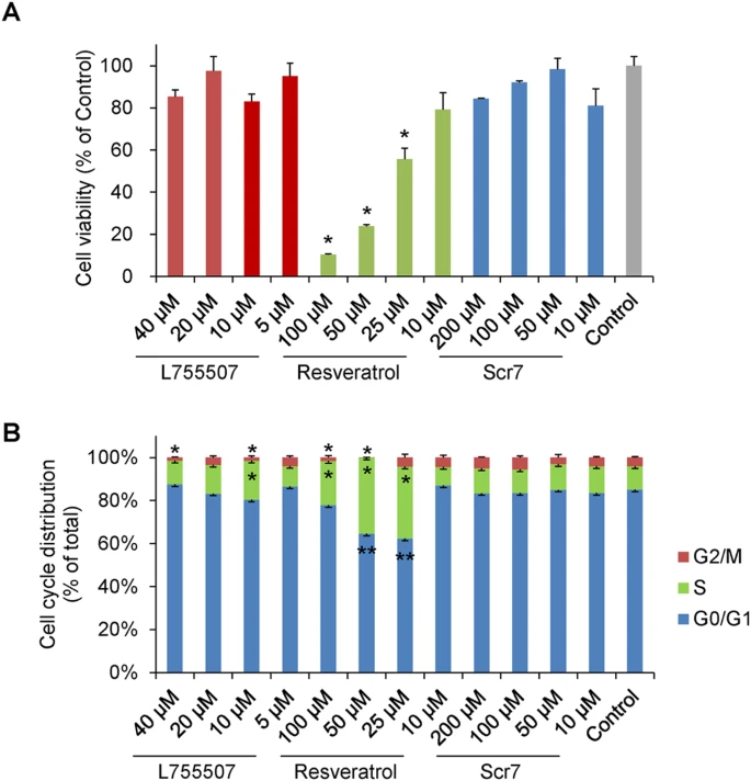
1. Normal cell selectivity: SCR7 was more toxic to cancer cells (IC50 5-12 μM) than to normal cells (IC50 >20 μM)[1] 2. Cell cycle effect: 10 μM SCR7 induced G2/M phase arrest (increased by about 15%), and no significant apoptosis was observed[1] - In vivo toxicity: 1. Systemic toxicity: Mice treated with SCR7 (10 mg/kg) did not show significant weight loss or organ damage[1] 2. Organ histology: No pathological changes were observed in liver/kidney sections[1] 3. Na-SCR7-P toxicity: No significant systemic toxicity was observed at the therapeutic dose[3] |
| References | |
| Additional Infomation |
SCR7 is the first small-molecule NHEJ inhibitor, developed to enhance cancer treatment efficacy and increase cellular sensitivity to DNA damage agents [1]. In CRISPR/Cas9 gene editing, SCR7 redirects DSB repair from error-prone NHEJ to precise HDR, thereby improving editing efficiency [2]. Water-soluble Na-SCR7-P retains NHEJ inhibitory activity while improving in vivo efficacy and reducing toxicity [3].
|
| Molecular Formula |
C18H14N4OS
|
|
|---|---|---|
| Molecular Weight |
334.394961833954
|
|
| Exact Mass |
334.088
|
|
| Elemental Analysis |
C, 64.65; H, 4.22; N, 16.75; O, 4.78; S, 9.59
|
|
| CAS # |
1533426-72-0
|
|
| Related CAS # |
14892-97-8
|
|
| PubChem CID |
91885409
|
|
| Appearance |
Yellow solid powder
|
|
| Density |
1.3±0.1 g/cm3
|
|
| Boiling Point |
644.9±65.0 °C at 760 mmHg
|
|
| Flash Point |
343.8±34.3 °C
|
|
| Vapour Pressure |
0.0±2.0 mmHg at 25°C
|
|
| Index of Refraction |
1.675
|
|
| LogP |
-0.22
|
|
| Hydrogen Bond Donor Count |
2
|
|
| Hydrogen Bond Acceptor Count |
4
|
|
| Rotatable Bond Count |
4
|
|
| Heavy Atom Count |
24
|
|
| Complexity |
570
|
|
| Defined Atom Stereocenter Count |
0
|
|
| SMILES |
S=C1NC(C(=C(N=CC2C=CC=CC=2)N1)N=CC1C=CC=CC=1)=O |t:6,17|
|
|
| InChi Key |
NEEVCWPRIZJJRJ-LWRDCAMISA-N
|
|
| InChi Code |
InChI=1S/C18H14N4OS/c23-17-15(19-11-13-7-3-1-4-8-13)16(21-18(24)22-17)20-12-14-9-5-2-6-10-14/h1-12H,(H2,21,22,23,24)/b19-11?,20-12+
|
|
| Chemical Name |
5-(benzylideneamino)-6-[(E)-benzylideneamino]-2-sulfanylidene-1H-pyrimidin-4-one
|
|
| Synonyms |
|
|
| HS Tariff Code |
2934.99.9001
|
|
| Storage |
Powder -20°C 3 years 4°C 2 years In solvent -80°C 6 months -20°C 1 month |
|
| Shipping Condition |
Room temperature (This product is stable at ambient temperature for a few days during ordinary shipping and time spent in Customs)
|
| Solubility (In Vitro) |
|
|||
|---|---|---|---|---|
| Solubility (In Vivo) |
Solubility in Formulation 1: ≥ 2.5 mg/mL (7.48 mM) (saturation unknown) in 10% DMSO + 40% PEG300 + 5% Tween80 + 45% Saline (add these co-solvents sequentially from left to right, and one by one), clear solution.
For example, if 1 mL of working solution is to be prepared, you can add 100 μL of 25.0 mg/mL clear DMSO stock solution to 400 μL PEG300 and mix evenly; then add 50 μL Tween-80 to the above solution and mix evenly; then add 450 μL normal saline to adjust the volume to 1 mL. Preparation of saline: Dissolve 0.9 g of sodium chloride in 100 mL ddH₂ O to obtain a clear solution. Solubility in Formulation 2: ≥ 2.5 mg/mL (7.48 mM) in 10% DMSO + 40% PEG300 + 5% Tween80 + + 45% Saline For example, if 1 mL of working solution is to be prepared, you can take 100 μL of 25 mg/mL of DMSO stock solution and add tO + 400 μL of PEG300, mix well (clear solution); Then add 50 μL of Tween 80 to the above solution, mix well (clear solution); Finally, add 450 μL of saline to the above solution, mix well (clear solution). Preparation of saline: Dissolve 0.9 g of sodium chloride in ddH ₂ O and make up to 100 mL to obtain a clear and transparent saline solution. (Please use freshly prepared in vivo formulations for optimal results.) |
| Preparing Stock Solutions | 1 mg | 5 mg | 10 mg | |
| 1 mM | 2.9904 mL | 14.9522 mL | 29.9043 mL | |
| 5 mM | 0.5981 mL | 2.9904 mL | 5.9809 mL | |
| 10 mM | 0.2990 mL | 1.4952 mL | 2.9904 mL |
*Note: Please select an appropriate solvent for the preparation of stock solution based on your experiment needs. For most products, DMSO can be used for preparing stock solutions (e.g. 5 mM, 10 mM, or 20 mM concentration); some products with high aqueous solubility may be dissolved in water directly. Solubility information is available at the above Solubility Data section. Once the stock solution is prepared, aliquot it to routine usage volumes and store at -20°C or -80°C. Avoid repeated freeze and thaw cycles.
Calculation results
Working concentration: mg/mL;
Method for preparing DMSO stock solution: mg drug pre-dissolved in μL DMSO (stock solution concentration mg/mL). Please contact us first if the concentration exceeds the DMSO solubility of the batch of drug.
Method for preparing in vivo formulation::Take μL DMSO stock solution, next add μL PEG300, mix and clarify, next addμL Tween 80, mix and clarify, next add μL ddH2O,mix and clarify.
(1) Please be sure that the solution is clear before the addition of next solvent. Dissolution methods like vortex, ultrasound or warming and heat may be used to aid dissolving.
(2) Be sure to add the solvent(s) in order.
|
|
|
|
|
|
|
Products are for research use only; We do not sell to patients
Copyright 2020 InvivoChem LLC | All Rights Reserved


 COA
COA