| Size | Price | Stock | Qty |
|---|---|---|---|
| 5mg |
|
||
| 10mg |
|
||
| 25mg |
|
||
| 50mg |
|
||
| 100mg |
|
||
| 250mg |
|
||
| 500mg | |||
| Other Sizes |


Purity: ≥98%
| Targets |
Stat3 (IC50 = 86 μM)
S3I-201 (NSC 74859) targets signal transducer and activator of transcription 3 (Stat3) with a Ki value of 82 μM for Stat3 DNA binding, and inhibits Stat3 phosphorylation (Tyr705) with IC50 values ranging from 40-70 μM in various tumor cells [1] S3I-201 (NSC 74859) exhibits high selectivity for Stat3, with no significant inhibition of Stat1, Stat5a, or Stat5b (IC50>200 μM) [1] |
|---|---|
| ln Vitro |
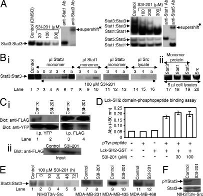
Stat3 DNA-binding activity is preferentially inhibited by NSC 74859 (S3I-201) over Stat1 (IC50 values: Stat3•Stat3, 86±33 μM; Stat1•Stat3, 160±43 μM; and Stat1•Stat1, >300 μM), while Stat5 DNA-binding activity is inhibited with an IC50 of 166±17 μM). The proliferation of transformed mouse fibroblasts NIH 3T3/v-Src and breast carcinoma cell lines (MDA-MB-231, MDA-MB-435, and MDA-MB-468) is inhibited and the number of viable cells is dramatically reduced by NSC 74859. NSC 74859 significantly promotes apoptosis in the constitutively active Stat3-containing human breast cancer cell lines MDA-MB-435 and NIH 3T3/v-Src at concentrations between 30 and 100 μM. The MDA-MB-435 cell line exhibits increased sensitivity to 30 μM NSC 74859. On the other hand, the normal mouse fibroblasts (NIH 3T3) and the human breast cancer MDA-MB-453 cells are less sensitive to NSC 74859 at 100 μM or less because they do not exhibit aberrant Stat3 activation. NSC 74859 produced non-specific, overall cytotoxicity at 300 μM or above, regardless of Stat3 activation status[1]. Huh-7 cells, independent of CD133+ status, are susceptible to STAT3 inhibition and do not express β2SP or TBGFR2. The IC50 for NSC 74859 is 100 μM. In Huh-7 and SNU-398 cells, the IC50 of NSC 74859 is 150 μM; in SNU-475 and SNU-182 cells, it is 15 μM. With an IC50 near 100 μM, NSC 74859 inhibits the MDA-MB-435, MDA-MB-453, and MDA-MB-231 cell lines that are used to treat breast cancer[2].
S3I-201 (NSC 74859) (20-100 μM) dose-dependently inhibited proliferation of human tumor cells (MDA-MB-468, A549, HCT116) with IC50 values of 45 μM, 62 μM, and 58 μM respectively [1] S3I-201 (NSC 74859) (50-100 μM) blocked Stat3 phosphorylation at Tyr705 in tumor cells, suppressed Stat3 dimerization, and inhibited Stat3-DNA binding (reduced by 75% at 100 μM via EMSA) [1] S3I-201 (NSC 74859) (50 μM) induced apoptosis in MDA-MB-468 cells: apoptotic rate increased by 42% (Annexin V staining), caspase-3/-7 activity enhanced by 2.8-fold, and anti-apoptotic proteins Bcl-2 and Survivin downregulated by 0.5-fold and 0.4-fold respectively [1] S3I-201 (NSC 74859) (30-80 μM) inhibited proliferation of hepatocellular carcinoma (HCC) cells with disrupted TGF-β signaling (Hep3B, PLC/PRF/5) with IC50 values of 52 μM and 65 μM [2] S3I-201 (NSC 74859) (50 μM) downregulated Stat3 target genes (Cyclin D1, c-Myc, Survivin) mRNA levels by 45-62% in Hep3B cells, and enhanced sensitivity to sorafenib (reduced sorafenib IC50 from 12 μM to 5 μM) [2] S3I-201 (NSC 74859) (50-100 μM) reduced growth hormone (GH) secretion by 32-45% in pituitary somatotroph adenoma cells (GH3, MMQ) [3] S3I-201 (NSC 74859) (100 μM) downregulated Stat3 phosphorylation (Tyr705) and target genes (c-Fos, c-Jun) mRNA expression by 58% and 42% respectively in GH3 cells [3] |
| ln Vivo |
For two weeks, NSC 74859 (S3I-201) or a vehicle is injected intravenously (i.v.) into human breast (MDA-MB-231) tumor-bearing mice every two or three days. Tumor measurements are obtained every two to three days. Mice treated with S3I-201 for human breast cancers show significant growth suppression as compared to control (vehicle-treated) tumors, which did not stop growing. Following treatment termination, an ongoing assessment of the treated mice reveals no return of tumor growth, indicating that S3I-201 may have a long-lasting effect on tumor growth[1]. For the duration of the trial, S3I-201 treatment of somatotroph tumor xenografts (n=15) dramatically reduced tumor development in comparison with vehicle-treated control tumors (n=15), which kept growing. As early as five days following NSC 74859 injection, tumors derived from rats treated with NSC 74859 are significantly less than those from the untreated group (220±16 mm3 vs. 287±16 mm3, P<0.01). Rats treated with NSC 74859 had an average tumor volume that is 64% smaller than that of controls fifteen days after treatment (449±40 mm3 vs. 708±83 mm3, P<0.01). Fifteen days after the start of treatment, the rats are killed and the tumors are extracted. Rats treated with NSC 74859 had an average tumor weight of 78±8 mg, whereas tumors from control rats weighed 114±13 mg (32% reduction; P<0.05)[3].
Furthermore, NSC 74859 treatment of Huh-7 xenografts in nude mice significantly retarded tumor growth, with an effective dose of only 5 mg/kg. Moreover, NSC 74859 inhibited tyrosine phosphorylation of STAT3 in HCC cells in vivo. [2] In addition, S3I-201 attenuated somatotroph tumor growth and GH secretion in a rat xenograft model. [3] S3I-201 (NSC 74859) (50 mg/kg, i.p., 5 times/week for 3 weeks) inhibited tumor growth in nude mice bearing MDA-MB-468 xenografts: tumor volume reduced by 60% and tumor weight decreased by 58% compared to the vehicle group [1] S3I-201 (NSC 74859) treatment in xenograft mice reduced Stat3 phosphorylation (Tyr705) by 65% and increased apoptotic index (TUNEL staining) by 3.2-fold in tumor tissues [1] S3I-201 (NSC 74859) (50 mg/kg, i.p., 5 times/week for 4 weeks) suppressed Hep3B xenograft growth in nude mice: tumor weight reduced by 55%, and Cyclin D1 and c-Myc protein levels in tumors downregulated by 0.4-fold and 0.5-fold [2] |
| Enzyme Assay |
Recombinant Stat3 protein was incubated with a biotin-labeled Stat3-specific DNA probe and serial concentrations of S3I-201 (NSC 74859) (20-200 μM) in binding buffer at room temperature for 30 minutes. The Stat3-DNA complex was separated by non-denaturing polyacrylamide gel electrophoresis, and the gel was transferred to a membrane for chemiluminescent detection. The inhibition rate of DNA binding was calculated relative to the vehicle control [1]
Cytosolic extracts from MDA-MB-468 cells (stimulated with EGF to activate Stat3) were incubated with S3I-201 (NSC 74859) (50-100 μM) for 1 hour. Stat3 dimers were immunoprecipitated with a Stat3 antibody, separated by SDS-PAGE, and detected by western blot. The level of dimerization was quantified by densitometry [1] |
| Cell Assay |
Transient Transfection of Cells and Treatment with Compound. [1]
Twelve to 24 h after seeding, cells were transfected with the appropriate plasmids. For detailed procedure information, see SI Materials and Methods. Twenty-four hours after transfection, cells were untreated (0.05% DMSO) or treated with S3I-201 (100 μM) for an additional 24 h and harvested, and cytosolic extracts were prepared for luciferase assay, as described, or cells were analyzed by annexin V binding and flow cytometry. Soft-Agar Colony-Formation Assay.[1] Colony formation assays were carried out in six-well dishes, as described previously. Treatment with S3I-201 was initiated 1 day after seeding cells by adding 100 μl of medium with or without S3I-201 and repeating every 3 days, until large colonies were evident. Measurement of Apoptosis by Flow Cytometry. [1] Proliferating cells were treated with or without S3I-201 for up to 48 h. In some cases, cells were first transfected with Stat3C, ST3-NT, or ST3-SH2 domain or mock-transfected for 24 h before treatment with compound for an additional 24–48 h. Cells were then detached and analyzed by annexin V binding according to the manufacturer's protocol and flow cytometry to quantify the percent apoptosis. Tumor cells (MDA-MB-468, A549, HCT116) were seeded in 96-well plates (5×10^3 cells/well) and treated with S3I-201 (NSC 74859) (20-100 μM) for 72 hours. Cell proliferation was assessed using a colorimetric assay, and IC50 values were calculated by fitting dose-response curves [1] MDA-MB-468 cells were seeded in 6-well plates and treated with S3I-201 (NSC 74859) (50-100 μM) for 24 hours. Cells were lysed, and protein extracts were subjected to western blot analysis using antibodies against phosphorylated Stat3 (Tyr705), total Stat3, Bcl-2, and Survivin [1] Hep3B and PLC/PRF/5 cells were seeded in 6-well plates and treated with S3I-201 (NSC 74859) (30-80 μM) for 48 hours. Total RNA was extracted for qPCR analysis of Cyclin D1, c-Myc, and Survivin mRNA expression (GAPDH as reference), and cell viability was measured by a cell counting kit [2] GH3 and MMQ cells were seeded in 24-well plates and treated with S3I-201 (NSC 74859) (50-100 μM) for 24 hours. Culture supernatants were collected to measure GH secretion by ELISA, and cell lysates were used for western blot detection of phosphorylated Stat3 and total Stat3 [3] MDA-MB-468 cells were treated with S3I-201 (NSC 74859) (50 μM) for 24 hours, stained with Annexin V-FITC/PI, and apoptotic cells were detected by flow cytometry. Caspase-3/-7 activity was measured using a luminescent assay kit [1] |
| Animal Protocol |
Dissolved in DMSO; ≤5 mg/kg; i.v. injection
Human breast cancer MDA-MB-231 cells are injected s.c. into the left flank of athymic nu/nu mice. Mice and in Vivo Tumor Studies. Six-week-old female athymic nude mice were purchased from Harlan (Indianapolis, IN) and maintained in the institutional animal facilities approved by the American Association for Accreditation of Laboratory Animal Care. Athymic nude mice were injected in the left flank area s.c. with 5 × 10⁶ human breast cancer MDA-MB-231 cells in 100 μl of PBS. After 5–10 days, tumors with a diameter of 3 mm were established. Animals were given S3I-201 i.v. at 5 mg/kg every 2 or 3 days for 2 weeks and monitored every 2 or 3 days. Animals were stratified so that the mean tumor sizes in all treatment were nearly identical. Tumor volume was calculated according to the formula V = 0.52 × a2× b, where a is the smallest superficial diameter and b is the largest superficial diameter.[1] Nude mice (6-8 weeks old) bearing MDA-MB-468 xenografts (1×10^6 cells injected subcutaneously) were randomly divided into vehicle and S3I-201 (NSC 74859) groups (n=6 per group). S3I-201 (NSC 74859) was dissolved in DMSO and normal saline (DMSO final concentration <1%) and administered via intraperitoneal injection at 50 mg/kg, 5 times per week for 3 weeks. Tumor volume was measured every 3 days using a caliper. Mice were euthanized, tumors were harvested for western blot (Stat3 phosphorylation) and TUNEL staining [1] Nude mice (6-8 weeks old) were subcutaneously injected with Hep3B cells (2×10^6 cells/mouse) to establish xenografts. Seven days after inoculation, mice were treated with S3I-201 (NSC 74859) (50 mg/kg, i.p., 5 times/week for 4 weeks) or vehicle. At the end of treatment, mice were sacrificed, tumors were weighed, and protein extracts were prepared for western blot analysis of Cyclin D1 and c-Myc [2] |
| Toxicity/Toxicokinetics |
In nude mice treated with S3I-201 (NSC 74859) (50 mg/kg, intraperitoneal injection, for 4 weeks), no significant weight loss or abnormal clinical symptoms (e.g., lethargy, diarrhea) were observed [1,2]. Serum levels of liver function markers (ALT, AST) and kidney function markers (BUN, Cr) in mice treated with S3I-201 (NSC 74859) were within the normal range and showed no significant difference compared to the control group [1,2].
|
| References |
|
| Additional Infomation |
S3I-201 is an aminobenzoic acid formed by the condensation of the carboxyl group of [(4-methylbenzene-1-sulfonyl)oxy]acetic acid with the amino group of 4-amino-2-hydroxybenzoic acid. It is a STAT3 inhibitor. It is a monohydroxybenzoic acid, toluenesulfonate, and aminobenzoic acid.
S3I-201 (NSC 74859) is a selective Stat3 inhibitor identified by structure-based virtual screening that targets the SH2 domain of Stat3 [1]. S3I-201 (NSC 74859) exerts its antitumor effect by blocking the phosphorylation, dimerization, and DNA binding of Stat3, thereby inhibiting the transcription of Stat3-dependent anti-apoptotic and pro-proliferative genes [1,2]. S3I-201 (NSC 74859) is effective against tumors with abnormal Stat3 activation, including triple-negative breast cancer, non-small cell lung cancer, colorectal cancer, and hepatocellular carcinoma with impaired TGF-β signaling pathway [1,2]. S3I-201 (NSC 74859) inhibits growth hormone secretion from pituitary growth hormone adenoma cells by downregulating the transcription of Stat3-mediated early genes (c-Fos, c-Jun) [3] |
| Molecular Formula |
C16H15NO7S
|
|
|---|---|---|
| Molecular Weight |
365.36
|
|
| Exact Mass |
365.056
|
|
| Elemental Analysis |
C, 52.60; H, 4.14; N, 3.83; O, 30.65; S, 8.78
|
|
| CAS # |
501919-59-1
|
|
| Related CAS # |
|
|
| PubChem CID |
252682
|
|
| Appearance |
White to gray solid powder
|
|
| Density |
1.5±0.1 g/cm3
|
|
| Boiling Point |
654.7±55.0 °C at 760 mmHg
|
|
| Flash Point |
349.8±31.5 °C
|
|
| Vapour Pressure |
0.0±2.1 mmHg at 25°C
|
|
| Index of Refraction |
1.642
|
|
| LogP |
3.08
|
|
| Hydrogen Bond Donor Count |
3
|
|
| Hydrogen Bond Acceptor Count |
7
|
|
| Rotatable Bond Count |
6
|
|
| Heavy Atom Count |
25
|
|
| Complexity |
578
|
|
| Defined Atom Stereocenter Count |
0
|
|
| SMILES |
OC1=CC(NC(COS(=O)(C2=CC=C(C=C2)C)=O)=O)=CC=C1C(O)=O
|
|
| InChi Key |
HWNUSGNZBAISFM-UHFFFAOYSA-N
|
|
| InChi Code |
InChI=1S/C16H15NO7S/c1-10-2-5-12(6-3-10)25(22,23)24-9-15(19)17-11-4-7-13(16(20)21)14(18)8-11/h2-8,18H,9H2,1H3,(H,17,19)(H,20,21)
|
|
| Chemical Name |
2-hydroxy-4-[[2-(4-methylphenyl)sulfonyloxyacetyl]amino]benzoic acid
|
|
| Synonyms |
|
|
| HS Tariff Code |
2934.99.9001
|
|
| Storage |
Powder -20°C 3 years 4°C 2 years In solvent -80°C 6 months -20°C 1 month Note: This product is not stable in solution, please use freshly prepared working solution for optimal results. |
|
| Shipping Condition |
Room temperature (This product is stable at ambient temperature for a few days during ordinary shipping and time spent in Customs)
|
| Solubility (In Vitro) |
DMSO: 73 mg/mL (199.8 mM)
Water: 1 mg/mL (Insoluble) Ethanol: 1 mg/mL |
|---|---|
| Solubility (In Vivo) |
Solubility in Formulation 1: ≥ 7.5 mg/mL (20.53 mM) (saturation unknown) in 10% DMSO + 40% PEG300 + 5% Tween80 + 45% Saline (add these co-solvents sequentially from left to right, and one by one), clear solution.
For example, if 1 mL of working solution is to be prepared, you can add 100 μL of 75.0 mg/mL clear DMSO stock solution to 400 μL PEG300 and mix evenly; then add 50 μL Tween-80 to the above solution and mix evenly; then add 450 μL normal saline to adjust the volume to 1 mL. Preparation of saline: Dissolve 0.9 g of sodium chloride in 100 mL ddH₂ O to obtain a clear solution. Solubility in Formulation 2: ≥ 7.5 mg/mL (20.53 mM) (saturation unknown) in 10% DMSO + 90% (20% SBE-β-CD in Saline) (add these co-solvents sequentially from left to right, and one by one), clear solution. For example, if 1 mL of working solution is to be prepared, you can add 100 μL of 75.0 mg/mL clear DMSO stock solution to 900 μL of 20% SBE-β-CD physiological saline solution and mix evenly. Preparation of 20% SBE-β-CD in Saline (4°C,1 week): Dissolve 2 g SBE-β-CD in 10 mL saline to obtain a clear solution. View More
Solubility in Formulation 3: ≥ 7.5 mg/mL (20.53 mM) (saturation unknown) in 10% DMSO + 90% Corn Oil (add these co-solvents sequentially from left to right, and one by one), clear solution. Solubility in Formulation 4: ≥ 2.5 mg/mL (6.84 mM) (saturation unknown) in 5% DMSO + 40% PEG300 + 5% Tween80 + 50% Saline (add these co-solvents sequentially from left to right, and one by one), clear solution. Preparation of saline: Dissolve 0.9 g of sodium chloride in 100 mL ddH₂ O to obtain a clear solution. Solubility in Formulation 5: ≥ 2.5 mg/mL (6.84 mM) (saturation unknown) in 5% DMSO + 95% (20% SBE-β-CD in Saline) (add these co-solvents sequentially from left to right, and one by one), clear solution. Preparation of 20% SBE-β-CD in Saline (4°C,1 week): Dissolve 2 g SBE-β-CD in 10 mL saline to obtain a clear solution. Solubility in Formulation 6: 5% DMSO, 95% PEG 300 :15 mg/mL |
| Preparing Stock Solutions | 1 mg | 5 mg | 10 mg | |
| 1 mM | 2.7370 mL | 13.6851 mL | 27.3703 mL | |
| 5 mM | 0.5474 mL | 2.7370 mL | 5.4741 mL | |
| 10 mM | 0.2737 mL | 1.3685 mL | 2.7370 mL |
*Note: Please select an appropriate solvent for the preparation of stock solution based on your experiment needs. For most products, DMSO can be used for preparing stock solutions (e.g. 5 mM, 10 mM, or 20 mM concentration); some products with high aqueous solubility may be dissolved in water directly. Solubility information is available at the above Solubility Data section. Once the stock solution is prepared, aliquot it to routine usage volumes and store at -20°C or -80°C. Avoid repeated freeze and thaw cycles.
Calculation results
Working concentration: mg/mL;
Method for preparing DMSO stock solution: mg drug pre-dissolved in μL DMSO (stock solution concentration mg/mL). Please contact us first if the concentration exceeds the DMSO solubility of the batch of drug.
Method for preparing in vivo formulation::Take μL DMSO stock solution, next add μL PEG300, mix and clarify, next addμL Tween 80, mix and clarify, next add μL ddH2O,mix and clarify.
(1) Please be sure that the solution is clear before the addition of next solvent. Dissolution methods like vortex, ultrasound or warming and heat may be used to aid dissolving.
(2) Be sure to add the solvent(s) in order.
|
|---|
|
|
|
|---|
|
|
Products are for research use only; We do not sell to patients
Copyright 2020 InvivoChem LLC | All Rights Reserved


 COA
COA