| Size | Price | Stock | Qty |
|---|---|---|---|
| 5mg |
|
||
| 25mg |
|
||
| 50mg |
|
||
| 100mg |
|
||
| 250mg |
|
||
| 500mg |
|
||
| 1g | |||
| Other Sizes |


Purity: ≥98%
| Targets |
Flt3 (Kd = 1.6±0.7 nM)
The target of Quizartinib (AC220; AC010220) is FMS-like tyrosine kinase 3 (FLT3). It exhibits potent inhibitory activity against FLT3 wild-type (FLT3-WT) with an IC50 of 1.6 nM, FLT3 internal tandem duplication (FLT3-ITD) mutation with an IC50 of 0.59 nM, and FLT3 D835V point mutation (a common resistance mutation) with an IC50 of 3.4 nM. For other related kinases, it shows high selectivity: IC50 for KIT is 16 nM, PDGFRα is 45 nM, and VEGFR2 is >1000 nM, indicating minimal off-target effects [1] |
|---|---|
| ln Vitro |
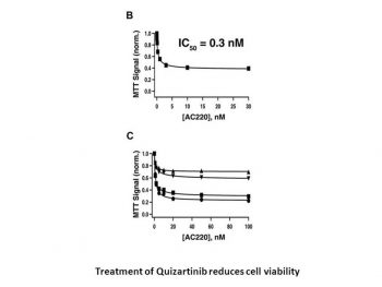
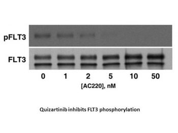
Quizartinib (AC220) is a novel substance designed specifically to inhibit FLT3 in the treatment of acute myeloid leukemia (AML). With an IC50 of 4.2±0.3 nM for FLT3-WT and 1.1±0.1 nM for FLT3-ITD, respectively, quizartinib inhibits FLT3-autophosphorylation. With an IC50 of 0.56±0.3 nM and >10,000 nM, respectively, quizartinib inhibits MV4-11 and A375 cells. When screened against most of the human protein kinome, quizartinib is highly selective and inhibits FLT3 with low nanomolar potency in cellular assays[1].
1. Antiproliferative activity: Quizartinib (AC220; AC010220) inhibits the proliferation of FLT3-ITD-positive AML cell lines (MV4-11, MOLM-13, MOLM-14) with IC50 values of 1.3 nM, 4.2 nM, and 3.8 nM, respectively. For FLT3-WT-positive AML cell lines (HL-60, THP-1), it shows much weaker activity (IC50 > 100 nM). For FLT3-negative cell lines (K562, Raji), no significant antiproliferative effect is observed even at concentrations up to 1000 nM [1] 2. Signaling pathway inhibition: Treatment with Quizartinib (AC220; AC010220) (10 nM for 2 hours) in MV4-11 cells significantly reduces the phosphorylation of FLT3 (p-FLT3) and its downstream signaling molecules, including STAT5 (p-STAT5), ERK1/2 (p-ERK1/2), and AKT (p-AKT). The inhibition of p-FLT3 is sustained for at least 24 hours after a single treatment [1] 3. Apoptosis induction: In MV4-11 cells, Quizartinib (AC220; AC010220) (10 nM) induces apoptosis in a time-dependent manner. After 24 hours of treatment, the apoptotic rate (Annexin V-positive cells) increases from 5.2% (control) to 42.3%; after 48 hours, it further rises to 68.7%. This is accompanied by the cleavage of caspase-3 and PARP, key markers of apoptosis [1] 4. Colony formation inhibition: In a methylcellulose colony formation assay, Quizartinib (AC220; AC010220) (1 nM) reduces the number of colonies formed by primary FLT3-ITD-positive AML blasts by 85% compared to the control. For primary FLT3-WT AML blasts, it only reduces colony formation by 12% at the same concentration [1] |
| ln Vivo |
Quizartinib (AC220) obliterates tumors in a FLT3-dependent mouse xenograft model at 10 mg/kg, potently inhibits FLT3 activity in primary patient cells, and significantly prolongs survival in a mouse model of FLT3-ITD AML at doses as low as 1 mg/kg when administered orally once daily. When comparing the oral and intravenous pharmacokinetics of quizartinib at 3 mg/kg in rats, the oral bioavailability was found to be roughly 40%. Mice are given a single oral gavage dose of quizartinib at a rate of 10 mg/kg, and they are killed twice after the dose in groups of four animals each. Time-dependent inhibition of FLT3 autophosphorylation was found in tumor samples when total FLT3 and phospho-FLT3 were quantified. After administration, FLT3 activity is reduced by 90% after two hours and 40% after twenty-four hours. Therefore, based on pharmacokinetic experiments, the degree of inhibition correlated favorably with the anticipated free Quizartinib plasma levels[1].
1. Xenograft tumor growth inhibition (subcutaneous model): Nude mice bearing subcutaneous MV4-11 tumors (FLT3-ITD-positive) are treated with Quizartinib (AC220; AC010220) via oral gavage at doses of 1 mg/kg, 3 mg/kg, and 10 mg/kg once daily for 14 days. The 3 mg/kg and 10 mg/kg groups show significant tumor growth inhibition: tumor volume at day 14 is 65% and 89% smaller than the vehicle control group, respectively. No significant tumor growth inhibition is observed in the 1 mg/kg group [1] 2. Xenograft survival extension (systemic model): SCID mice are injected intravenously with MV4-11 cells to establish a systemic AML model. Treatment with Quizartinib (AC220; AC010220) (3 mg/kg, oral, once daily) starting 3 days after cell injection extends the median survival time from 21 days (vehicle control) to 48 days, representing a 128% increase [1] 3. Target inhibition in tumor tissues: In the subcutaneous MV4-11 tumor model, oral administration of Quizartinib (AC220; AC010220) (3 mg/kg) for 6 hours reduces p-FLT3, p-STAT5, and p-ERK1/2 levels in tumor tissues by 78%, 82%, and 75%, respectively, compared to the vehicle control [1] |
| Enzyme Assay |
Kinase binding experiments using KinomeScan are conducted. The kinase construct used in the FLT3 assay spanned only the catalytic domain (amino acids 592 to 969). The juxtamembrane domain is absent from this construct, which is intended to quantify the intrinsic binding affinity of inhibitors to the open FLT3 active site[1].
1. FLT3 kinase activity assay: Recombinant human FLT3 protein (wild-type or mutant) is incubated with Quizartinib (AC220; AC010220) at various concentrations (0.01 nM to 1000 nM) in a reaction buffer containing ATP (10 μM, [γ-32P]ATP labeled) and a synthetic peptide substrate (corresponding to the FLT3 autophosphorylation site). The reaction is carried out at 30°C for 60 minutes, then terminated by adding 50% trichloroacetic acid. The phosphorylated peptide is captured on a P81 phosphocellulose filter, and the radioactivity is measured using a scintillation counter. The IC50 value is calculated by plotting the percentage of kinase activity (relative to the vehicle control) against the logarithm of drug concentration and fitting with a four-parameter logistic model [1] 2. Kinase selectivity assay: The inhibitory activity of Quizartinib (AC220; AC010220) (100 nM) against a panel of 60 human kinases (including KIT, PDGFRα, VEGFR2, EGFR, SRC) is evaluated using the same kinase assay protocol as above. The percentage of inhibition for each kinase is determined, and kinases with inhibition >50% are further tested to calculate their IC50 values [1] |
| Cell Assay |
The cells MV4-11 and RS4;11 are cultivated in Iscove media supplemented with 10% fetal bovine serum (FBS) and RPMI complete with 10% FBS, respectively. In order to perform proliferation assays, cells are seeded at a density of 40,000 cells per well in a 96-well plate after being cultured for an entire night in low serum media (0.5% FBS). The cells are supplemented with inhibitors (such as quizartinib) and incubated for 72 hours at 37°C. The Cell Titer-Blue Cell Viability Assay is used to measure cell viability. Cells are cultured in low serum medium (0.5% FBS) overnight, and the next day, they are seeded at a density of 400 000 cells per well in a 96-well plate to measure the inhibition of FLT3 autophosphorylation. Inhibitors, such as quizartinib, are cultured in the cells for two hours at 37°C. The 2-hour compound incubation is followed by a 15-minute addition of 100 ng/mL FLT3 ligand to cause FLT3 autophosphorylation in RS4;11 cells. Prepared cell lysates are incubated in 96-well plates that have been coated with a total FLT3 capture antibody beforehand. Either a biotinylated FLT3 antibody or an anti-phosphotyrosine antibody is used to incubate on the coated plates in order to detect total FLT3 or FLT3 autophosphorylation. For electrochemiluminescence detection on the Meso Scale Discovery platform, a SULFO-tagged streptavidin secondary antibody is utilized in both situations[1].
1. Cell proliferation assay (MTT method): AML cell lines (MV4-11, MOLM-13, HL-60, etc.) are seeded in 96-well plates at a density of 5×10³ cells/well and incubated overnight. Quizartinib (AC220; AC010220) is added at concentrations ranging from 0.1 nM to 1000 nM, and the cells are cultured for 72 hours. MTT reagent (5 mg/mL) is added to each well (10 μL/well), and incubation continues for 4 hours. The medium is removed, and 150 μL of DMSO is added to dissolve the formazan crystals. The absorbance is measured at 570 nm using a microplate reader. The IC50 is calculated as the drug concentration that inhibits cell proliferation by 50% relative to the vehicle control [1] 2. Western blot analysis: MV4-11 cells are treated with Quizartinib (AC220; AC010220) (0.1 nM to 100 nM) for 2 hours or 24 hours. Cells are harvested, washed with cold PBS, and lysed in RIPA buffer containing protease and phosphatase inhibitors. The protein concentration is determined using a BCA assay. Equal amounts of protein (30 μg/lane) are separated by SDS-PAGE (10% gel) and transferred to PVDF membranes. Membranes are blocked with 5% non-fat milk in TBST for 1 hour, then incubated with primary antibodies against p-FLT3, FLT3, p-STAT5, STAT5, p-ERK1/2, ERK1/2, p-AKT, AKT, cleaved caspase-3, PARP, or GAPDH (loading control) overnight at 4°C. After washing with TBST, membranes are incubated with horseradish peroxidase-conjugated secondary antibodies for 1 hour at room temperature. Signals are detected using an enhanced chemiluminescence (ECL) reagent, and band intensity is quantified using ImageJ software [1] 3. Apoptosis assay (Annexin V/PI staining): MV4-11 cells are treated with Quizartinib (AC220; AC010220) (10 nM) for 24 hours or 48 hours. Cells are harvested, washed with cold PBS, and resuspended in binding buffer. Annexin V-FITC and propidium iodide (PI) are added to the cell suspension, which is then incubated in the dark for 15 minutes at room temperature. The apoptotic rate is analyzed using a flow cytometer, with Annexin V-positive/PI-negative cells considered early apoptotic and Annexin V-positive/PI-positive cells considered late apoptotic [1] 4. Colony formation assay: Primary AML blasts (isolated from patient bone marrow) are resuspended in methylcellulose medium containing cytokines (IL-3, GM-CSF, SCF). Quizartinib (AC220; AC010220) (0.1 nM to 10 nM) is added, and the cell suspension is plated in 35 mm dishes (1×10⁴ cells/dish). Dishes are incubated at 37°C in a 5% CO2 incubator for 14 days. Colonies (≥50 cells) are counted under an inverted microscope, and the percentage of colony inhibition is calculated relative to the vehicle control [1] |
| Animal Protocol |
Mice: The mice used are female nu/NU or severe combined immunodeficient mice. Quizartinib (hydrochloride salt) is formulated in 22% hydroxypropyl-β-cyclodextrin, CEP-701 is formulated in 20% gelucire 44/14 in water (vol/vol), MLN-518 and SU 11248 are formulated in 10 mM sodium citrate (pH 3.5), PKC-412 is formulated in 3:1 gelucire 44/14-propylene glycol (vol/vol), and Bay 43-9006 is formulated in 80% PEG-400. Compound concentrations are selected in a volume of 10 mL/kg to deliver the intended dose. Oral gavage is used to administer compounds, and plasma samples are taken 0,25,0.5,1,2,4,6, and 24 hours after dosing. In order to obtain three independent plasma concentration time courses, eye bleeds (150 μL) are obtained semilongitudinally using three groups of three animals each, taking two to three time points per animal. Using four volumes of acetonitrile containing an internal standard, plasma samples and controls (25 μL) are extracted, and liquid chromatography tandem mass spectrometry is used for analysis.
Pharmacokinetic studies[1] Female NU/NU or severe combined immunodeficient mice were purchased from Charles River Laboratories or Harlan. AC220 (hydrochloride salt) was formulated in 22% hydroxypropyl-β-cyclodextrin, CEP-701 was formulated in 20% gelucire 44/14 in water (vol/vol), MLN-518 and sunitinib were formulated in 10 mM sodium citrate (pH 3.5), PKC-412 was formulated in 3:1 gelucire 44/14–propylene glycol (vol/vol), and sorafenib (toluene sulfonate salt) was formulated in 80% PEG-400. Compound concentrations were chosen to deliver the desired dose in a volume of 10 mL/kg. Compounds were administered by oral gavage and plasma samples collected 0.25, 0.5, 1, 2, 4, 6, and 24 hours after dosing. To collect plasma samples, eye bleeds (150 μL) were taken semilongitudinally using 3 groups of 3 animals each, taking 2 to 3 time points per animal to obtain a total of 3 independent plasma concentration time courses. Plasma samples and controls (25 μL) were extracted with 4 volumes of acetonitrile containing an internal standard and analyzed by liquid chromatography tandem mass spectrometry. Pharmacokinetic parameters were obtained by fitting the normalized liquid chromatography tandem mass spectrometry peak areas to a noncompartmental model using the linear trapezoidal estimation method in the WinNonlin software package. Mouse studies at Ambit complied with the recommendations of the “Guide for Care and Use of Laboratory Animals”45 with respect to restraint, husbandry, surgical procedures, feed and fluid regulation, and veterinary care. Animal efficacy studies[1] Subcutaneous xenograft model.[1] This model was performed at Ambit to measure in vivo inhibition of FLT3, and by Piedmont Research Center LLC to determine antitumor efficacy, following published procedures. Compounds were formulated and administered as described for pharmacokinetic studies. To measure FLT3 inhibition, tumors were harvested at 2 or 24 hours after compound administration, weighed, and lysed by mechanical dissociation. Tumor lysates were cleared of protein and tissue fragments by centrifugation at 835g for 15 minutes. Cleared lysates were assayed for total and phosphorylated FLT3 using the electrochemiluminescence-based enzyme-linked immunoassay (ELISA) described in “Cellular assays.” Bone marrow engraftment model.[1] The model was performed according to published procedures.20 For intravenous bone marrow engraftment, nonobese diabetic/severe combined immunodeficient mice were acclimated for 2 weeks before pretreatment with 150 mg/kg cyclophosphamide delivered intraperitoneally once a day for 2 days. After a 48-hour rest period, animals were given an intravenous injection of 5 × 10⁶ MV4-11 cells into the tail vein. AC220 was formulated and delivered as described for pharmacokinetic studies. 1. Subcutaneous xenograft model: Female nude mice (6-8 weeks old) are anesthetized with isoflurane. MV4-11 cells (5×10⁶ cells in 0.2 mL of PBS mixed with Matrigel at a 1:1 ratio) are injected subcutaneously into the right flank of each mouse. When tumors reach a volume of ~100 mm³, mice are randomly divided into 4 groups (n=6/group): vehicle control (0.5% methylcellulose + 0.2% Tween 80 in water), Quizartinib (AC220; AC010220) 1 mg/kg, 3 mg/kg, and 10 mg/kg. Drugs are administered via oral gavage once daily for 14 days. Tumor volume is measured every 2 days using a caliper, and calculated as (length × width²)/2. Body weight is recorded weekly to monitor toxicity [1] 2. Systemic xenograft model: Female SCID mice (6-8 weeks old) are injected intravenously via the tail vein with MV4-11 cells (1×10⁶ cells in 0.2 mL of PBS). Three days after cell injection, mice are divided into 2 groups (n=8/group): vehicle control and Quizartinib (AC220; AC010220) 3 mg/kg. Drugs are given orally once daily. Mice are monitored daily for signs of morbidity (weight loss >20%, lethargy, hunched posture), and the date of death is recorded to calculate median survival time [1] 3. Tissue collection and analysis: At the end of the subcutaneous xenograft experiment, mice are euthanized by CO2 inhalation. Tumors are excised, weighed, and divided into two parts: one part is fixed in 10% formalin for histopathological analysis, and the other part is snap-frozen in liquid nitrogen for Western blot analysis (to detect p-FLT3, p-STAT5, etc.) [1] |
| ADME/Pharmacokinetics |
Absorption, Distribution and Excretion
In healthy subjects, the mean (standard deviation) absolute bioavailability of quinzartinib in tablet form was 71% (±7%). Following oral administration on an empty stomach, the median time to peak concentration (Tmax) of quinzartinib and AC886 in healthy subjects was approximately 4 hours (range 2 to 8 hours) and 5 to 6 hours (range 4 to 120 hours), respectively. In newly diagnosed acute myeloid leukemia patients, after once-daily administration of 35.4 mg quinzartinib, the Cmax and AUC0-24h during induction therapy were 140 ng/mL (71%) and 2,680 ng·h/mL (85%), respectively, while those during consolidation therapy were 204 ng/mL (64%) and 3,930 ng·h/mL (78%), respectively. During induction therapy, the Cmax and AUC0-24h of metabolite AC886 were estimated to be 163 ng/mL (52%) and 3,590 ng·h/mL (51%), respectively; during consolidation therapy, they were estimated to be 172 ng/mL (47%) and 3,800 ng·h/mL (46%), respectively. Increasing the once-daily dose of quezartinib to 53 mg resulted in Cmax and AUC0-24h of quezartinib increasing to 529 ng/mL (60%) and 10,200 ng·h/mL (75%) at steady state, respectively. The Cmax and AUC0-24h of metabolite AC886 also increased to 262 ng/mL (48%) and 5,790 ng·h/mL (46%), respectively. No clinically significant differences in quinzartinib pharmacokinetics were observed when administered concurrently with a high-fat, high-calorie meal. Following a single 53 mg dose of radiolabeled quinzartinib in healthy subjects, 76.3% of the total radioactive material was recovered in feces (4% unchanged) and 1.64% in urine. The estimated steady-state volume of distribution in healthy subjects was 275 L (17%). The estimated total clearance of quinzartinib in healthy subjects was 2.23 L/hour (29%). Metabolism/Metabolites: In vitro studies have shown that quinzartinib is primarily metabolized via CYP3A4/5 oxidation, while AC886 is generated and metabolized by CYP3A4/5. Biological Half-Life Mean (Standard Deviation) Effective Half-Life During maintenance therapy, the half-lives (t1/2) of quizartinib and AC886 in newly diagnosed acute myeloid leukemia (AML) patients were 81 hours (±73) and 136 hours (±113), respectively. 1. Oral Pharmacokinetics in Mice: Male C57BL/6 mice (n=3 at each time point) were orally administered quizartinib (AC220; AC010220) at a dose of 10 mg/kg (dissolved in 0.5% methylcellulose + 0.2% Tween 80). Blood samples were collected at 0.25, 0.5, 1, 2, 4, 8, 12, and 24 hours after administration. Plasma was separated by centrifugation (4°C, 3000 rpm, 10 min) and analyzed using a validated LC-MS/MS method. The main pharmacokinetic parameters were: peak plasma concentration (Cmax) = 892 ng/mL, time to peak concentration (Tmax) = 1 h, area under the plasma concentration-time curve (AUC0-24h) from 0 to 24 hours = 5640 ng·h/mL, elimination half-life (t1/2) = 6.8 h, and oral bioavailability = 42% [1] 2. Tissue distribution in mice: Mice were sacrificed at 2 hours (Tmax) and 8 hours after oral administration of Quizartinib (AC220; AC010220) (10 mg/kg). Tissues (brain, heart, liver, spleen, kidney, lung, bone marrow) were collected, homogenized, and analyzed by LC-MS/MS. Two hours later, the highest drug concentration was found in the liver (3240 ng/g), followed by the spleen (2860 ng/g) and bone marrow (2150 ng/g). The lowest drug concentration was found in the brain tissue (45 ng/g), indicating poor blood-brain barrier penetration [1]. 3. Plasma protein binding rate: The plasma protein binding rate of Quizartinib (AC220; AC010220) was determined by ultrafiltration. The drug was added to the plasma of mice, rats, dogs and humans at concentrations of 10 ng/mL and 1000 ng/mL, respectively. After incubation at 37°C for 1 hour, the plasma was centrifuged at 3000 rpm for 30 minutes using an ultrafiltration device (molecular weight cutoff of 30 kDa). The drug concentration in the filtrate (free drug) and the original plasma (total drug) was determined by liquid chromatography-tandem mass spectrometry (LC-MS/MS). The protein binding rate was greater than 99% at all species and concentrations [1]. |
| Toxicity/Toxicokinetics |
Hepatotoxicity
In premarketing clinical trials of quizartinib in patients with acute myeloid leukemia (AML), elevated alanine aminotransferase (ALT) levels occurred in 10% to 16% of patients, with 1% to 3% experiencing ALT elevations exceeding five times the upper limit of normal (ULN). However, similar ALT elevations have been reported in chemotherapy-naïve quizartinib-naïve patients, and in most cases, these elevations are transient, asymptomatic, and unrelated to serum bilirubin elevation. Intermittent liver enzyme elevations are not uncommon in treatment-naïve AML patients due to bacterial, viral, and opportunistic infections. While occasional cases of acute liver injury and liver failure have been observed in quizartinib registration trials, all cases were attributed to other comorbidities and factors (multi-organ failure) unrelated to quizartinib. No clinically significant liver injury cases related to quizartinib treatment have been reported since its approval in the United States. Probability Score: E (Unlikely to be the cause of clinically significant liver injury). Effects during pregnancy and lactation> ◉ Overview of use during lactation There is currently no information on the clinical use of quizartinib during lactation. Because quizartinib binds to plasma proteins at a rate exceeding 99%, its concentration in breast milk may be very low. However, the manufacturer recommends discontinuing breastfeeding during quizartinib treatment and for one month after the last dose. ◉ Effects on breastfed infants No published information found as of the revision date. ◉ Effects on lactation and breast milk No published information found as of the revision date. Protein binding> The in vitro plasma protein binding rates of quizartinib and AC886 are both 99% or higher. The in vitro blood-to-plasma concentration ratios (Quizartinib and AC886) are 0.79–1.30 and 1.36–3.19, respectively. 1. Acute toxicity in mice: Female and male C57BL/6 mice (n=3 per sex per dose group) were administered quizartinib (AC220; AC010220) orally at doses of 30 mg/kg, 60 mg/kg and 100 mg/kg, respectively. Mice were monitored for mortality and clinical symptoms for 14 days. No deaths were observed in the 30 mg/kg and 60 mg/kg dose groups. At the 100 mg/kg dose, 2 out of 6 mice died within 48 hours. The surviving mice showed transient lethargy and weight loss (maximum weight loss of 12% on day 3) and recovered on day 7 [1] 2. Subacute toxicity in mice: Mice were treated with quizartinib (AC220; AC010220) (1 mg/kg, 3 mg/kg, 10 mg/kg, orally, once daily) for 28 days. No significant changes in body weight, food intake, or clinical chemical parameters (ALT, AST, creatinine, blood urea nitrogen) were observed in the 1 mg/kg and 3 mg/kg dose groups. A slight increase in ALT (1.5 times higher than the control group) was observed in the 10 mg/kg dose group, but no histopathological changes in liver tissue were detected [1]. 3. Hematologic toxicity: In the 28-day subacute toxicity study, the white blood cell count (18% lower than the control group) and platelet count (15% lower than the control group) in the 10 mg/kg dose group were slightly decreased, but these changes were reversible within 7 days of drug withdrawal [1]. |
| References |
|
| Additional Infomation |
Pharmacodynamics
In a mouse model of FLT3-ITD-dependent leukemia, quinzartinib demonstrated antitumor activity. In vitro studies have shown that quinzartinib is a major inhibitor of slow delayed rectified potassium currents (IKs). In patients with acute myeloid leukemia (AML) treated with quinzartinib, the daily dose was 90 mg for women and 135 mg for men for 28 days. Following treatment, the median levels of phosphorylated FLT3 (pFLT3) and total FLT3 (tFLT3) decreased from 3312 RLU and 5639 RLU on day 1 to 1235 RLU and 142 RLU on day 8, respectively. Furthermore, pFLT3 levels were significantly higher in ITD-positive patients than in non-ITD-positive patients on day 1 (p < 0.0001, Mann-Whitney test). However, pFLT3 levels decreased to similar levels regardless of whether the patient carried an ITD mutation. Exposure-response analysis predicted that during maintenance therapy, at doses of 26.5 mg and 53 mg, the median QTcF interval at the median steady-state peak plasma concentration (Cmax) would be prolonged by 18 and 24 ms in a concentration-dependent manner [upper limit of two-sided 90% confidence interval (CI): 21 and 27 ms]. 1. Treatment Background: Quizatinib (AC220; AC010220) is a small molecule tyrosine kinase inhibitor specifically designed for the treatment of acute myeloid leukemia (AML) harboring FLT3 mutations, particularly FLT3-ITD. FLT3-ITD mutations are present in approximately 30% of AML patients and are associated with poor prognosis [1]. 2. Mechanism of Action: Quizatinib (AC220; AC010220) exerts its anti-AML effect through competitive binding. Quizartinib (AC220; AC010220) binds to the ATP-binding pocket of FLT3, thereby inhibiting FLT3 autophosphorylation and activation of downstream signaling pathways (JAK-STAT, RAS-ERK, PI3K-AKT). This leads to inhibition of AML cell proliferation, induction of apoptosis, and suppression of the self-renewal capacity of leukemia stem cells [1]. 3. Selectivity advantage: Compared with first-generation FLT3 inhibitors (such as sorafenib and midotulin), quizartinib has higher selectivity for FLT3 and potent activity against FLT3 D835V (a common mutation that leads to resistance to first-generation inhibitors), making it a promising drug for the treatment of relapsed/refractory FLT3-mutant AML [1]. |
| Molecular Formula |
C29H32N6O4S
|
|---|---|
| Molecular Weight |
560.67
|
| Exact Mass |
560.22
|
| Elemental Analysis |
C, 62.13; H, 5.75; N, 14.99; O, 11.41; S, 5.72
|
| CAS # |
950769-58-1
|
| Related CAS # |
1132827-21-4 (HCl);950769-58-1;
|
| PubChem CID |
24889392
|
| Appearance |
White to light yellow solid powder
|
| Density |
1.4±0.1 g/cm3
|
| Index of Refraction |
1.691
|
| LogP |
4.03
|
| Hydrogen Bond Donor Count |
2
|
| Hydrogen Bond Acceptor Count |
8
|
| Rotatable Bond Count |
8
|
| Heavy Atom Count |
40
|
| Complexity |
849
|
| Defined Atom Stereocenter Count |
0
|
| SMILES |
O=C(NC1C=CC(C2=CN3C(SC4C3=CC=C(OCCN3CCOCC3)C=4)=N2)=CC=1)NC1C=C(C(C)(C)C)ON=1
|
| InChi Key |
CVWXJKQAOSCOAB-UHFFFAOYSA-N
|
| InChi Code |
InChI=1S/C29H32N6O4S/c1-29(2,3)25-17-26(33-39-25)32-27(36)30-20-6-4-19(5-7-20)22-18-35-23-9-8-21(16-24(23)40-28(35)31-22)38-15-12-34-10-13-37-14-11-34/h4-9,16-18H,10-15H2,1-3H3,(H2,30,32,33,36)
|
| Chemical Name |
1-(5-tert-butyl-1,2-oxazol-3-yl)-3-[4-[6-(2-morpholin-4-ylethoxy)imidazo[2,1-b][1,3]benzothiazol-2-yl]phenyl]urea
|
| Synonyms |
Quizartinib; AC220 or AC010220; AC 220; Quizartinib; 950769-58-1; AC220; Quizartinib (AC220); 1-(5-(tert-butyl)isoxazol-3-yl)-3-(4-(7-(2-morpholinoethoxy)benzo[d]imidazo[2,1-b]thiazol-2-yl)phenyl)urea; Quizartinib HCl; AC-220; AC-010220; AC 010220;Vanflyta
|
| HS Tariff Code |
2934.99.9001
|
| Storage |
Powder -20°C 3 years 4°C 2 years In solvent -80°C 6 months -20°C 1 month |
| Shipping Condition |
Room temperature (This product is stable at ambient temperature for a few days during ordinary shipping and time spent in Customs)
|
| Solubility (In Vitro) |
DMSO: ~33.2 mg/mL (~59.2 mM)
Water: 1 mg/mL (Insoluble) Ethanol: 1 mg/mL |
|---|---|
| Solubility (In Vivo) |
Solubility in Formulation 1: ≥ 1 mg/mL (1.78 mM) (saturation unknown) in 10% DMF 40% PEG300 + 5% Tween80 + 45% Saline (add these co-solvents sequentially from left to right, and one by one), clear solution.
Preparation of saline: Dissolve 0.9 g of sodium chloride in 100 mL ddH₂ O to obtain a clear solution. Solubility in Formulation 2: ≥ 1 mg/mL (1.78 mM) (saturation unknown) in 10% DMF 90% (20% SBE-β-CD in Saline) (add these co-solvents sequentially from left to right, and one by one), clear solution. Preparation of 20% SBE-β-CD in Saline (4°C,1 week): Dissolve 2 g SBE-β-CD in 10 mL saline to obtain a clear solution. View More
Solubility in Formulation 3: 1 mg/mL (1.78 mM) in 10% DMF 90% Corn Oil (add these co-solvents sequentially from left to right, and one by one), suspension solution; with ultrasonication. Solubility in Formulation 4: 15% Captisol: 30mg/mL |
| Preparing Stock Solutions | 1 mg | 5 mg | 10 mg | |
| 1 mM | 1.7836 mL | 8.9179 mL | 17.8358 mL | |
| 5 mM | 0.3567 mL | 1.7836 mL | 3.5672 mL | |
| 10 mM | 0.1784 mL | 0.8918 mL | 1.7836 mL |
*Note: Please select an appropriate solvent for the preparation of stock solution based on your experiment needs. For most products, DMSO can be used for preparing stock solutions (e.g. 5 mM, 10 mM, or 20 mM concentration); some products with high aqueous solubility may be dissolved in water directly. Solubility information is available at the above Solubility Data section. Once the stock solution is prepared, aliquot it to routine usage volumes and store at -20°C or -80°C. Avoid repeated freeze and thaw cycles.
Calculation results
Working concentration: mg/mL;
Method for preparing DMSO stock solution: mg drug pre-dissolved in μL DMSO (stock solution concentration mg/mL). Please contact us first if the concentration exceeds the DMSO solubility of the batch of drug.
Method for preparing in vivo formulation::Take μL DMSO stock solution, next add μL PEG300, mix and clarify, next addμL Tween 80, mix and clarify, next add μL ddH2O,mix and clarify.
(1) Please be sure that the solution is clear before the addition of next solvent. Dissolution methods like vortex, ultrasound or warming and heat may be used to aid dissolving.
(2) Be sure to add the solvent(s) in order.
Link: https://clinicaltrials.gov/ct2/show/NCT04493138
Conditions:Chronic Myelomonocytic Leukemia|Myelodysplastic Syndrome|Myeloproliferative Neoplasm|Recurrent Chronic Myelomonocytic Leukemia|Recurrent Myelodysplastic Syndrome|Recurrent Myeloproliferative NeoplasmLink: https://clinicaltrials.gov/ct2/show/NCT06578247
Conditions:LeukemiaLink: https://clinicaltrials.gov/ct2/show/NCT04128748
Conditions:Acute Myeloid Leukemia|Blasts More Than 10 Percent of Bone Marrow Nucleated Cells|High Risk Myelodysplastic Syndrome|Recurrent Acute Myeloid Leukemia|Recurrent Myelodysplastic Syndrome|Refractory Acute Myeloid Leukemia|Refractory Myelodysplastic Syndrome
Title:Assessment of Quizartinib Pharmacokinetic in Subjects With Severe Hepatic Impairment
Status:Recruiting
updateDate:2026-04-01
Ctid:NCT06740799
Link: https://clinicaltrials.gov/ct2/show/NCT06740799
Conditions:Hepatic ImpairmentLink: https://clinicaltrials.gov/ct2/show/NCT03793478
Conditions:Acute Myeloid LeukemiaLink: https://clinicaltrials.gov/ct2/show/NCT04107727
Conditions:Acute Myeloid LeukemiaLink: https://clinicaltrials.gov/ct2/show/NCT07478991
Conditions:Acute Myeloid Leukemia, AdultLink: https://clinicaltrials.gov/ct2/show/NCT05735184
Conditions:Acute Myeloid Leukemia|Mixed Lineage Leukemia Gene Mutation|Refractory AML|AML With Mutated NPM1|Acute Myeloid Leukemia Recurrent|Acute Myeloid Leukemia, in Relapse|NPM1 Mutation|KMT2Ar|Myeloid Sarcoma|Nucleophosmin 1-mutated Acute Myeloid LeukemiaLink: https://clinicaltrials.gov/ct2/show/NCT06769490
Conditions:Acute Myeloid LeukemiaLink: https://clinicaltrials.gov/ct2/show/NCT03661307
Conditions:Acute Myeloid Leukemia|Myelodysplastic Syndrome|Recurrent Acute Myeloid Leukemia|Recurrent Myelodysplastic Syndrome|Refractory Acute Myeloid LeukemiaLink: https://clinicaltrials.gov/ct2/show/NCT04047641
Conditions:Acute Myeloid Leukemia|Blasts 20 Percent or More of Bone Marrow Nucleated Cells|High Risk Myelodysplastic Syndrome|Recurrent Acute Biphenotypic Leukemia|Recurrent Acute Myeloid Leukemia|Recurrent High Risk Myelodysplastic Syndrome|Refractory Acute Myeloid Leukemia|Refractory High Risk Myelodysplastic SyndromeLink: https://clinicaltrials.gov/ct2/show/NCT06824168
Conditions:Acute Myeloid Leukemia|LeukemiaLink: https://clinicaltrials.gov/ct2/show/NCT06262438
Conditions:Acute Myeloid Leukemia in ChildrenLink: https://clinicaltrials.gov/ct2/show/NCT06772246
Conditions:Healthy SubjectsLink: https://clinicaltrials.gov/ct2/show/NCT01892371
Conditions:FLT3 Gene Mutation Negative|FLT3 Internal Tandem Duplication Positive|Recurrent Acute Myeloid Leukemia|Recurrent Chronic Myelomonocytic Leukemia|Recurrent Myelodysplastic Syndrome|Refractory Acute Myeloid Leukemia|Refractory Chronic Myelomonocytic Leukemia|Refractory Myelodysplastic SyndromeLink: https://clinicaltrials.gov/ct2/show/NCT02834390
Conditions:Acute Myeloid LeukemiaLink: https://clinicaltrials.gov/ct2/show/NCT06740825
Conditions:Healthy SubjectsLink: https://clinicaltrials.gov/ct2/show/NCT03735875
Conditions:Acute Myeloid Leukemia With FLT3/ITD Mutation|Recurrent Acute Myeloid Leukemia|Refractory Acute LeukemiaLink: https://clinicaltrials.gov/ct2/show/NCT02668653
Conditions:Acute Myeloid Leukemia|LeukemiaLink: https://clinicaltrials.gov/ct2/show/NCT04209725
Conditions:Leukemia, Myeloid, AcuteLink: https://clinicaltrials.gov/ct2/show/NCT04473664
Conditions:Hepatic Impairment|Moderate Impaired Hepatic FunctionLink: https://clinicaltrials.gov/ct2/show/NCT04687761
Conditions:Leukemia, Myeloid, Acute|De Novo|Age More 60yrLink: https://clinicaltrials.gov/ct2/show/NCT03723681
Conditions:Acute Myeloid Leukemia (AML)Link: https://clinicaltrials.gov/ct2/show/NCT04459598
Conditions:Healthy Subjects|Drug-drug Interaction|Pharmacokinetics|QuizartinibLink: https://clinicaltrials.gov/ct2/show/NCT04676243
Conditions:Acute Myeloid LeukemiaLink: https://clinicaltrials.gov/ct2/show/NCT03552029
Conditions:Acute Myeloid LeukemiaLink: https://clinicaltrials.gov/ct2/show/NCT01411267
Conditions:Lymphoblastic Leukemia, Acute, Childhood|Myelogenous Leukemia, Acute, ChildhoodLink: https://clinicaltrials.gov/ct2/show/NCT04112589
Conditions:Acute Myeloid LeukemiaLink: https://clinicaltrials.gov/ct2/show/NCT04459585
Conditions:Healthy Subjects|Drug-drug Interaction|Pharmacokinetics|QuizartinibLink: https://clinicaltrials.gov/ct2/show/NCT02039726
Conditions:AMLLink: https://clinicaltrials.gov/ct2/show/NCT03135054
Conditions:AML|FLT3-ITD MutationLink: https://clinicaltrials.gov/ct2/show/NCT00462761
Conditions:Acute Myeloid Leukemia|Leukemia|Myelodysplastic Syndrome|AML|MDSLink: https://clinicaltrials.gov/ct2/show/NCT02984995
Conditions:Leukemia, Myeloid, AcuteLink: https://clinicaltrials.gov/ct2/show/NCT01565668
Conditions:Leukemia, Myeloid, AcuteLink: https://clinicaltrials.gov/ct2/show/NCT01468467
Conditions:Leukemia, Myeloid, AcuteLink: https://clinicaltrials.gov/ct2/show/NCT02675478
Conditions:Relapsed AML|Refractory AMLLink: https://clinicaltrials.gov/ct2/show/NCT01390337
Conditions:Leukemia, Myeloid, AcuteLink: https://clinicaltrials.gov/ct2/show/NCT01576185
Conditions:Childhood Acute Myeloid Leukemia/Other Myeloid MalignanciesLink: https://clinicaltrials.gov/ct2/show/NCT01236144
Conditions:Acute Myeloid Leukaemia|High Risk Myelodysplastic SyndromeLink: https://www.clinicaltrialsregister.eu/ctr-search/search?query=2013-004890-28
Condition:Refractory or Relapsed FLT3-ITD Positive Acute Myeloid Leukemia (AML)Link: https://www.clinicaltrialsregister.eu/ctr-search/search?query=2011-005408-13
Condition:Acute Myeloid LeukemiaLink: https://www.clinicaltrialsregister.eu/ctr-search/search?query=2010-021444-18
Condition:Acute Myeloid Leukaemia and High Risk Myelodysplastic SyndromeLink: https://www.clinicaltrialsregister.eu/ctr-search/search?query=2009-013093-41
Condition:Acute Myeloid LeukemiaLink: https://www.clinicaltrialsregister.eu/ctr-search/search?query=2016-002919-18
Condition:Relapsed or Refractory acute myeloid leukemia (AML) in subjects aged ≥1 month to ≤21 years with feline McDonough sarcoma-like tyrosine kinase 3 (FLT3)-internal tandem duplication (ITD) mutations following failure of front-line intensive chemotherapy
|
 |
Products are for research use only; We do not sell to patients
Copyright 2020 InvivoChem LLC | All Rights Reserved


 COA
COA