| Size | Price | Stock | Qty |
|---|---|---|---|
| 5mg |
|
||
| 10mg |
|
||
| 25mg |
|
||
| 50mg |
|
||
| 100mg |
|
||
| Other Sizes |
Purity: ≥98%
| Targets |
MGL/monoacylglycerol lipase (IC50 = 93 nM)
Pristimerin targets multiple cellular pathways. It inhibits the NF-κB signaling pathway, reducing the expression of anti-apoptotic and pro-inflammatory genes. It also inhibits the STAT3 pathway, which is involved in cell proliferation and survival. Pristimerin modulates the PI3K/Akt/mTOR pathway, induces reactive oxygen species (ROS) generation, and activates caspase-dependent apoptosis. It also inhibits the expression of matrix metalloproteinases (MMPs), reducing cancer cell invasion and metastasis. |
|---|---|
| ln Vitro |
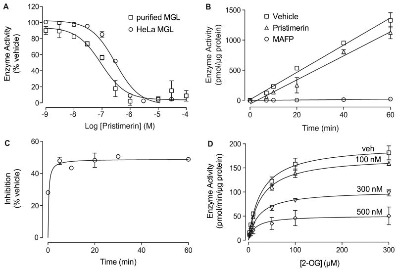
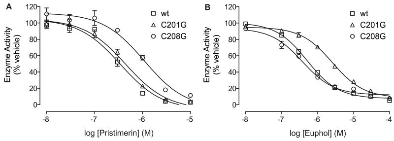
Pristimerin has been shown to suppress the activity of both pure and non-purified MGL (transfected HeLa cell cell lysates) at an IC50 of 93±8 nM and 398±68 nM, respectively. Pristimerin works as a fast, reversible, and non-competitive inhibitor of MGL. By forming a polar contact with a regulating cysteine, perhaps Cys208, prismerin's binding to MGL may be enhanced[1]. Pristimerin has a dose- and time-dependent effect on the viability of HFLS-RA and HUVEC cells. Pristimerin lowers the autophosphorylation of VEGFR2 caused by VEGF and lessens the activation of the VEGFR2-mediated signaling pathway generated by VEGF [2].
In vitro, pristimerin demonstrates potent antiproliferative activity against various cancer cell lines, including breast, prostate, lung, colon, and leukemia cells. It induces apoptosis through both intrinsic and extrinsic pathways, with IC50 values typically in the nanomolar to low micromolar range depending on the cell line. Pristimerin also exhibits anti-inflammatory activity by reducing the production of pro-inflammatory cytokines (TNF-α, IL-6, IL-1β) and inhibiting iNOS and COX-2 expression in stimulated macrophages. |
| ln Vivo |
Tumor angiogenesis and inflammation are both inhibited by pramimerin. Pristimerin considerably lowers the expression of pro-angiogenic factors in sera, such as TNF-α, Ang-1, and MMP-9, and significantly reduces vessel density in the synovial membrane tissues of inflamed joints[2].
Pristimerin inhibits inflammation and tumor angiogenesis. The present study focused on the inhibition of angiogenesis by Pristimerin in adjuvant-induced arthritic rats and the underlying molecular mechanisms. The results clearly demonstrate for the first time that Pristimerin significantly reduces vessel density in synovial membrane tissues of inflamed joints and reduces the expression of pro-angiogenic factors in sera, including TNF-α, Ang-1, and MMP-9. Pristimerin also decreased the expression of VEGF and p-VEGFR2 in the synovial membrane, whereas the total amount of VEGFR2 remained unchanged. Pristimerin suppressed the sprouting vessels of the aortic ring and inhibited VEGF-induced HFLS-RA migration in vitro. Pristimerin also inhibited VEGF-induced proliferation, migration and tube formation by HUVECs, blocked the autophosphorylation of VEGF-induced VEGFR2 and consequently downregulated the signaling pathways of activated PI3K, AKT, mTOR, ERK1/2, JNK, and p38 in VEGF-induced HUVECs. Our results indicate that Pristimerin suppressed synovial angiogenesis in our rat model and in vitro by interrupting the targeting of VEGFR2 activation. Therefore, Pristimerin has potential as an angiogenesis inhibitor in the treatment of rheumatoid arthritis. In vivo, pristimerin has shown significant antitumor activity in various xenograft models. It inhibits tumor growth, reduces metastasis, and induces apoptosis in tumor tissues. The compound also exhibits anti-inflammatory effects in animal models of inflammation, including carrageenan-induced paw edema and LPS-induced sepsis. Its antioxidant properties contribute to its protective effects against oxidative stress-related diseases. Detailed dosing regimens and efficacy outcomes are available in the scientific literature. |
| Enzyme Assay |
Screening for MGL Inhibitors[1]
The Spectrum Collection is composed of 2,000 compounds, supplied in a 10 mM dimethyl sulfoxide (DMSO) solution. We pooled groups of 10 compounds and performed a primary screen at a concentration of 1 μM. Individual compounds from positive groups (≥50% MGL inhibition) were subjected to a secondary screen at 10 μM. Full concentration-inhibition curves were generated using new lots of dry compounds. Enzyme Assays[1] Purified recombinant rat MGL was prepared and enzyme activity was assayed as described previously (King et al., 2007). Full-length rat ABHD6 or ABHD12 was subcloned into a pEF-V5/His vector by TOPO cloning and verified by DNA sequencing. HeLa cells were transiently transfected with pEF6 vector, ABHD6-V5-pEF6 or ABHD12-V5-pEF6 using Superfect reagent. 48 hours after transfection, cells were harvested and the homogenates were prepared in 50 mM Tris-Cl, pH 8.0, containing 0.32 M sucrose. ABHD activity was measured using a modified MGL assay procedure (pH 7.5, 3 μg protein per reaction, 30 min at 37°C) (King et al., 2007). Samples were extracted (King et al., 2007) and analyzed by liquid chromatography/mass spectrometry (LC/MS) (see below). 2-AG-hydrolase activity (in pmol/min/mg protein) in mock-transfected HeLa cells was 1.4±0.05, in pEF6-transfected (vector only) was 1.3±0.01, and in ABHD6-transfected cells was 6.9±0.2 (n = 3). FAAH and DGL activities were measured in rat brain homogenates as described. Recombinant NAAA protein was prepared from HEK293 cells stably transfected with pCMV-Flag-rNAAA using SuperFect reagent and screened with G418 (0.3 mg/ml). Cells were harvested and sonicated in 20 mM Tris-HCl (pH 7.5) with 0.32 M sucrose, centrifuged at 800 × g for 15 min at 4°C, and supernatant was centrifuged at 12,000 × g for 30 min at 4°C. The pellet was suspen ded in phosphate-buffered saline (PBS) and subjected to 2 freeze-thaw cycles at -80°C. The suspension was centrifuged at 105,000 × g for 1 hr at 4°C, and the supernatant containing rNAAA was kept at -80°C until use. NAAA activity was measured by combining substrate (50 μM heptadecenoylethanolamide) with protein (10 μg) in assay buffer (50 mM sodium hydrogen phosphate buffer, pH 5.0, 0.1% Triton X-100, 3 mM DTT) in a final volume of 0.2 ml for 30 min at 37°C. Reactions were stopped by addition of 0.2 ml cold methanol containing 1 nmol of heptadecanoic acid. For MGL, ABHD and NAAA assays samples were analyzed by LC/MS on an XDB Eclipse C18 column (2.1×30 mm i.d., 1.8 μm,) at 0.6 ml/min for 0.6 min with a solvent mixture of 95% methanol and 5% water, both containing 0.25% acetic acid and 5 mM ammonium acetate. The column temperature was 50°C. Samples were analyzed by electrospray ionization in the negative mode. Capillary voltage was 4 kV, fragmentor voltage was 100 V, nebulizer pressure was 60 psi. N2 was used as drying gas at a flow rate of 13 liters/min and a temperature of 350°C. We monitored the appropriate enzyme activity product (MGL and ABHD, m/z = 281, NAAA, m/z = 267) in the selected ion monitoring (SIM) mode using heptadecanoic acid as standard (m/z = 269). The in vitro activity of pristimerin is assessed using various biochemical and cell-based assays. For NF-κB inhibition, cells are transfected with an NF-κB reporter plasmid and treated with pristimerin, and luciferase activity is measured. For apoptosis studies, cells are treated with various concentrations of pristimerin, and caspase activity is measured using fluorogenic substrates, or apoptosis is assessed by flow cytometry using Annexin V/PI staining. Cell viability is assessed using MTT or CellTiter-Glo assays. |
| Cell Assay |
HFLS-RA (5 × 10³ cells/mL) or HUVECs (1 × 10⁴ cells/well) are seeded in 96-well plates and cultured in normal growth medium for 24 h. The cells are then incubated with different Pristimerin concentrations (0, 0.125, 0.25, 0.5 μM). The effects of Pristimerin on HUVECs viability are determined under VEGF-induced conditions. Cell viability is quantified by MTT assay. At 4 h before the end of the culture period, 30 μL of MTT solution (5.0 mg/mL) is added to each well. Cells without Pristimerin or VEGF served as a vehicle control[2]. |
| Animal Protocol |
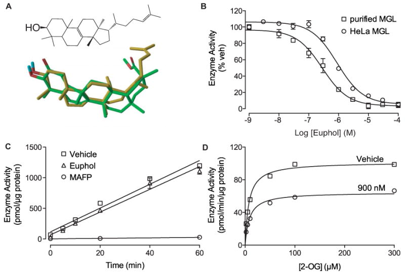
Primary cortical neuron cultures were prepared from embryonic day 18-20 Wistar rats (Stella et al., 2001). Cultures were maintained for 10 days at 37°C with 5% CO2 before treatment with pristimerin (1 μM), euphol (10 μM), NAM (1 μM) or vehicle (0.1% DMSO in Dulbecco's Modified Eagle Medium (DMEM)) for 30 min at 37°C. Reactions were stopped by washing with ice-cold PBS and cells were harvested in 2 ml 50% methanol. Lysates were vortexed for 10 s, protein concentrations were measured by bicinchoninic acid (BCA) protein assay (Pierce, Rockford, IL), and samples were extracted in 4 ml of ice-cold methanol/chloroform/water (1:2:1, vol:vol:vol) containing 0.5 nmol of 2-[2H8]-AG, and 10 pmol each of [2H4]-PEA, added as internal standard. Organic phases were recovered, evaporated under N2, reconstituted in 50 μl chloroform/methanol (1:3, vol:vol) and analyzed by LC/MS as described (Astarita and Piomelli, 2009).[1]
Pristimerin prevents arthritis progression and decreases the severity of arthritis in AA rats[2] To examine the effect of pristimerin on arthritis, the clinical and histopathological characteristics of the SD rat model of Mtb-induced arthritis, which shares immunological and pathological features with human, were examined RA. AA rats were injected daily intraperitoneally with Pristimerin from day 11 to day 24 after initial immunization; the control rats received DMSO (0.4%). The arthritis severity scores and back metatarsal volume of vehicle-treated rats were significantly increased... In vivo, pristimerin is typically administered intraperitoneally or orally to mice bearing tumor xenografts. Doses typically range from 0.5-5 mg/kg administered daily or every other day. Tumor growth is monitored by measuring tumor volume. At the end of the study, tumors are excised and analyzed for apoptosis (TUNEL staining), proliferation (Ki-67 staining), and expression of target proteins (Western blotting). For anti-inflammatory studies, pristimerin is administered before or after induction of inflammation, and inflammatory markers are measured in serum and tissues. |
| ADME/Pharmacokinetics |
Pristimerin has a molecular weight of 464.64 g/mol and a molecular formula of C30H40O4. It is a lipophilic compound with limited water solubility but good solubility in organic solvents such as DMSO and ethanol. The compound should be stored at -20°C under appropriate conditions to maintain stability. Pharmacokinetic parameters such as bioavailability and half-life vary depending on the route of administration and formulation.
|
| Toxicity/Toxicokinetics |
Oral LD50 in mice: 8 g/kg
Intraperitoneal LD50 in mice: 200 mg/kg Subcutaneous LD50 in mice: 400 mg/kg Pristimerin has shown cytotoxic effects on various cancer cell lines at nanomolar to low micromolar concentrations. In animal studies, the compound is generally well-tolerated at therapeutic doses, but dose-dependent toxicity may occur at higher doses. The compound should be used with caution due to its potent biological activity. Comprehensive toxicological studies are required to establish its full safety profile for potential therapeutic applications. |
| References | |
| Additional Infomation |
Pristimerin is a carboxylic acid ester. Pristimerin is a quinone methyl triterpenoid compound that has been studied for its anticancer potential. Pristimerin has been reported to exist in Celastrus orbiculatus, Celastraceae plants, and other organisms with relevant data.
Pristimerin is a natural triterpenoid with potent anticancer, anti-inflammatory, and antioxidant activities. It has been extensively studied in preclinical models for its potential therapeutic applications in cancer and inflammatory diseases. The compound's multiple mechanisms of action—including NF-κB and STAT3 inhibition, ROS generation, and caspase activation—make it a promising candidate for drug development. Pristimerin is not approved for clinical use and is intended for research purposes only. It is available from chemical suppliers for research applications. |
| Molecular Formula |
C30H40O4
|
|---|---|
| Molecular Weight |
464.65
|
| Exact Mass |
464.292
|
| Elemental Analysis |
C, 77.55; H, 8.68; O, 13.77
|
| CAS # |
1258-84-0
|
| Related CAS # |
Celastrol;34157-83-0
|
| PubChem CID |
159516
|
| Appearance |
Orange to reddish brown solid
|
| Density |
1.2±0.1 g/cm3
|
| Boiling Point |
607.7±55.0 °C at 760 mmHg
|
| Melting Point |
219.5°C
|
| Flash Point |
195.1±25.0 °C
|
| Vapour Pressure |
0.0±3.9 mmHg at 25°C
|
| Index of Refraction |
1.582
|
| Source |
Originated from plants: Celastraceae Tripterygium wilfordii Hook. f.
|
| LogP |
7.54
|
| Hydrogen Bond Donor Count |
1
|
| Hydrogen Bond Acceptor Count |
4
|
| Rotatable Bond Count |
2
|
| Heavy Atom Count |
34
|
| Complexity |
1120
|
| Defined Atom Stereocenter Count |
6
|
| SMILES |
C[C@](C1=CC=C(C(C)=C2O)C3=CC2=O)(CC[C@]4(C)[C@@]5([H])C[C@@](C(OC)=O)(C)CC4)[C@]5(CC[C@]13C)C
|
| InChi Key |
JFACETXYABVHFD-WXPPGMDDSA-N
|
| InChi Code |
InChI=1S/C30H40O4/c1-18-19-8-9-22-28(4,20(19)16-21(31)24(18)32)13-15-30(6)23-17-27(3,25(33)34-7)11-10-26(23,2)12-14-29(22,30)5/h8-9,16,23,32H,10-15,17H2,1-7H3/t23-,26-,27-,28+,29-,30+/m1/s1
|
| Chemical Name |
methyl (2R,4aS,6aR,6aS,14aS,14bR)-10-hydroxy-2,4a,6a,6a,9,14a-hexamethyl-11-oxo-1,3,4,5,6,13,14,14b-octahydropicene-2-carboxylate
|
| Synonyms |
UNII-28ZK7PR57S; Celastrol-methylether; Celastrol methyl ester; Celastrol-methylether; Pristimerine; GNF-PF-476; CHEBI:8416; MFCD01711331; Pristimerin
|
| HS Tariff Code |
2934.99.9001
|
| Storage |
Powder -20°C 3 years 4°C 2 years In solvent -80°C 6 months -20°C 1 month |
| Shipping Condition |
Room temperature (This product is stable at ambient temperature for a few days during ordinary shipping and time spent in Customs)
|
| Solubility (In Vitro) |
DMF : 25 mg/mL (~53.81 mM)
DMSO : ~20 mg/mL (~43.04 mM) |
|---|---|
| Solubility (In Vivo) |
Solubility in Formulation 1: ≥ 2 mg/mL (4.30 mM) (saturation unknown) in 10% DMSO + 90% Corn Oil (add these co-solvents sequentially from left to right, and one by one), clear solution.
For example, if 1 mL of working solution is to be prepared, you can add 100 μL of 20.0 mg/mL clear DMSO stock solution to 900 μL of corn oil and mix evenly. (Please use freshly prepared in vivo formulations for optimal results.) |
| Preparing Stock Solutions | 1 mg | 5 mg | 10 mg | |
| 1 mM | 2.1522 mL | 10.7608 mL | 21.5216 mL | |
| 5 mM | 0.4304 mL | 2.1522 mL | 4.3043 mL | |
| 10 mM | 0.2152 mL | 1.0761 mL | 2.1522 mL |
*Note: Please select an appropriate solvent for the preparation of stock solution based on your experiment needs. For most products, DMSO can be used for preparing stock solutions (e.g. 5 mM, 10 mM, or 20 mM concentration); some products with high aqueous solubility may be dissolved in water directly. Solubility information is available at the above Solubility Data section. Once the stock solution is prepared, aliquot it to routine usage volumes and store at -20°C or -80°C. Avoid repeated freeze and thaw cycles.
Calculation results
Working concentration: mg/mL;
Method for preparing DMSO stock solution: mg drug pre-dissolved in μL DMSO (stock solution concentration mg/mL). Please contact us first if the concentration exceeds the DMSO solubility of the batch of drug.
Method for preparing in vivo formulation::Take μL DMSO stock solution, next add μL PEG300, mix and clarify, next addμL Tween 80, mix and clarify, next add μL ddH2O,mix and clarify.
(1) Please be sure that the solution is clear before the addition of next solvent. Dissolution methods like vortex, ultrasound or warming and heat may be used to aid dissolving.
(2) Be sure to add the solvent(s) in order.
|
|
|
Products are for research use only; We do not sell to patients
Copyright 2020 InvivoChem LLC | All Rights Reserved


 COA
COA