

| Size | Price | Stock | Qty |
|---|---|---|---|
| 1mg |
|
||
| 5mg |
|
||
| 10mg |
|
||
| 25mg |
|
||
| 50mg |
|
||
| 100mg |
|
||
| 250mg |
|
||
| 500mg | |||
| Other Sizes |
Purity: ≥98%
| Targets |
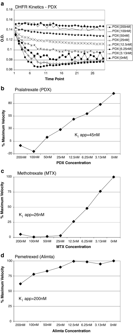
Pralatrexate (racemic) primarily targets dihydrofolate reductase (DHFR, Ki = 0.015 μM) and thymidylate synthase (TS, Ki = 0.12 μM), two key folate-dependent enzymes in nucleotide biosynthesis. These Ki values were determined via in vitro enzyme inhibition assays using purified human enzymes [4]
- Pralatrexate (racemic) inhibits DHFR to block tetrahydrofolate production, thereby suppressing nucleotide synthesis [1] |
|---|---|
| ln Vitro |
When administered to different T lymphoma cell lines, pralatrexate (100 pM-200 μM; 48-72 h) demonstrates cytotoxicity that is dependent on both concentration and time. The following are the IC50 values at 48 and 72 hours: 1.1 and 2.5 nM for H9 cells; 1.7 and 2.4 nM for P12 cells; 3.2 and 4.2 nM for CEM cells; 5.5 and 2.7 nM for PF-382 cells; 1 and 1.7 nM for KOPT-K1 cells; 97.4 and 1.2 nM for DND-41 cells; and 247.8 nM and 0.77 nM for HPB-ALL cells. After 48 hours of treatment, HH cells showed some resistance, with an IC50 of 2.8 nM at 72 hours [1]. Treatment with pralidoxate (2-5.5 nM; 48-72 h; H9, HH, P12, and PF382 cells) results in strong apoptosis and caspase-8 and caspase-9 activation [1]. Treatment of H9 and P12 cells with pralitrexate (3 nM; 16–48 hours) dramatically raises p27 levels and promotes the accumulation of inducible folate carrier type 1 (RFC-1) in the cells [1].
In human T-cell lymphoma cell lines (Jurkat, Karpas 299, Hut-78): Pralatrexate (racemic) alone exhibited antiproliferative activity with IC50 values of 0.008 μM (Jurkat), 0.012 μM (Karpas 299), and 0.015 μM (Hut-78) (MTT assay, 72-hour incubation). When combined with bortezomib (0.005 μM), the combination index (CI) was 0.4-0.6 (CI < 1 indicating synergism), and apoptotic cells (Annexin V-positive) increased from 15%-20% (single drug) to 45%-55% (combination). Western blot showed that the combination enhanced cleavage of caspase-3 and PARP (apoptosis markers) by 2-3 folds compared to Pralatrexate (racemic) alone [1] - In human solid tumor cell lines (HCT-116 colorectal cancer, A549 NSCLC, MDA-MB-231 breast cancer): Pralatrexate (racemic) inhibited cell viability with IC50 values of 0.006 μM (HCT-116), 0.009 μM (A549), and 0.011 μM (MDA-MB-231) (MTT assay, 72-hour incubation). PCR analysis revealed that 0.01 μM Pralatrexate (racemic) downregulated TS mRNA expression by 40%-50% in HCT-116 cells [4] |
| ln Vivo |
In comparison to either drug alone, the combination of Bortezomib (0.5 mg/kg) and Pralatrexate (15 mg/kg; intraperitoneal injection; on days 1, 4, 8, and 11; SCID-beige mice) increased effectiveness [1].
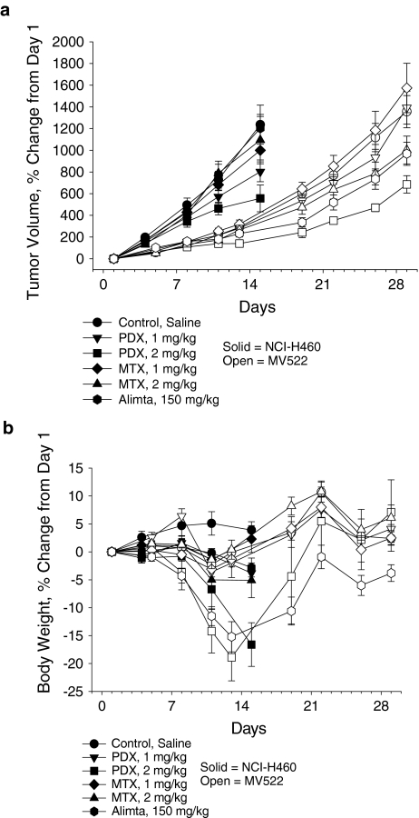
In nude mice bearing Karpas 299 (T-cell lymphoma) xenografts: Mice were divided into 4 groups (n=6/group): control (vehicle), Pralatrexate (racemic) alone (10 mg/kg, intraperitoneal injection, twice weekly), bortezomib alone (0.5 mg/kg, intraperitoneal injection, twice weekly), and combination group. After 3 weeks of treatment, the tumor volume inhibition rate was 45%-50% (single Pralatrexate (racemic)), 30%-35% (single bortezomib), and 75%-80% (combination). Immunohistochemistry of tumor tissues showed that the combination reduced Ki-67 (proliferation marker) positive cells by 60%-70%, compared to 30%-35% with Pralatrexate (racemic) alone [1] - In nude mice bearing HCT-116 (colorectal cancer) or MDA-MB-231 (breast cancer) xenografts: Pralatrexate (racemic) was administered intravenously at 15 mg/kg once weekly for 4 weeks. The tumor weight inhibition rate was 60%-65% (HCT-116) and 55%-60% (MDA-MB-231). Serum thymidine (a TS substrate) levels decreased by 50%-55% in treated mice, confirming in vivo inhibition of TS activity [4] - In the PROPEL study (clinical phase II): Pralatrexate (racemic) (30 mg/m², intravenous infusion, weekly for 6 weeks, followed by 2-week rest) was administered to patients with relapsed/refractory transformed mycosis fungoides. The overall response rate (ORR) was 28% (95% CI: 16%-43%), with a median progression-free survival (PFS) of 4.5 months. Among patients with skin-only disease, the ORR was 35%, higher than that in patients with extracutaneous involvement (18%) [2] - In a randomized phase 2b study (NSCLC patients post-platinum failure): Patients received either Pralatrexate (racemic) (60 mg/m², intravenous infusion, every 2 weeks) or erlotinib (150 mg/day, oral). The median PFS was 2.6 months (Pralatrexate) vs. 2.8 months (erlotinib) (p=0.78), and the ORR was 5% (Pralatrexate) vs. 8% (erlotinib). [3] |
| Enzyme Assay |
DHFR Activity Assay: The reaction was conducted in 50 mM Tris-HCl buffer (pH 7.4) containing 10 mM MgCl2, 0.1 mM NADPH, and 0.05 mM dihydrofolate (substrate). Purified human DHFR was pre-incubated with Pralatrexate (racemic) (0.001-0.1 μM) at 37°C for 10 minutes, then substrate was added to initiate the reaction. The decrease in absorbance at 340 nm (due to NADPH oxidation) was recorded every minute for 20 minutes. The Ki value was calculated by fitting the inhibition curve to the Lineweaver-Burk plot [4]
- TS Activity Assay: The assay used 50 mM potassium phosphate buffer (pH 7.2) with 5 mM MgCl2, 0.1 mM [3H]-dUMP (substrate), and 0.2 mM N5,N10-methylenetetrahydrofolate (cofactor). Purified human TS was incubated with Pralatrexate (racemic) (0.01-1 μM) at 37°C for 15 minutes, followed by substrate addition. After 30 minutes, the reaction was terminated with 10% trichloroacetic acid, and the radioactive precipitate ([3H]-dTMP) was measured via liquid scintillation counting. The Ki value was determined based on the dose-dependent inhibition of TS activity [4] |
| Cell Assay |
Cell Cytotoxicity Assay[1]
Cell Types: T-lymphoma cell lines Tested Concentrations: 100 pM-200 µM Incubation Duration: 48 hrs (hours), 72 hrs (hours) Experimental Results: demonstrated concentration- and time-dependent cytotoxicity against a broad panel of T-lymphoma cell lines. Apoptosis Analysis[1] Cell Types: H9, HH, P12 and PF382 cells Tested Concentrations: 2 nM, 3 nM, 4 nM, 5.5 nM Incubation Duration: 48 hrs (hours), 72 hrs (hours) Experimental Results: Induced potent apoptosis and caspase activation. Western Blot Analysis[1] Cell Types: H9 and P12 cells Tested Concentrations: 3 nM Incubation Duration: 16 hrs (hours), 24 hrs (hours), 48 hrs (hours) Experimental Results: Clearly increased p27 levels and increased the accumulation of RFC-1 in cells. MTT Antiproliferation Assay (T-cell lymphoma cells): Jurkat, Karpas 299, and Hut-78 cells were seeded in 96-well plates at 2×10⁴ cells/well in RPMI 1640 medium (10% fetal bovine serum). Pralatrexate (racemic) was added at concentrations of 0.001-0.1 μM, and cells were incubated at 37°C (5% CO2) for 72 hours. 20 μL MTT solution (5 mg/mL in PBS) was added, and incubation continued for 4 hours. The supernatant was removed, 150 μL dimethyl sulfoxide was added to dissolve formazan crystals, and absorbance at 570 nm was measured. IC50 was calculated as the drug concentration inhibiting 50% cell viability [1] - Annexin V/PI Apoptosis Assay (Karpas 299 cells): Cells were seeded in 6-well plates at 1×10⁶ cells/well and treated with Pralatrexate (racemic) (0.01 μM) alone or with bortezomib (0.005 μM) for 48 hours. Cells were harvested, washed with cold PBS, and resuspended in binding buffer. Annexin V-FITC and PI were added, and cells were incubated in the dark for 20 minutes at room temperature. Apoptotic cells were analyzed via flow cytometry [1] - Clone Formation Assay (HCT-116 cells): Cells were seeded in 6-well plates at 100 cells/well and allowed to attach for 24 hours. Pralatrexate (racemic) was added at 0.002, 0.005, 0.01 μM, and cells were cultured for 14 days (medium changed every 3 days). Colonies were fixed with 4% paraformaldehyde (15 minutes) and stained with 0.1% crystal violet (30 minutes). Colonies with >50 cells were counted, and the clone formation rate was (colonies in treatment/colonies in control) × 100% [4] |
| Animal Protocol |
Animal/Disease Models: SCID-beige mice (5-7weeks old) injected with HH cells[1]
Doses: 15 mg/kg Route of Administration: intraperitoneal (ip)injection; on days 1, 4, 8, and 11 Experimental Results: demonstrated superior efficacy in T-cell malignancies. Karpas 299 Xenograft Model (nude mice): Female nude mice (6-8 weeks old, 18-22 g) were subcutaneously injected with 2×10⁶ Karpas 299 cells (suspended in 0.2 mL PBS/Matrigel 1:1) into the right flank. When tumors reached 100-150 mm³, mice were randomized into 4 groups: (1) Control: 0.9% saline + 0.1% dimethyl sulfoxide (intraperitoneal injection, twice weekly); (2) Pralatrexate (racemic): dissolved in 0.9% saline + 0.1% dimethyl sulfoxide, 10 mg/kg, intraperitoneal injection, twice weekly; (3) Bortezomib: dissolved in 0.9% saline, 0.5 mg/kg, intraperitoneal injection, twice weekly; (4) Combination: same doses and schedule as single drugs. Tumor volume (length × width² / 2) and body weight were measured twice weekly for 3 weeks. At study end, mice were euthanized, and tumors were collected for immunohistochemistry [1] - HCT-116/MDA-MB-231 Xenograft Models (nude mice): Male nude mice (6-8 weeks old, 20-24 g) were subcutaneously implanted with 5×10⁶ HCT-116 or 3×10⁶ MDA-MB-231 cells (0.2 mL PBS/Matrigel 1:1) into the left flank. When tumors grew to 200-250 mm³, mice were divided into 2 groups (n=5/group): control (5% glucose solution) and Pralatrexate (racemic) (dissolved in 5% glucose solution, 15 mg/kg, intravenous injection, once weekly). Treatment lasted 4 weeks, with tumor volume and body weight measured weekly. Mice were sacrificed at study end, and tumors/serum were collected for analysis [4] |
| ADME/Pharmacokinetics |
Absorption, Distribution and Excretion
Following intravenous injection, pralatrexate has 100% bioavailability. Pralatrexate exhibits dose-proportional linear pharmacokinetic characteristics within a dose range of 30–325 mg/m². In the first dose of the first treatment cycle, after an intravenous bolus of 30 mg/m² racemic pralatrexate (starting dose), non-compartmental pharmacokinetic analysis estimated its Cmax and AUC0-∞ to be 5,815 ng/mL and 267,854 ng/mL·min, respectively. Both diastereomers of pralatrexate exhibited a multiphasic decrease in plasma concentration, with a rapid initial decrease followed by a slow terminal decrease. The initial decrease in plasma concentration is thought to reflect clearance of pralatrexate via renal and non-renal mechanisms, while the slow terminal phase may represent the return of pralatrexate to plasma concentrations from deep intracellular compartments, enterohepatic circulation, or after deglutamate conversion. Following a single administration of FOLOTYN at a dose of 30 mg/m², approximately 34% of pralatrexate is excreted unchanged in the urine. Following administration of radiolabeled pralatrexate, 39% (CV = 28%) of the dose is recovered unchanged in the urine, and 34% (CV = 88%) is recovered unchanged in the feces and/or its metabolites. Within 24 hours, 10% (CV = 95%) of the dose is exhaled. The steady-state volumes of distribution for the S- and R-diasteriomers of pralatrexate are 105 L and 37 L, respectively. The total systemic clearance of the diasteriomers of pralatrexate is 417 mL/min (S-diasteriomer) and 191 mL/min (R-diasteriomer), respectively. In 10 patients with PTCL, the pharmacokinetics of pralatrexate monotherapy (30 mg/m², once weekly, intravenous bolus over 3–5 minutes, for 6 weeks, 7 weeks per cycle) were evaluated. The total systemic clearance of the diastereomers of pralatrexate was 417 mL/min (S-diastereomer) and 191 mL/min (R-diastereomer). The total systemic exposure (AUC) and maximum plasma concentration (Cmax) of pralatrexate increased proportionally with dose (dose range 30–325 mg/m², including pharmacokinetic data from high-dose solid tumor clinical studies). The pharmacokinetics of pralatrexate did not change significantly across multiple treatment cycles, and no accumulation of pralatrexate was observed. The steady-state volumes of distribution of the diastereomers of pralatrexate were 105 L (S-diastereomer) and 37 L (R-diastereomer). In vitro studies have shown that pralatrexate binds to approximately 67% of plasma proteins. For more complete data on the absorption, distribution, and excretion of pralatrexate (7 components), please visit the HSDB record page. Metabolism/Metabolites Although the liver has been shown to metabolize pralatrexate to some extent, in vitro studies have shown that pralatrexate is not significantly metabolized by any CYP450 isoenzymes or glucuronidases. In vitro studies using human hepatocytes, liver microsomes, and S9 cells have shown that pralatrexate is primarily metabolized by cells. Component analysis and recombinant human CYP450 isoenzyme analysis indicate that pralatrexate is not significantly metabolized by phase I hepatic CYP450 isoenzymes or phase II hepatic glucuronidases. Biological Half-Life The terminal elimination half-life of pralatrexate is 12–18 hours (coefficient of variation [CV] = 62–120%). The terminal elimination half-life of pralatrexate is 12–18 hours (coefficient of variation (CV) = 62–120%). In nude mice treated with pralatrexate (racemic mixture) (15 mg/kg, intravenously): plasma concentration-time curves conformed to a two-compartment model. The terminal half-life (t1/2β) was 2.8 ± 0.3 hours, AUC0-∞ was 18.5 ± 2.1 μg·h/mL, and clearance (CL) was 0.8 ± 0.1 mL/h/g. Approximately 70%–75% of the dose is excreted unchanged in the urine within 24 hours [4] - In patients with transformed mycosis fungoides (PROPEL study): after intravenous infusion of pralatrexate (racemic) (30 mg/m²), the mean plasma half-life was 1.8 ± 0.5 hours, and the steady-state volume of distribution (Vdss) was 12.6 ± 3.2 L/m². The drug is primarily excreted by the kidneys, with 65%–70% of the dose recovered in the urine within 48 hours [2] |
| Toxicity/Toxicokinetics |
Hepatotoxicity
Platrexate treatment may cause elevated serum enzymes, but these abnormalities are usually mild and resolve spontaneously, increasing to more than 5 times the upper limit of normal only in 2% to 6% of patients, and rarely requiring dose adjustment. No clinically significant acute liver injury caused by pralatrexate has been reported in the literature, but monitoring for hepatotoxicity is recommended. While pralatrexate has not been definitively linked to hepatic sinusoidal obstruction syndrome, it is rarely used at high doses in pretreatment regimens for neoplastic diseases or bone marrow transplantation, where alkylating agents are commonly associated with this complication. Probability Score: E (Unlikely, but suspected as a rare cause of liver injury). Protein Binding Platrexate has an in vitro protein binding rate of approximately 67%. Interactions A Phase I clinical study investigated the effects of co-administration with the uricosuric drug probenecid on the pharmacokinetics of pralatrexate. Concomitant administration of escalating doses of probenecid resulted in delayed clearance of pralatrexate, accompanied by a corresponding increase in exposure. Because it is excreted by the kidneys (accounting for approximately 34% of the total clearance of pralatrexate), concomitant administration of drugs that are primarily cleared by the kidneys (e.g., nonsteroidal anti-inflammatory drugs, trimethoprim/sulfamethoxazole) may result in delayed clearance of pralatrexate. In nude mice (Karpas 299 model): pralatrexate (racemic) (10 mg/kg, twice weekly) caused a transient 8%–10% decrease in body weight during the first week, which recovered during the second week. No significant changes in serum ALT, AST, or creatinine were observed compared to the control group [1] -In the PROPEL study (patients): The most common grade 3/4 adverse events (AEs) pralatrexate (racemic) adverse reactions included mucositis (25%), thrombocytopenia (20%), and neutropenia (15%). Grade 1/2 adverse reactions included fatigue (40%), nausea (30%), and diarrhea (20%). Plasma protein binding of pralatrexate (racemic) is 90%-95% (in vitro human plasma assay) [2] - In a stage 2b non-small cell lung cancer study: Grade 3/4 adverse events of pralatrexate (racemic) (60 mg/m², every 2 weeks) included anemia (18%), mucositis (12%) and elevated liver enzymes (10%) [3] - In nude mice (HCT-116 model): pralatrexate (racemic) (15 mg/kg, once a week) did not cause significant organ toxicity (no abnormal lesions were found in liver and kidney histology) [4] |
| References |
|
| Additional Infomation |
Therapeutic Uses
Aminopterin/analogs and derivatives; folic acid antagonists Platrexate is indicated for the treatment of patients with relapsed or refractory peripheral T-cell lymphoma (PTCL). This indication is based on overall response rate. Clinical benefits such as improved progression-free survival or overall survival have not been demonstrated. /US product label includes/ T-cell lymphoma (TCL) is characterized by poor response to chemotherapy and generally poor prognosis. While molecular profiling has identified different biological subtypes and therapeutic targets in B-cell lymphomas, progress in the molecular characterization of TCL has been slow. Markers expressed on the surface of malignant T cells, such as CD2, CD3, CD4, CD25, and CD52, were the first TCL-specific therapeutic targets identified. However, because these receptors are also present on normal T cells, TCL treatment based on monoclonal antibodies (mAbs) or immunotoxins (ITs) inevitably leads to varying degrees of immunosuppression. Therefore, although some mAbs/ITs have shown significant efficacy in specific subtypes of TCL, more specific drugs targeting preferentially activated signaling pathways in malignant T cells are still needed. Histone deacetylase (HDAC) inhibitors are one such new class of drugs. These molecules selectively induce apoptosis in a variety of transformed cells, including malignant T cells, both in vitro and in vivo. Some HDAC inhibitors have been studied in TCL with encouraging results and have recently been approved for clinical use. Immunomodulatory drugs, such as interferons and Toll-like receptor (TLR) agonists, have shown significant clinical efficacy in TCL, playing a particularly important role in the treatment of primary cutaneous T-cell lymphoma (CTCL). Although most classical cytotoxic drugs have limited efficacy against T-cell lymphoma (TCL), drugs that inhibit purine and pyrimidine metabolism (called nucleoside analogs) and novel antifolate drugs (such as pralatrexate) have high activity against TCL. As the molecular profiling of TCL continues to deepen, new drugs targeting TCL are being discovered at an increasingly rapid pace. Clinical trials are currently underway, and these drugs are being integrated into combination therapy regimens for TCL, for relapsed/refractory TCL and as first-line treatment. Drug Warnings FOLOTYN can suppress bone marrow function, manifesting as thrombocytopenia, neutropenia, and anemia. Dosage should be adjusted based on absolute neutrophil count (ANC) and platelet count before each administration. FOLOTYN treatment may cause mucositis.If grade 2 or higher mucositis is observed, the dosage should be adjusted. Patients should be instructed to take folic acid and vitamin B12 to reduce the incidence of treatment-related hematologic toxicity and mucositis. …Patients should take a low dose of folic acid orally daily. Folic acid should be started 10 days before the first dose of FOLOTYN and continued throughout the course of treatment and for 30 days after the last dose of FOLOTYN. Patients should also receive an intramuscular injection of vitamin B12 no more than 10 weeks before the first dose of FOLOTYN, followed by injections every 8–10 weeks. Subsequent vitamin B12 injections can be administered on the same day as FOLOTYN treatment. Although FOLOTYN has not been formally tested in patients with renal impairment, caution is advised when using FOLOTYN in patients with moderate to severe renal impairment. Patients should be monitored for renal function and systemic toxicity due to increased drug exposure. For more complete data on drug warnings for pralatrexate (10 in total), please visit the HSDB records page. Pharmacodynamics Platlatrexate is a folic acid analog that inhibits folic acid metabolism, thereby hindering the synthesis of amino acids and nucleic acids. Furthermore, pralatrexate competes with folic acid for enzymatic processing by folate polyglutamate synthase (FPGS), increasing cellular retention. Compared to methotrexate, pralatrexate exhibits a 10-fold higher binding affinity to reduced folate carrier protein-1 (RFC-1), making it more readily taken up by cells and a more effective substrate for FPGS. The Km values for pralatrexate and methotrexate to RFC-1 are 0.3 μmol/L and 4.8 μmol/L, respectively, while their Km values to FPGS are 5.9 μmol/L and 32.3 μmol/L, respectively. Therefore, pralatrexate demonstrates stronger cytotoxicity and better retention in cancer cells. Due to its antifolate activity, the main toxicity of pralatrexate is mucositis, which may require discontinuation of treatment or dose reduction. In five patients with non-small cell lung cancer who received a supertherapeutic dose of pralatrexate at 230 mg/m², the mean change in QTcF interval at the end of infusion was 6.1 ms (90% CI: -0.6, 12.7) from baseline, and 7.8 ms (90% CI: 3.0, 12.6) at 1 hour post-infusion. However, the QTcF interval did not exceed 470 ms in any patient, and the absolute increase in QTcF interval from baseline did not exceed 30 ms. Furthermore, this study dose was significantly higher than the target dose for patients with peripheral T-cell lymphoma, and pralatrexate does not inhibit human ether-a-go-go-related gene (hERG) potassium channels. Therefore, pralatrexate use is unlikely to cause delayed cardiac repolarization. Pralatrexate (racemic mixture) exerts a synergistic antitumor effect with bortezomib in T-cell lymphoma by enhancing apoptosis: bortezomib inhibits the proteasome degradation of pro-apoptotic proteins, while pralatrexate (racemic mixture) inhibits nucleotide synthesis, thereby producing a \"metabolic stress + apoptosis sensitization\" effect [1]. The PROPEL study confirmed that pralatrexate (racemic mixture) is an effective treatment option for relapsed/refractory transformed mycosis fungoides, especially suitable for patients with only skin involvement (high objective response rate). This study used prophylactic folic acid and vitamin B12 supplementation to reduce mucositis and myelosuppression [2] - In a phase 2b non-small cell lung cancer (NSCLC) study, pralatrexate (racemate) showed similar efficacy to erlotinib in patients after platinum-based chemotherapy, but with different adverse reaction profiles (pralatrexate was more likely to cause mucositis, while erlotinib mainly caused rash/diarrhea), suggesting that pralatrexate may be an alternative treatment option for patients intolerant to EGFR inhibitors [3] - Pralatrexate (racemate) is a structural analog of 10-deazoaminopterin with enhanced affinity for dihydrofolate reductase (DHFR) (10 times higher than methotrexate) and better tumor penetration, which makes it more effective in vivo against human xenograft tumors [4] |
| Molecular Formula |
C23H23N7O5
|
|---|---|
| Molecular Weight |
477.47
|
| Exact Mass |
477.176
|
| CAS # |
146464-95-1
|
| Related CAS # |
(R)-Pralatrexate;1320211-70-8
|
| PubChem CID |
148121
|
| Appearance |
Light yellow to yellow solid powder
|
| Density |
1.5±0.1 g/cm3
|
| Melting Point |
215 °C(dec.)
|
| Index of Refraction |
1.704
|
| LogP |
0.23
|
| Hydrogen Bond Donor Count |
5
|
| Hydrogen Bond Acceptor Count |
11
|
| Rotatable Bond Count |
10
|
| Heavy Atom Count |
35
|
| Complexity |
809
|
| Defined Atom Stereocenter Count |
1
|
| SMILES |
C#CCC(CC1=CN=C2C(=N1)C(=NC(=N2)N)N)C3=CC=C(C=C3)C(=O)N[C@@H](CCC(=O)O)C(=O)O
|
| InChi Key |
OGSBUKJUDHAQEA-WMCAAGNKSA-N
|
| InChi Code |
InChI=1S/C23H23N7O5/c1-2-3-14(10-15-11-26-20-18(27-15)19(24)29-23(25)30-20)12-4-6-13(7-5-12)21(33)28-16(22(34)35)8-9-17(31)32/h1,4-7,11,14,16H,3,8-10H2,(H,28,33)(H,31,32)(H,34,35)(H4,24,25,26,29,30)/t14?,16-/m0/s1
|
| Chemical Name |
N -(4-{1-[(2,4-diaminopteridin-6-yl)methyl]but-3-yn-1-yl}benzoyl)-L-glutamic acid
|
| Synonyms |
PDX; Pralatrexate; 10-Propargyl-10-deazaaminopterin; trade name: Folotyn.
|
| HS Tariff Code |
2934.99.9001
|
| Storage |
Powder -20°C 3 years 4°C 2 years In solvent -80°C 6 months -20°C 1 month |
| Shipping Condition |
Room temperature (This product is stable at ambient temperature for a few days during ordinary shipping and time spent in Customs)
|
| Solubility (In Vitro) |
DMSO: 28 mg/mL (58.6 mM)
Water: 1 mg/mL (Insoluble) Ethanol: 1 mg/mL |
|---|---|
| Solubility (In Vivo) |
Solubility in Formulation 1: ≥ 2.5 mg/mL (5.24 mM) (saturation unknown) in 10% DMSO + 40% PEG300 + 5% Tween80 + 45% Saline (add these co-solvents sequentially from left to right, and one by one), clear solution.
For example, if 1 mL of working solution is to be prepared, you can add 100 μL of 25.0 mg/mL clear DMSO stock solution to 400 μL PEG300 and mix evenly; then add 50 μL Tween-80 to the above solution and mix evenly; then add 450 μL normal saline to adjust the volume to 1 mL. Preparation of saline: Dissolve 0.9 g of sodium chloride in 100 mL ddH₂ O to obtain a clear solution. Solubility in Formulation 2: ≥ 2.5 mg/mL (5.24 mM) (saturation unknown) in 10% DMSO + 90% (20% SBE-β-CD in Saline) (add these co-solvents sequentially from left to right, and one by one), clear solution. For example, if 1 mL of working solution is to be prepared, you can add 100 μL of 25.0 mg/mL clear DMSO stock solution to 900 μL of 20% SBE-β-CD physiological saline solution and mix evenly. Preparation of 20% SBE-β-CD in Saline (4°C,1 week): Dissolve 2 g SBE-β-CD in 10 mL saline to obtain a clear solution. (Please use freshly prepared in vivo formulations for optimal results.) |
| Preparing Stock Solutions | 1 mg | 5 mg | 10 mg | |
| 1 mM | 2.0944 mL | 10.4719 mL | 20.9437 mL | |
| 5 mM | 0.4189 mL | 2.0944 mL | 4.1887 mL | |
| 10 mM | 0.2094 mL | 1.0472 mL | 2.0944 mL |
*Note: Please select an appropriate solvent for the preparation of stock solution based on your experiment needs. For most products, DMSO can be used for preparing stock solutions (e.g. 5 mM, 10 mM, or 20 mM concentration); some products with high aqueous solubility may be dissolved in water directly. Solubility information is available at the above Solubility Data section. Once the stock solution is prepared, aliquot it to routine usage volumes and store at -20°C or -80°C. Avoid repeated freeze and thaw cycles.
Calculation results
Working concentration: mg/mL;
Method for preparing DMSO stock solution: mg drug pre-dissolved in μL DMSO (stock solution concentration mg/mL). Please contact us first if the concentration exceeds the DMSO solubility of the batch of drug.
Method for preparing in vivo formulation::Take μL DMSO stock solution, next add μL PEG300, mix and clarify, next addμL Tween 80, mix and clarify, next add μL ddH2O,mix and clarify.
(1) Please be sure that the solution is clear before the addition of next solvent. Dissolution methods like vortex, ultrasound or warming and heat may be used to aid dissolving.
(2) Be sure to add the solvent(s) in order.
| NCT Number | Recruitment | interventions | Conditions | Sponsor/Collaborators | Start Date | Phases |
| NCT02594267 | Completed | Drug: Pralatrexate Injection | Peripheral T-Cell Lymphoma (PTCL) | Acrotech Biopharma Inc. | November 10, 2015 | Phase 1 |
| NCT03355768 | Withdrawn | Drug: Romidepsin Drug: Pralatrexate |
Lymphoma, T-Cell, Peripheral | Jennifer Amengual | September 1, 2018 | Phase 3 |
| NCT03598998 | Active, not recruiting | Biological: Pembrolizumab Drug: Pralatrexate |
Anaplastic Large Cell Lymphoma Nodal Peripheral T-Cell Lymphoma With TFH Phenotype |
City of Hope Medical Center | February 4, 2019 | Phase 1 Phase 2 |
| NCT03240211 | Recruiting | Drug: Pembrolizumab Drug: Pralatrexate |
PTCL CTCL |
University of Virginia | February 2, 2022 | Phase 1 |
 |
|---|
 |
 |
Products are for research use only; We do not sell to patients
Copyright 2020 InvivoChem LLC | All Rights Reserved


 COA
COA