| Size | Price | Stock | Qty |
|---|---|---|---|
| 10mg |
|
||
| 25mg |
|
||
| 50mg |
|
||
| 100mg |
|
||
| 250mg |
|
||
| 500mg |
|
||
| 1g | |||
| Other Sizes |


Purity: ≥98%
| Targets |
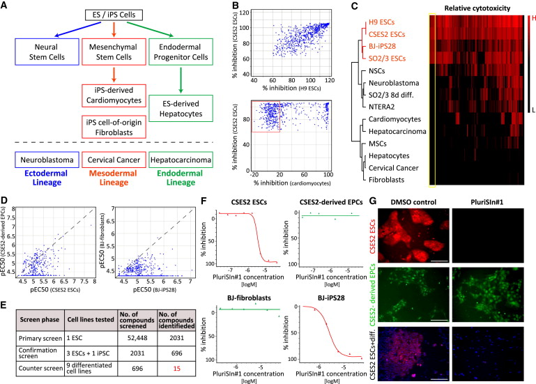
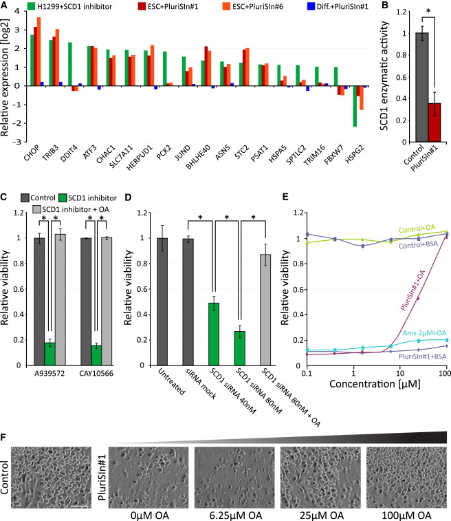
Stearoyl-CoA desaturase (SCD, mainly SCD1 subtype): PluriSIn-1 (NSC 14613) selectively inhibits SCD, the enzyme that catalyzes the conversion of saturated fatty acids (e.g., stearic acid) to monounsaturated fatty acids (e.g., oleic acid). In enzymatic assays using purified human SCD1, the IC50 of PluriSIn-1 for SCD activity was approximately 1.2 μM [1]
|
|---|---|
| ln Vitro |
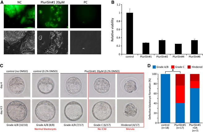
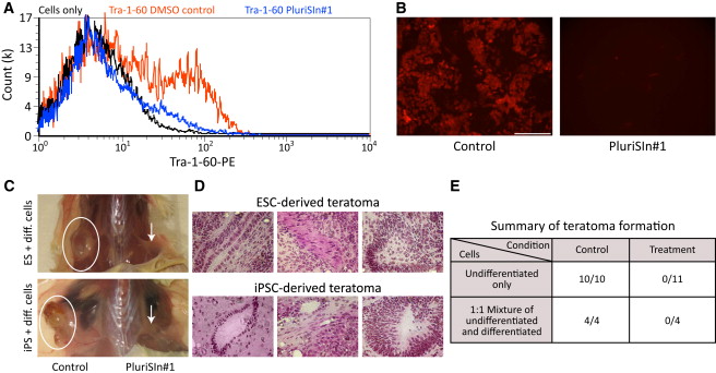
Stearoyl-coA desaturase (SCD) on induced pluripotent stem cells (iPSCs)-derived cardiomyocytes (CM) is inhibited by PluriSIn 1, a small chemical. Notably, we show that treatment with PluriSIn 1 at 20 µM for one day dramatically causes the apoptosis of Nanog-positive induced pluripotent stem cells (iPSDs). Treatment with PluriSIn 1 also considerably reduces the mRNA and protein level of Nanog, a marker for both cell pluripotency and tumor growth. Furthermore, 4 days of 20 µM PluriSIn 1 treatment reduced Nanog-positive stem cells in cultured iPSD but did not increase apoptosis of iPS-derived CM. We intramyocardially injected PluriSIn 1- or DMSO-treated iPSD in a mouse model of myocardial infarction (MI) to examine whether therapy with PluriSIn 1 inhibits tumorigenicity of iPSD following cell transplantation. Two weeks after injection, DMSO-treated iPSD easily developed Nanog-expressing tumors, which is avoided by administering PluriSIn 1. Furthermore, PluriSIn 1 therapy does not alter the expression of cardiac differentiation markers cTnI, α-MHC, or MLC-2v (P>0.05, n=4). Crucially, iPS-derived CM treated with PluriSIn 1 shows the capacity to engraft and endure in the infarcted myocardium[1].
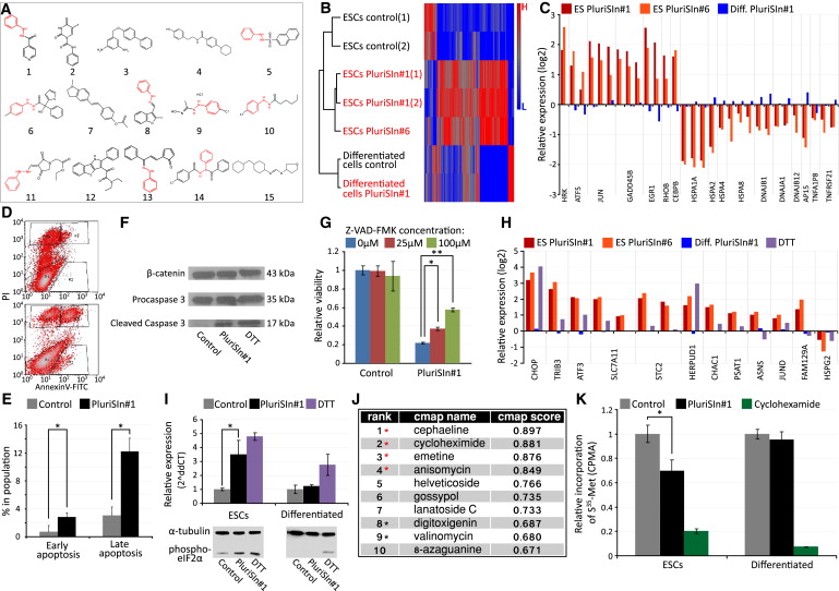
Inhibition of Nanog-positive tumorigenic cells (derived from iPS cells): - Cytotoxicity against Nanog-positive cells: PluriSIn-1 (0.5-5 μM) dose-dependently reduced the viability of Nanog-positive iPS-derived tumorigenic cells. The IC50 for cell viability (MTT assay, 72 h incubation) was 1.8 μM. At 2 μM, the viability of Nanog-positive cells decreased by 70% compared to the control group, while the viability of Nanog-negative non-tumorigenic cells remained >90% [1] - SCD activity inhibition and lipid metabolism disruption: Treatment with PluriSIn-1 (1-3 μM) for 48 h inhibited SCD activity in Nanog-positive cells by 65-80%, as measured by reduced conversion of [14C]-stearic acid to [14C]-oleic acid. Gas chromatography-mass spectrometry (GC-MS) analysis showed a 40-50% decrease in intracellular monounsaturated fatty acids (oleic acid, palmitoleic acid) and a 25-30% increase in saturated fatty acids (stearic acid, palmitic acid) [1] - Apoptosis induction in Nanog-positive cells: PluriSIn-1 (2 μM) induced apoptosis in Nanog-positive cells, with the apoptotic rate (Annexin V⁺/PI⁺ cells) increasing from 5% (control) to 45% (48 h treatment) as detected by flow cytometry. Western blot analysis revealed increased cleaved caspase-3 (3.5-fold) and cleaved PARP (2.8-fold) expression, and decreased anti-apoptotic protein Bcl-2 (0.4-fold) expression [1] - Effect on normal cells: PluriSIn-1 (up to 5 μM) had no significant inhibitory effect on the viability of normal human cardiomyocytes (HCMs) or normal mouse embryonic fibroblasts (MEFs), with cell viability >85% (vs. control). No obvious apoptosis or lipid metabolism disruption was observed in these normal cells [1] |
| ln Vivo |
Nude mouse model of iPS cell-derived myocardial transplantation:
- Model establishment: Male nude mice (6-8 weeks old) were anesthetized, and 5×10⁵ Nanog-positive iPS cells were injected into the left ventricular myocardium to establish a tumorigenic transplantation model [1] - Drug treatment and efficacy: Mice were randomly divided into two groups (n=8/group): control group (vehicle: 10% DMSO in PBS, i.p.) and PluriSIn-1 treatment group (10 mg/kg, i.p., once daily for 14 days). After 4 weeks of transplantation, the tumor formation rate in the control group was 60% (5 out of 8 mice developed myocardial tumor nodules), while the tumor formation rate in the treatment group was only 15% (1 out of 8 mice). The average volume of tumor nodules in the control group was 120 ± 25 mm³, and no detectable tumor nodules were found in the treatment group [1] - Myocardial tissue safety: Immunohistochemical staining of myocardial tissue showed that PluriSIn-1 treatment reduced the number of Nanog-positive cells (from 25 ± 5 cells/mm² to 3 ± 1 cells/mm²) without causing obvious myocardial damage (no increase in myocardial fibrosis or necrosis). Hematoxylin-eosin (HE) staining showed normal myocardial cell morphology in the treatment group, consistent with the control group [1] |
| Enzyme Assay |
SCD activity assay (purified human SCD1):
1. Reagent preparation: Prepare reaction buffer containing 50 mM Tris-HCl (pH 7.4), 10 mM MgCl₂, 2 mM ATP, 0.1 mM CoA, 0.2 mM NADPH, and 50 μg/mL purified human SCD1 protein. Prepare [14C]-stearic acid substrate (0.1 μCi/μL) dissolved in ethanol [1] 2. Reaction setup: Add 80 μL of reaction buffer to a 1.5 mL centrifuge tube, then add 10 μL of PluriSIn-1 (final concentration: 0.1-10 μM) or vehicle (10% DMSO). Incubate at 37℃ for 10 min to pre-incubate the drug with the enzyme. Add 10 μL of [14C]-stearic acid substrate to initiate the reaction, and incubate at 37℃ for 60 min [1] 3. Reaction termination and product separation: Add 200 μL of 1 M HCl to terminate the reaction, then add 500 μL of chloroform/methanol (2:1, v/v) to extract lipids. Centrifuge at 3,000 × g for 10 min at room temperature, collect the lower organic phase, and evaporate to dryness under nitrogen gas. Resuspend the residue in 20 μL of chloroform, and separate the substrate (stearic acid) and product (oleic acid) by thin-layer chromatography (TLC) using a solvent system of hexane/ethyl ether/acetic acid (80:20:1, v/v/v) [1] 4. Activity quantification: Visualize the fatty acid bands on the TLC plate using iodine vapor, scrape the bands corresponding to stearic acid and oleic acid, and transfer them to scintillation vials. Add 5 mL of scintillation fluid, and measure the radioactivity using a liquid scintillation counter. SCD activity is calculated as the ratio of [14C]-oleic acid radioactivity to the total radioactivity of [14C]-stearic acid and [14C]-oleic acid. The inhibition rate is [(activity of control - activity of treatment)/activity of control] × 100% [1] |
| Cell Assay |
Cell viability assay (MTT method):
1. Cell seeding: Nanog-positive iPS-derived tumorigenic cells, Nanog-negative non-tumorigenic cells, or normal human cardiomyocytes (HCMs) were seeded into 96-well plates at a density of 5×10³ cells/well. Incubate at 37℃, 5% CO₂ for 24 h to allow cell adhesion [1] 2. Drug treatment: Replace the medium with fresh medium containing PluriSIn-1 at different concentrations (0.1-10 μM), with 6 replicate wells per concentration. The control group was treated with vehicle (10% DMSO in medium). Continue incubation for 72 h [1] 3. MTT reaction: Add 20 μL of MTT solution (5 mg/mL in PBS) to each well, and incubate at 37℃ for 4 h. Aspirate the supernatant carefully, add 150 μL of DMSO to each well to dissolve the formazan crystals, and shake the plate for 10 min at room temperature to ensure complete dissolution [1] 4. Absorbance measurement: Use a microplate reader to measure the absorbance at 570 nm. Cell viability is calculated as (absorbance of treatment group / absorbance of control group) × 100%. The IC50 value is determined by fitting the dose-response curve using GraphPad Prism software [1] - Apoptosis assay (Annexin V-FITC/PI double staining): 1. Cell preparation: Seed Nanog-positive cells into 6-well plates at a density of 2×10⁵ cells/well, incubate for 24 h, then treat with PluriSIn-1 (2 μM) or vehicle for 48 h [1] 2. Staining: Collect the cells by trypsinization (without EDTA), wash twice with cold PBS, and resuspend in 1× binding buffer at a concentration of 1×10⁶ cells/mL. Add 5 μL of Annexin V-FITC and 5 μL of PI to 100 μL of cell suspension, incubate in the dark at room temperature for 15 min [1] 3. Flow cytometry analysis: Add 400 μL of 1× binding buffer to the cell suspension, and analyze using a flow cytometer within 1 h. The apoptotic cells are divided into early apoptosis (Annexin V⁺/PI⁻) and late apoptosis (Annexin V⁺/PI⁺), and the total apoptotic rate is calculated [1] - Lipid metabolism analysis (GC-MS): 1. Lipid extraction: Treat Nanog-positive cells with PluriSIn-1 (1-3 μM) for 48 h, collect 1×10⁷ cells, wash with PBS, and add 1 mL of methanol/chloroform (2:1, v/v) containing internal standard (heptadecanoic acid). Homogenize the cells on ice, incubate at room temperature for 30 min, then add 0.4 mL of water, vortex, and centrifuge at 12,000 × g for 10 min. Collect the lower chloroform phase [1] 2. Fatty acid methylation: Evaporate the chloroform phase to dryness under nitrogen, add 1 mL of 14% boron trifluoride-methanol solution, incubate at 60℃ for 30 min to methylate fatty acids. Cool to room temperature, add 1 mL of hexane and 1 mL of water, vortex, centrifuge at 3,000 × g for 5 min, and collect the upper hexane phase [1] 3. GC-MS detection: Inject 1 μL of the hexane phase into a gas chromatograph-mass spectrometer. The chromatographic column is a DB-23 capillary column (30 m × 0.25 mm × 0.25 μm), with helium as the carrier gas. The column temperature program is: initial temperature 150℃, hold for 2 min, then increase to 250℃ at 5℃/min, hold for 10 min. The mass spectrometer is operated in electron impact ionization mode (70 eV), and fatty acid methyl esters are identified by comparing with standard spectra. The relative content of each fatty acid is calculated by the area normalization method [1] |
| Animal Protocol |
Nude mouse myocardial transplantation model of Nanog-positive iPS cells:
1. Animal preparation: Male nude mice (6-8 weeks old, body weight 20-22 g) were housed under specific pathogen-free (SPF) conditions with a 12 h light/dark cycle, free access to food and water. Mice were acclimated for 1 week before the experiment [1] 2. Model establishment: Mice were anesthetized with isoflurane (induction: 5%, maintenance: 2%). After opening the chest (left lateral thoracotomy), 5×10⁵ Nanog-positive iPS cells (suspended in 10 μL of PBS mixed with 10% Matrigel) were injected into the left ventricular myocardium using a 30G needle. The chest was closed layer by layer, and mice were placed in a warm environment to recover from anesthesia [1] 3. Grouping and drug administration: Twenty-four hours after cell transplantation, mice were randomly divided into two groups (n=8/group): - Control group: Intraperitoneal injection of vehicle (10% DMSO in PBS) once daily for 14 days. - PluriSIn-1 treatment group: Intraperitoneal injection of PluriSIn-1 (10 mg/kg) once daily for 14 days. PluriSIn-1 was prepared by dissolving in DMSO to make a 10 mg/mL stock solution, then diluting with PBS to the final concentration (DMSO content ≤1%) before administration [1] 4. Sample collection and detection: - Tumor monitoring: Mice were monitored for general condition (activity, food intake, body weight) every 3 days. Four weeks after transplantation, mice were euthanized by cervical dislocation. The heart was removed, and the number and volume of tumor nodules on the myocardial surface were counted (tumor volume was calculated as length × width² / 2) [1] - Myocardial tissue analysis: A portion of the myocardial tissue was fixed in 4% paraformaldehyde for 24 h, embedded in paraffin, and sectioned (5 μm thick) for HE staining and immunohistochemical staining (primary antibody: anti-Nanog, secondary antibody: HRP-conjugated goat anti-rabbit IgG). The number of Nanog-positive cells was counted under a light microscope (5 high-power fields per section) [1] - Safety evaluation: The liver, kidney, lung, and spleen of mice were collected, fixed in 4% paraformaldehyde, sectioned, and stained with HE to observe pathological changes. Serum was collected before euthanasia to detect alanine transaminase (ALT), aspartate transaminase (AST), blood urea nitrogen (BUN), and creatinine levels [1] |
| Toxicity/Toxicokinetics |
In vitro toxicity:
- Normal cells:PluriSIn-1 (concentration up to 5 μM) showed no significant cytotoxicity to normal human cardiomyocytes (HCM), normal mouse embryonic fibroblasts (MEF), or normal human umbilical vein endothelial cells (HUVEC). Cell viability (MTT assay) was 85% higher than that of the control group, and no obvious apoptosis (Annexin V/PI staining) or lipid metabolism disorder (GC-MS) was observed [1] - Non-tumor cells: PluriSIn-1 (3 μM) had no inhibitory effect on the viability of Nanog negative non-tumor iPS-derived cells, and the cell viability was 90% higher than that of the control group [1] - In vivo toxicity: - General toxicity: During the 14-day administration of PluriSIn-1 (10 mg/kg, intraperitoneal injection), the weight of nude mice remained stable (treatment group: 21.5 ± 1.2 g, control group: 22.1 ± 1.0 g, at the end of treatment), and no abnormal behaviors (e.g., somnolence, anorexia, diarrhea) were observed [1] - Organ toxicity: HE staining of the major organs (liver, kidney, lung, spleen) in the treatment group showed no obvious pathological damage (no hepatocellular necrosis, renal tubular damage, lung inflammation or spleen atrophy). Serum biochemical indicators were all within the normal range: ALT (28 ± 5 U/L vs. control group 30 ± 4 U/L), AST (45 ± 6 U/L vs. control group 48 ± 5 U/L), BUN (14 ± 2 mg/dL vs. control group 15 ± 2 mg/dL), creatinine (0.7 ± 0.1 mg/dL vs. control group 0.8 ± 0.1 mg/dL) [1] |
| References | |
| Additional Infomation |
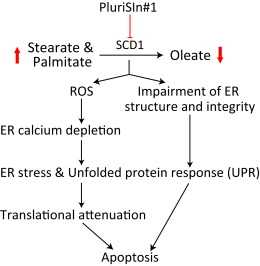
PluriSIn-1 (NSC 14613) is a small molecule compound designed to improve the safety of induced pluripotent stem cell (iPS) transplantation. Its core function is to selectively eliminate Nanog-positive tumorigenic cells in iPS cell populations, thereby reducing the risk of tumor formation after transplantation [1]. The mechanism of action of PluriSIn-1 is based on the high dependence of Nanog-positive tumorigenic cells on SCD-mediated monounsaturated fatty acid synthesis. By inhibiting SCD activity, PluriSIn-1 disrupts the balance of intracellular fatty acids (reducing monounsaturated fatty acids and accumulating saturated fatty acids), thereby specifically triggering endoplasmic reticulum stress and caspase-dependent apoptosis pathways in Nanog-positive cells [1]. In the field of myocardial transplantation, PluriSIn-1 not only reduces the tumorigenic risk of iPS cell transplantation, but also does not damage normal myocardial tissue or affect the survival and function of non-tumorigenic iPS-derived cardiomyocytes. This makes it a potential safe adjuvant for cell therapy in cardiovascular diseases [1]. Nanog is a key transcription factor that maintains the pluripotency and tumorigenicity of stem cells. PluriSIn-1 selectively eliminates Nanog-positive cells, avoiding the non-specific toxicity of traditional chemotherapy drugs, and provides a targeted strategy to improve the safety of stem cell transplantation [1].
|
| Molecular Formula |
C12H11N3O
|
|
|---|---|---|
| Molecular Weight |
213.24
|
|
| Exact Mass |
213.09
|
|
| CAS # |
91396-88-2
|
|
| Related CAS # |
|
|
| PubChem CID |
225362
|
|
| Appearance |
Light yellow to khaki solid powder
|
|
| Density |
1.3±0.1 g/cm3
|
|
| Boiling Point |
335.7±15.0 °C at 760 mmHg
|
|
| Melting Point |
177-178℃
|
|
| Flash Point |
156.9±20.4 °C
|
|
| Vapour Pressure |
0.0±0.7 mmHg at 25°C
|
|
| Index of Refraction |
1.655
|
|
| LogP |
1.34
|
|
| Hydrogen Bond Donor Count |
2
|
|
| Hydrogen Bond Acceptor Count |
3
|
|
| Rotatable Bond Count |
3
|
|
| Heavy Atom Count |
16
|
|
| Complexity |
220
|
|
| Defined Atom Stereocenter Count |
0
|
|
| SMILES |
O=C(C1=CC=NC=C1)NNC2=CC=CC=C2
|
|
| InChi Key |
HUDWXDLBWRHCKO-UHFFFAOYSA-N
|
|
| InChi Code |
InChI=1S/C12H11N3O/c16-12(10-6-8-13-9-7-10)15-14-11-4-2-1-3-5-11/h1-9,14H,(H,15,16)
|
|
| Chemical Name |
N-phenylisonicotinohydrazide
|
|
| Synonyms |
|
|
| HS Tariff Code |
2934.99.9001
|
|
| Storage |
Powder -20°C 3 years 4°C 2 years In solvent -80°C 6 months -20°C 1 month |
|
| Shipping Condition |
Room temperature (This product is stable at ambient temperature for a few days during ordinary shipping and time spent in Customs)
|
| Solubility (In Vitro) |
DMSO: 43 mg/mL (201.7 mM)
Water: 1 mg/mL (Insoluble) Ethanol: 1 mg/mL |
|---|---|
| Solubility (In Vivo) |
Solubility in Formulation 1: ≥ 2.5 mg/mL (11.72 mM) (saturation unknown) in 10% DMSO + 40% PEG300 + 5% Tween80 + 45% Saline (add these co-solvents sequentially from left to right, and one by one), clear solution.
For example, if 1 mL of working solution is to be prepared, you can add 100 μL of 25.0 mg/mL clear DMSO stock solution to 400 μL PEG300 and mix evenly; then add 50 μL Tween-80 to the above solution and mix evenly; then add 450 μL normal saline to adjust the volume to 1 mL. Preparation of saline: Dissolve 0.9 g of sodium chloride in 100 mL ddH₂ O to obtain a clear solution. Solubility in Formulation 2: ≥ 2.5 mg/mL (11.72 mM) (saturation unknown) in 10% DMSO + 90% (20% SBE-β-CD in Saline) (add these co-solvents sequentially from left to right, and one by one), clear solution. For example, if 1 mL of working solution is to be prepared, you can add 100 μL of 25.0 mg/mL clear DMSO stock solution to 900 μL of 20% SBE-β-CD physiological saline solution and mix evenly. Preparation of 20% SBE-β-CD in Saline (4°C,1 week): Dissolve 2 g SBE-β-CD in 10 mL saline to obtain a clear solution. (Please use freshly prepared in vivo formulations for optimal results.) |
| Preparing Stock Solutions | 1 mg | 5 mg | 10 mg | |
| 1 mM | 4.6896 mL | 23.4478 mL | 46.8955 mL | |
| 5 mM | 0.9379 mL | 4.6896 mL | 9.3791 mL | |
| 10 mM | 0.4690 mL | 2.3448 mL | 4.6896 mL |
*Note: Please select an appropriate solvent for the preparation of stock solution based on your experiment needs. For most products, DMSO can be used for preparing stock solutions (e.g. 5 mM, 10 mM, or 20 mM concentration); some products with high aqueous solubility may be dissolved in water directly. Solubility information is available at the above Solubility Data section. Once the stock solution is prepared, aliquot it to routine usage volumes and store at -20°C or -80°C. Avoid repeated freeze and thaw cycles.
Calculation results
Working concentration: mg/mL;
Method for preparing DMSO stock solution: mg drug pre-dissolved in μL DMSO (stock solution concentration mg/mL). Please contact us first if the concentration exceeds the DMSO solubility of the batch of drug.
Method for preparing in vivo formulation::Take μL DMSO stock solution, next add μL PEG300, mix and clarify, next addμL Tween 80, mix and clarify, next add μL ddH2O,mix and clarify.
(1) Please be sure that the solution is clear before the addition of next solvent. Dissolution methods like vortex, ultrasound or warming and heat may be used to aid dissolving.
(2) Be sure to add the solvent(s) in order.
|
|---|
|
|
|
|---|
|
|
Products are for research use only; We do not sell to patients
Copyright 2020 InvivoChem LLC | All Rights Reserved


 COA
COA