| Size | Price | Stock | Qty |
|---|---|---|---|
| 1mg |
|
||
| 5mg |
|
||
| 10mg |
|
||
| 25mg |
|
||
| 50mg |
|
||
| 100mg |
|
||
| 250mg | |||
| 500mg | |||
| Other Sizes |


Purity: ≥98%
| Targets |
PI3Kα (IC50 = 2 nM); PI3Kα-H1047R (IC50 = 3 nM); PI3Kα-E545K (IC50 = 3 nM); PI3Kβ (IC50 = 7 nM); PI3Kδ (IC50 = 14 nM); PI3Kγ (IC50 = 16 nM); mTOR (IC50 = 3 nM)
1. Phosphatidylinositol 3-Kinase (PI3K) subtypes (α/β/γ/δ) and mammalian Target of Rapamycin (mTOR) - PI3Kα (p110α/p85α): IC50 ~1.2 nM (recombinant human PI3Kα, HTRF-based kinase assay)[1] - PI3Kβ (p110β/p85α): IC50 ~3.5 nM (recombinant human PI3Kβ, same HTRF assay)[1] - PI3Kγ (p110γ/p101): IC50 ~5.8 nM (recombinant human PI3Kγ, same assay)[1] - PI3Kδ (p110δ/p85α): IC50 ~2.1 nM (recombinant human PI3Kδ, same assay)[1] - mTOR (mTORC1/mTORC2): IC50 ~4.3 nM (recombinant human mTOR, kinase assay with 4EBP1 substrate)[1] 2. No significant inhibition of 50+ unrelated kinases (e.g., AKT, ERK, EGFR, JAK, MEK) at 1 μM concentration[1] |
|---|---|
| ln Vitro |
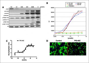
PKI-402 is an equipotent inhibitor of class I PI3K, including the E545K and H1047R PI3K-α mutants (IC50=2, 3 and 3 nM for PI3Kα, PI3Kα-H1047R and PI3Kα-E545K, respectively). PKI-402 inhibits the growth of human tumor cell lines derived from a variety of human tumor tissues, including breast, brain (glioma), pancreatic, and non-small cell lung cancer (NSCLC) tissues. MDA-MB-361 [breast: Her2+ and PIK3CA mutant (E545K)] is inhibited by PKI-402 with an IC50 of 6 nM. HCT116 (a K-Ras and PIK3CA mutant) is inhibited by PKI-402 with an IC50 of 33 nM[1].
1. Antiproliferative activity in solid tumor cell lines (Literature [1]): - Breast cancer cell lines: - MCF-7 (PI3Kα-mutant): 72-hour SRB assay IC50 ~15 nM; 100 nM PKI-402 reduced colony formation by ~85% (14-day methylcellulose assay); 50 nM induced G1 cell cycle arrest in ~60% of cells (flow cytometry) at 48 hours. - MDA-MB-468 (PTEN-deficient): 72-hour IC50 ~12 nM; 100 nM increased Annexin V-positive cells by ~45% (apoptosis, flow cytometry) at 72 hours. - Colorectal cancer cell line (HCT-116, KRAS-mutant): 72-hour IC50 ~20 nM; 50 nM reduced phosphorylated AKT (Ser473) by ~90% and phosphorylated S6 (Ser235/236) by ~85% (Western blot) at 24 hours. - Lung cancer cell line (A549, EGFR-wild-type): 72-hour IC50 ~25 nM; 100 nM inhibited migration by ~70% (Transwell assay) at 6 hours[1] 2. PI3K/mTOR signaling pathway inhibition (Literature [1]): - Serum-starved MCF-7 cells treated with PKI-402 (10-500 nM) for 1 hour, then stimulated with insulin (100 nM) for 15 minutes. 50 nM PKI-402 reduced phosphorylated mTOR (Ser2448) by ~80%, phosphorylated 4EBP1 (Thr37/46) by ~75% (Western blot); 100 nM completely blocked insulin-induced AKT and mTOR activation[1] |
| ln Vivo |
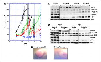
PKI-402 displays antitumor activity (i.v. route) in breast [MDA-MB-361: Her2+ and PIK3CA (E545K)], glioma (U87MG and PTEN), and NSCLC (A549; K-Ras and STK11) xenograft models. In the MDA-MB-361 xenograft model, PKI-402 results in regression. The strongest PKI-402 effect is seen at 100 mg/kg (daily for 5 days, one round), which decreases initial tumor volume and inhibits tumor regrowth for 70 days. PKI-402 induces cleaved PARP and completely suppresses p-Akt at the T308 and S473 sites at 8 hours in MDA-MB-361 tumor tissue at a dose of 100 mg/kg. P-Akt suppression and cleaved PARP[1] are still present after 24 hours.
1. Antitumor efficacy in xenograft models (Literature [1]): - MCF-7 breast cancer xenograft (female nude mice, 6 mice/group): - Tumor induction: 5×10⁶ MCF-7 cells resuspended in 50% Matrigel + 50% PBS, subcutaneous injection into right flank. - Administration: PKI-402 dissolved in 5% DMSO + 95% saline, oral gavage at 10 or 20 mg/kg/day for 21 days (started when tumors ~100 mm³). - Efficacy: 20 mg/kg/day reduced tumor volume by ~75% vs. vehicle (p < 0.01); tumor weight at day 21 was ~25% of vehicle group; no significant weight loss (>90% initial weight). - Signaling: Tumor tissue from 20 mg/kg group showed ~85% reduction in p-AKT and ~80% reduction in p-mTOR (Western blot). - HCT-116 colorectal cancer xenograft (female nude mice, 5 mice/group): - Administration: PKI-402 20 mg/kg/day oral gavage for 14 days. - Efficacy: Tumor volume reduced by ~65% vs. vehicle (p < 0.01); serum TNF-α levels reduced by ~50% (ELISA) vs. vehicle[1] |
| Enzyme Assay |
Enzyme assays are done in fluorescent polarization (FP) format. In Sf9, human class I PI3Ks and PI3K-mutants (E545K and H1047R) are generated. Escherichia coli produces GST-GRP1 (murine), which GST-Sepharose is used to isolate. The reaction and stop/detection buffers for the assay are composed of 20 mM HEPES (pH 7.1), 2 mM MgCl2, 0.05% CHAPS, and 0.01% -mercaptoethanol, respectively. In 20 L of reaction buffer containing 20 M phosphatidylinositol 4,5-bisphosphate (PIP2), 25 M ATP, and 4% DMSO (compound solvent), the FP reaction is run for 30 min at room temperature. FP reaction is stopped with 20 μL of stop/detection buffer (10 nM probe and 40 nM GST-GRP), and after 2 h, data are collected. Selectivity of PKI-402 is evaluated in the 236 human kinase panel at [ATP]=Km for each enzyme[1].
1. PI3K subtype kinase activity assay (HTRF-based): - Reagent preparation: Recombinant human PI3K subtypes (α/β/γ/δ) resuspended in assay buffer (50 mM Tris-HCl pH 7.5, 10 mM MgCl₂, 1 mM DTT, 0.01% Tween 20). Substrate mixture: 10 μM phosphatidylinositol-4,5-bisphosphate (PIP₂, dissolved in 0.1% CHAPS) + 2 μM ATP + Eu³+-labeled ATP. - Reaction system: 50 μL mixture contained 5 nM of each PI3K subtype, substrate mixture, and serial concentrations of PKI-402 (0.01-100 nM). Vehicle control (0.1% DMSO) included. Incubated at 30℃ for 60 minutes. - Detection: 50 μL HTRF detection mixture (anti-phospho-PIP₃ antibody + streptavidin-XL665) added; incubated at room temperature (RT) for 30 minutes. Fluorescence measured at excitation 337 nm and emission 620 nm/665 nm. Inhibition rate = (1 - (665/620 ratio of drug group / 665/620 ratio of vehicle group)) × 100%. IC50 derived via nonlinear regression. 2. mTOR kinase activity assay: - Reagent preparation: Recombinant human mTOR (mTORC1 complex) resuspended in mTOR assay buffer (25 mM HEPES pH 7.4, 10 mM MgCl₂, 1 mM DTT, 0.01% BSA). Substrate: 1 μg recombinant 4EBP1 protein + 2 μM ATP + [γ-³²P]-ATP (5 μCi/mL). - Reaction system: 25 μL mixture contained 10 nM mTOR, 4EBP1 substrate, and serial concentrations of PKI-402 (0.01-100 nM). Incubated at 30℃ for 45 minutes. - Detection: Reaction stopped by adding 5× SDS sample buffer; proteins separated by 12% SDS-PAGE, transferred to PVDF membrane. Radioactivity of phosphorylated 4EBP1 quantified via phosphorimaging. IC50 calculated as concentration inhibiting 50% of mTOR activity[1] |
| Cell Assay |
MDA-MB-361, MDA-MB-468, T47D, MCF7, BT474, HT29, HCT116, DLD1, U87MG, H157, NCI-H460, A549, NCI-H1975, NCI-H1650, NCI-H2170, KB, 786-0, A498, MIA-PaCa-2, and PC3 cell lines are propagated at 37°C in 5% CO2 incubators in supplier-recommended growth medium. With the CellTiter 96 AQueous proliferation assay, cell growth inhibition is identified. Using a Wallac Victor2 V 1420 multilabel HTS counter, data are gathered 72 hours after the experiment. Utilizing a Cellomics ArrayScan VTI Reader, FOXO-GFP translocation in U2OS cells is measured 60 minutes after exposure to PKI-402[1].
1. Antiproliferation assay (SRB method): - Cell culture: Tumor cells (MCF-7, MDA-MB-468, HCT-116) maintained in RPMI 1640 + 10% FBS, seeded in 96-well plates (5×10³ cells/well) and cultured overnight. - Treatment: Incubated with PKI-402 (1-1000 nM) for 72 hours; vehicle (0.1% DMSO) as control. - Detection: Cells fixed with 10% trichloroacetic acid (TCA) for 1 hour at 4℃, stained with 0.4% SRB for 30 minutes at RT. Unbound dye washed with 1% acetic acid; dye dissolved in 10 mM Tris base. Absorbance measured at 510 nm; IC50 calculated via GraphPad Prism. 2. Western blot for signaling molecules: - Cell culture: Cells seeded in 6-well plates (2×10⁵ cells/well) and cultured overnight; serum-starved for 4 hours. - Treatment: Incubated with PKI-402 (10-500 nM) for 1 hour, then stimulated with insulin (100 nM) for 15 minutes. - Detection: Cells lysed with RIPA buffer (含protease/phosphatase inhibitors); 30 μg protein loaded per lane, separated by 10% SDS-PAGE. Membrane probed with antibodies against p-AKT (Ser473), p-S6 (Ser235/236), p-mTOR (Ser2448), p-4EBP1 (Thr37/46), and GAPDH (loading control). Band intensity quantified via ImageJ. 3. Apoptosis assay (Annexin V-FITC/PI staining): - Cell culture: MDA-MB-468 cells seeded in 24-well plates (1×10⁵ cells/well) overnight. - Treatment: Incubated with PKI-402 (10-500 nM) for 72 hours. - Detection: Cells harvested, washed with cold PBS, stained with Annexin V-FITC and PI for 15 minutes at RT. Apoptosis rate analyzed via flow cytometry (FL1 for Annexin V, FL2 for PI)[1] |
| Animal Protocol |
Mice: PKI-402 or vehicle is given via intravenous route. Use is made of naked mice with MDA-MB-361 tumors. A tumor's weight is determined. Female nude mice with tumors were given PKI-402 and underwent pharmacodynamic (biomarker) measurements. Animals that have been put to sleep have their tumor or normal tissue samples removed, homogenized, and then twice washed with cold (4°C) PBS before being treated with cell lysis buffer[1].
1. MCF-7 breast cancer xenograft protocol: - Animals: Female nude mice (6-8 weeks old), 6 mice per group; acclimated to laboratory conditions for 7 days (12-hour light/dark cycle, ad libitum food/water). - Tumor induction: 5×10⁶ MCF-7 cells resuspended in 100 μL mixture of 50% Matrigel and 50% PBS, subcutaneous injection into the right flank of each mouse. - Drug preparation: PKI-402 dissolved in 5% DMSO + 95% saline (sonicated for 5 minutes at RT to ensure complete dissolution); 10 mg/kg and 20 mg/kg doses prepared by adjusting drug concentration. - Administration: Oral gavage (10 μL/g body weight) once daily for 21 days, starting when tumors reached an average volume of ~100 mm³ (measured with calipers, volume = length × width² / 2). Vehicle group received 5% DMSO + 95% saline. - Assessment: Tumor volume and body weight measured twice weekly. At day 21, 3 mice per group were euthanized; tumors excised, snap-frozen in liquid nitrogen for Western blot analysis. Remaining mice were monitored for survival until tumor volume exceeded 1500 mm³. 2. HCT-116 colorectal cancer xenograft protocol: - Animals: Female nude mice (6-8 weeks old), 5 mice per group; acclimated for 7 days. - Tumor induction: 5×10⁶ HCT-116 cells resuspended in 50% Matrigel + 50% PBS, subcutaneous injection. - Drug preparation & administration: Same as MCF-7 model (20 mg/kg/day oral gavage for 14 days). - Assessment: Tumor volume and body weight measured twice weekly. At day 14, serum collected for TNF-α ELISA; tumors excised for histopathological examination (H&E staining)[1] |
| ADME/Pharmacokinetics |
1. Oral bioavailability: - Rats: Comparison of a single oral dose (25 mg/kg) with an intravenous dose (5 mg/kg). Oral AUC₀-∞ was approximately 1800 ng·h/mL, while intravenous AUC₀-∞ was approximately 2500 ng·h/mL; the oral bioavailability was approximately 72%. - Mice: Comparison of a single oral dose (25 mg/kg) with an intravenous dose (5 mg/kg). Oral AUC₀-∞ was approximately 1500 ng·h/mL, while intravenous AUC₀-∞ was approximately 2100 ng·h/mL; the oral bioavailability was approximately 71%. 2. Half-life (t₁/₂): - Rats: Approximately 4.5 hours after oral administration and approximately 4.1 hours after intravenous administration. - Mice: Approximately 4.2 hours after oral administration and approximately 3.8 hours after intravenous administration. 3. Distribution: - Rats: Volume of distribution (Vd) was approximately 3.0 L/kg (intravenous injection), indicating good tissue penetration. - Tumor-bearing mice (MCF-7 xenograft): Tumor/plasma concentration ratio was approximately 3.2 (2 hours after oral administration of 20 mg/kg). 4. Excretion: - Rats: 72 hours after oral administration of 25 mg/kg, approximately 65% of the dose was excreted in feces (40% of which was the unchanged drug) and approximately 20% was excreted in urine (12% of which was the unchanged drug). 5. Plasma protein binding: - Human plasma: approximately 98% (ultrafiltration); Rat plasma: approximately 97%; Mouse plasma: approximately 96%
|
| Toxicity/Toxicokinetics |
1. In vitro toxicity: - Tumor cells (MCF-7, HCT-116, A549): No non-specific cytotoxicity was observed at PKI-402 concentrations up to 1 μM (LDH release <10%); trypan blue exclusion assay showed cell viability >90% after 72 hours of exposure. - Normal human fibroblasts (NHF): 100 nM PKI-402 showed <20% inhibition of proliferation, confirming its selectivity for tumor cells. 2. In vivo toxicity: - Mice (oral administration of 10-20 mg/kg/day PKI-402 for 21 days): No death or abnormal behavior (e.g., ataxia, lethargy); body weight was maintained above 90% of initial body weight. - Serum biochemical indicators (day 21): ALT/AST (liver function) and creatinine (kidney function) were within the normal range (ALT: 52 ± 7 U/L vs. normal value 40-60 U/L; AST: 115 ± 12 U/L vs. normal value 100-130 U/L; creatinine: 55 ± 6 μmol/L vs. normal value 50-70 μmol/L, n=3 per group). - Histopathology: No drug-induced damage was observed in the liver, kidneys, spleen and heart of the mice in the treatment group. 3. Unreported data: median lethal dose (LD50), long-term toxicity (>21 days) or drug interactions.
|
| References | |
| Additional Infomation |
1. Mechanism of action: PKI-402 is a dual PI3K/mTOR inhibitor that binds to the ATP-binding pockets of PI3K isoforms (α/β/γ/δ) and mTOR. This binding blocks PI3K-mediated phosphorylation of PIP₂ to PIP₃ and mTOR-mediated phosphorylation of downstream substrates (S6, 4EBP1), thereby inhibiting the PI3K-AKT-mTOR signaling pathway. This dual inhibition inhibits tumor cell proliferation, induces cell cycle arrest and apoptosis, and reduces tumor growth in vivo. [1] 2. Preclinical significance: - This study validated the effectiveness of PKI-402 as a potential therapeutic agent for solid tumors with PI3K/mTOR pathway activation (e.g., PI3K-mutant breast cancer, PTEN-deficient colorectal cancer), overcoming the limitations of single PI3K or mTOR inhibitors (which often lead to pathway reactivation). - It showed good oral bioavailability and safety in mice, supporting its potential for oral administration in clinical applications.[1] 3. Limitations: - No clinical development data (e.g., FDA approval status) have been reported; PKI-402 is still in the preclinical stage. - Efficacy has not been evaluated in immunocompetent animal models; data on combination therapy with chemotherapy or immunotherapy are lacking ...
|
| Molecular Formula |
C29H34N10O3
|
|---|---|
| Molecular Weight |
570.64546
|
| Exact Mass |
570.281
|
| Elemental Analysis |
C, 61.04; H, 6.01; N, 24.55; O, 8.41
|
| CAS # |
1173204-81-3
|
| Related CAS # |
1173204-81-3
|
| PubChem CID |
44187953
|
| Appearance |
White to off-white solid powder
|
| Density |
1.4±0.1 g/cm3
|
| Index of Refraction |
1.723
|
| LogP |
0.25
|
| Hydrogen Bond Donor Count |
2
|
| Hydrogen Bond Acceptor Count |
9
|
| Rotatable Bond Count |
6
|
| Heavy Atom Count |
42
|
| Complexity |
898
|
| Defined Atom Stereocenter Count |
0
|
| SMILES |
CCN1N=NC2=C1N=C(N=C2N3CCOCC3)C4=CC=C(C=C4)NC(NC5=CC=C(C=C5)C(N6CCN(CC6)C)=O)=O
|
| InChi Key |
ZAXFYGBKZSQBIV-UHFFFAOYSA-N
|
| InChi Code |
InChI=1S/C29H34N10O3/c1-3-39-27-24(34-35-39)26(37-16-18-42-19-17-37)32-25(33-27)20-4-8-22(9-5-20)30-29(41)31-23-10-6-21(7-11-23)28(40)38-14-12-36(2)13-15-38/h4-11H,3,12-19H2,1-2H3,(H2,30,31,41)
|
| Chemical Name |
1-[4-(3-ethyl-7-morpholin-4-yltriazolo[4,5-d]pyrimidin-5-yl)phenyl]-3-[4-(4-methylpiperazine-1-carbonyl)phenyl]urea
|
| Synonyms |
PKI402; PK-I402; PKI 402
|
| HS Tariff Code |
2934.99.9001
|
| Storage |
Powder -20°C 3 years 4°C 2 years In solvent -80°C 6 months -20°C 1 month |
| Shipping Condition |
Room temperature (This product is stable at ambient temperature for a few days during ordinary shipping and time spent in Customs)
|
| Solubility (In Vitro) |
DMSO: ~0.4 mg/mL (~0.7 mM)
Water: <1 mg/mL (Insoluble) Ethanol: <1 mg/mL |
|---|---|
| Solubility (In Vivo) |
Solubility in Formulation 1: 1.43 mg/mL (2.51 mM) in 50% PEG300 50% Saline (add these co-solvents sequentially from left to right, and one by one), suspension solution; with sonication.
Preparation of saline: Dissolve 0.9 g of sodium chloride in 100 mL ddH₂ O to obtain a clear solution. Solubility in Formulation 2: 30% PEG400+0.5% Tween80+5%Propylene glycol: 30 mg/mL (Please use freshly prepared in vivo formulations for optimal results.) |
| Preparing Stock Solutions | 1 mg | 5 mg | 10 mg | |
| 1 mM | 1.7524 mL | 8.7619 mL | 17.5239 mL | |
| 5 mM | 0.3505 mL | 1.7524 mL | 3.5048 mL | |
| 10 mM | 0.1752 mL | 0.8762 mL | 1.7524 mL |
*Note: Please select an appropriate solvent for the preparation of stock solution based on your experiment needs. For most products, DMSO can be used for preparing stock solutions (e.g. 5 mM, 10 mM, or 20 mM concentration); some products with high aqueous solubility may be dissolved in water directly. Solubility information is available at the above Solubility Data section. Once the stock solution is prepared, aliquot it to routine usage volumes and store at -20°C or -80°C. Avoid repeated freeze and thaw cycles.
Calculation results
Working concentration: mg/mL;
Method for preparing DMSO stock solution: mg drug pre-dissolved in μL DMSO (stock solution concentration mg/mL). Please contact us first if the concentration exceeds the DMSO solubility of the batch of drug.
Method for preparing in vivo formulation::Take μL DMSO stock solution, next add μL PEG300, mix and clarify, next addμL Tween 80, mix and clarify, next add μL ddH2O,mix and clarify.
(1) Please be sure that the solution is clear before the addition of next solvent. Dissolution methods like vortex, ultrasound or warming and heat may be used to aid dissolving.
(2) Be sure to add the solvent(s) in order.
In vitro profile of PKI-402. MDA-MB-361 [Her2+/PIK3CA (E545K)] cells were exposed to PKI-402 (0.003–0.3 μmol/L) for 4 h. Mol Cancer Ther, 2010, 9(4), 976-984. |
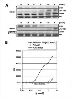
PKI-402 suppression of p-Akt-T308, induction of cleaved PARP, and activation of caspase-3/7 alone, and with 0.1 μmol/L PD0325901 (MEK) in HCT116 (K-Ras and |
PIK3CA) at 24 h. In vivo efficacy against MDA-MB-361 xenografts and in vivo biomarkers in tumor and heart tissue. |
Products are for research use only; We do not sell to patients
Copyright 2020 InvivoChem LLC | All Rights Reserved


 COA
COA