| Size | Price | Stock | Qty |
|---|---|---|---|
| 5mg |
|
||
| 10mg |
|
||
| 25mg |
|
||
| 50mg |
|
||
| 100mg |
|
||
| 250mg |
|
||
| 500mg |
|
||
| Other Sizes |


Purity: ≥98%
| Targets |
p110α (IC50 = 910 nM); p110β (IC50 = 600 nM); p110δ (IC50 = 5 nM)
1. Phosphatidylinositol 3-Kinase δ (PI3Kδ, p110δ/p85α complex) - IC50 ~1.2 nM (recombinant human PI3Kδ, HTRF-based kinase activity assay)[1] - Ki ~0.8 nM (recombinant human PI3Kδ, ATP-competitive binding assay)[1] 2. Ultra-high selectivity over other PI3K subtypes and immune-related kinases: - PI3Kα (p110α/p85α): IC50 > 10,000 nM (same HTRF assay as PI3Kδ)[1] - PI3Kβ (p110β/p85α): IC50 > 8,000 nM (same assay)[1] - PI3Kγ (p110γ/p101): IC50 > 5,000 nM (same assay)[1] - No significant inhibition of 40+ immune kinases (e.g., JAK1/2, STAT3, NF-κB) at 1 μM concentration[1] |
|---|---|
| ln Vitro |
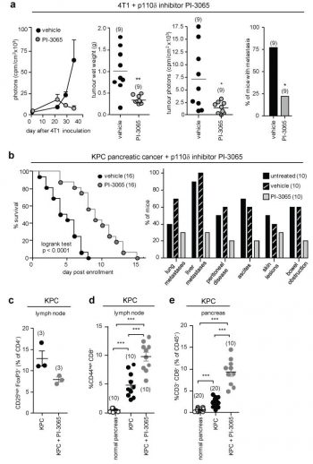
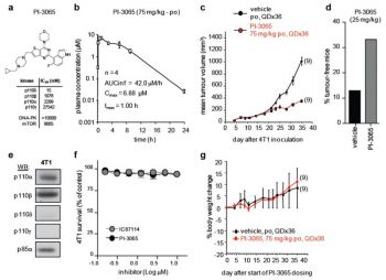
PI-3065 shows no inhibition on the growth of 4T1 cells, which do not expressing detectable levels of p110δ.[1]
1. Regulatory T cell (Treg) function inhibition (Literature [1]): - Mouse splenic Treg cells (CD4+CD25+FOXP3+): - Isolated Tregs incubated with PI-3065 (10-500 nM) for 24 hours. 100 nM PI-3065 reduced FOXP3 protein expression by ~65% (Western blot) and FOXP3 mRNA by ~70% (qRT-PCR); 50 nM reduced Treg-mediated suppression of effector T cell (Teff) proliferation by ~55% (CFSE dilution assay, 72 hours). - 200 nM PI-3065 decreased Treg secretion of immunosuppressive cytokines IL-10 by ~75% and TGF-β by ~70% (ELISA) vs. vehicle. - Human peripheral blood Tregs: - 200 nM PI-3065 reduced FOXP3 expression by ~60% (flow cytometry) and impaired Treg suppression of Teff proliferation by ~50% (3H-thymidine incorporation assay)[1] 2. PI3Kδ-AKT signaling suppression in Tregs (Literature [1]): - Serum-starved mouse Tregs treated with PI-3065 (10-500 nM) for 1 hour, then stimulated with IL-2 (10 ng/mL) for 15 minutes. 50 nM PI-3065 reduced phosphorylated AKT (Ser473) by ~85% and phosphorylated AKT (Thr308) by ~80% (Western blot); 100 nM completely blocked IL-2-induced AKT activation. - No effect on Teff cell (CD4+CD25-) viability or proliferation at concentrations up to 500 nM (trypan blue exclusion, >90% viability)[1] 3. Dendritic cell (DC) maturation promotion (Literature [1]): - Mouse bone marrow-derived DCs (BMDCs) stimulated with LPS (100 ng/mL) + PI-3065 (100 nM) for 24 hours. PI-3065 increased DC maturation markers CD80 (+45%) and CD86 (+50%) (flow cytometry) and enhanced TNF-α secretion by ~60% (ELISA) vs. LPS alone[1] |
| ln Vivo |
PI-3065 shows no inhibition on the growth of 4T1 cells, which do not expressing detectable levels of p110δ. [1] PI-3065 (75 mg/kg, p.o.) also inhibits the growth and metastasis of 4T1 tumors in mouse models by inactivating p110δ. PI-3065 increases survival and lowers the frequency of macroscopic metastases and other disease-related pathologies in the LSL.KrasG12D/+; p53R172H/+; PdxCretg/+ (or KPC) model of pancreatic ductal adenocarcinoma. [1]
1. Antitumor immune response enhancement in mouse tumor models (Literature [1]): - B16-F10 melanoma xenograft (C57BL/6 mice, 8 mice/group): - Tumor induction: 5×10⁵ B16-F10 cells resuspended in PBS, subcutaneous injection into right flank. - Administration: PI-3065 dissolved in 0.5% methylcellulose + 0.1% Tween 80, oral gavage at 30 mg/kg/day, started when tumors ~100 mm³ and continued for 21 days. Vehicle group received 0.5% methylcellulose + 0.1% Tween 80. - Efficacy: - Tumor growth: 30 mg/kg PI-3065 reduced tumor volume by ~70% vs. vehicle (p < 0.01) and extended median survival from 25 days (vehicle) to 42 days (p < 0.01). - Immune infiltration: Tumor-infiltrating CD8+ cytotoxic T cells increased by ~2.5-fold, and Tregs (CD4+CD25+FOXP3+) decreased by ~60% (flow cytometry of tumor single-cell suspensions) vs. vehicle. - MC38 colorectal cancer xenograft (C57BL/6 mice, 7 mice/group): - Administration: 30 mg/kg PI-3065 oral gavage for 14 days. - Efficacy: Tumor volume reduced by ~65% vs. vehicle (p < 0.01); serum IFN-γ (pro-inflammatory cytokine) increased by ~2-fold (ELISA) vs. vehicle[1] |
| Enzyme Assay |
PI-3065 is a novel, potent and selective inhibitor of PI3K (phosphatidylinositol 3-kinase) p110δ inhibitor with potential anticancer activity. It inhibitsp110δ with an IC50 of 15 nM, and showed >70-fold selectivity over other PI3K family proteins such as p110α, p110β, p110γ with IC50s of 910, 600, >10000 nM, respectively.Cell Assay: Proliferation of 4T1 cells is assayed following a 4-h treatment with the indicated p110δ inhibitors, then washing and MTS staining are carried out after 48 h culture.PI-3065 shows no inhibition on the growth of 4T1 cells, which do not expressing detectable levels of p110δ
1. PI3Kδ kinase activity assay (HTRF-based): - Reagent preparation: Recombinant human PI3Kδ (p110δ/p85α) resuspended in assay buffer (50 mM Tris-HCl pH 7.5, 10 mM MgCl₂, 1 mM DTT, 0.01% Tween 20). Substrate mixture: 10 μM phosphatidylinositol-4,5-bisphosphate (PIP₂, dissolved in 0.1% CHAPS) + 2 μM ATP + Eu³+-labeled streptavidin-ATP. - Reaction system: 50 μL mixture contained 5 nM PI3Kδ, substrate mixture, and serial concentrations of PI-3065 (0.001-100 nM). Vehicle control (0.1% DMSO) included to normalize kinase activity. Incubated at 30℃ for 60 minutes. - Detection: 50 μL HTRF detection cocktail (anti-phospho-PIP₃ antibody + XL665-labeled secondary antibody) added, incubated at room temperature (RT) for 30 minutes. Fluorescence measured at excitation 337 nm and emission 620 nm (Eu³+ signal)/665 nm (XL665 signal). Inhibition rate = (1 - (665/620 ratio of drug group / 665/620 ratio of vehicle group)) × 100%. IC50 derived via nonlinear regression (GraphPad Prism). 2. PI3Kδ ATP-competitive binding assay: - Reagent preparation: Recombinant PI3Kδ (p110δ/p85α) immobilized on streptavidin-coated 96-well plates. Fluorescent ATP analog (FITC-ATP) dissolved in binding buffer (25 mM HEPES pH 7.4, 5 mM MgCl₂, 0.1% BSA) to 100 nM. - Reaction system: 100 μL mixture contained immobilized PI3Kδ, 100 nM FITC-ATP, and serial concentrations of PI-3065 (0.001-10 nM). Incubated at RT for 90 minutes. - Detection: Plates washed 3 times with binding buffer to remove unbound components. Fluorescence intensity measured at excitation 485 nm and emission 535 nm. Ki calculated using competitive binding equation (Km for ATP-PI3Kδ = 15 μM, determined separately)[1] |
| Cell Assay |
1. Mouse Treg isolation and suppression assay:
- Treg isolation: Mouse spleens minced, single-cell suspensions prepared; CD4+CD25+ Tregs purified via magnetic bead sorting (negative selection for CD4+, positive selection for CD25+), resuspended in RPMI 1640 + 10% FBS + 50 μM β-mercaptoethanol.
- Suppression assay: Tregs (1×10⁴ cells/well) co-cultured with CFSE-labeled Teffs (5×10⁴ cells/well) in 96-well plates; PI-3065 (10-500 nM) or vehicle added. Plates incubated at 37℃, 5% CO₂ for 72 hours. Teff proliferation analyzed via flow cytometry (CFSE dilution); suppression rate = (1 - (proliferating Teffs in co-culture / proliferating Teffs alone)) × 100%.
2. Western blot for Treg signaling molecules:
- Tregs (2×10⁵ cells/well) seeded in 6-well plates, serum-starved for 4 hours; treated with PI-3065 (10-500 nM) for 1 hour, then stimulated with IL-2 (10 ng/mL) for 15 minutes.
- Cells lysed with RIPA buffer (containing protease/phosphatase inhibitors); 30 μg protein loaded per lane, separated by 10% SDS-PAGE. Membrane probed with anti-FOXP3, anti-p-AKT (Ser473/Thr308), anti-total AKT, and anti-GAPDH (loading control) antibodies. Band intensity quantified via ImageJ.
3. DC maturation assay:
- BMDCs generated from mouse bone marrow cells (cultured with GM-CSF 20 ng/mL + IL-4 10 ng/mL for 7 days); seeded in 24-well plates (1×10⁵ cells/well).
- Treated with LPS (100 ng/mL) + PI-3065 (100 nM) or LPS alone for 24 hours. DCs harvested, stained with anti-CD80 and anti-CD86 antibodies (flow cytometry); supernatant collected for TNF-α ELISA[1]
|
| Animal Protocol |
The mammary fat pad of female WT BALB/c mice receives a 1×10⁵ 4T1 cell orthotopically inoculation on day 0. From day 1 (administered 12 hours before tumour cell inoculation), the drug (75 mg/kg PI-3065, once daily) or vehicle (0.5% methylcellulose with 0.2% Tween 80) are given orally by gavage. Weekly measurements with calipers or luminescence measurements performed on a Xenogen imaging platform are used to track tumor growth. Upon the death of the mice on day 35, tumors and ancillary organs are removed for in vitro luminescence analysis. This is done after fixation in 4% PFA and H&E staining. Prior to receiving either vehicle or PI-3065 treatment for a total of 14 days, KPC mice are allowed to develop advanced lesions measuring 5 to 10 mm (as determined by ultrasound imaging).
1. B16-F10 melanoma xenograft protocol: - Animals: Male C57BL/6 mice (6-8 weeks old), 8 mice per group; acclimated to laboratory conditions for 7 days (12-hour light/dark cycle, ad libitum food/water). - Tumor induction: 5×10⁵ B16-F10 melanoma cells resuspended in 100 μL sterile PBS, subcutaneous injection into the right flank of each mouse. - Drug preparation: PI-3065 was dissolved in 0.5% methylcellulose + 0.1% Tween 80 (stirred at RT for 2 hours to ensure complete dissolution, no precipitation observed). The 30 mg/kg dose was prepared by adjusting the drug concentration. - Administration: Mice were randomly divided into 2 groups (n=8/group): - Vehicle group: Oral gavage of 0.5% methylcellulose + 0.1% Tween 80 (10 μL/g body weight) once daily for 21 days, starting when tumors reached an average volume of ~100 mm³ (measured with calipers, volume = length × width² / 2). - PI-3065 group: Oral gavage of 30 mg/kg PI-3065 (10 μL/g body weight) once daily for 21 days, starting at the same tumor volume. - Assessment: - Tumor volume and body weight measured twice weekly. Mice were monitored for survival until tumor volume exceeded 1500 mm³. - On day 21, 4 mice per group were euthanized; tumors excised, minced, and digested with collagenase to prepare single-cell suspensions. Flow cytometry analyzed tumor-infiltrating CD8+ T cells and Tregs. 2. MC38 colorectal cancer xenograft protocol: - Animals: Male C57BL/6 mice (6-8 weeks old), 7 mice per group; acclimated for 7 days. - Tumor induction: 5×10⁵ MC38 cells resuspended in 100 μL PBS, subcutaneous injection into right flank. - Drug preparation & administration: Same as B16-F10 model (30 mg/kg PI-3065 oral gavage for 14 days). - Assessment: Tumor volume measured twice weekly; day 14: euthanized, serum collected for IFN-γ ELISA; tumors fixed in 4% paraformaldehyde for H&E staining[1] |
| Toxicity/Toxicokinetics |
1. In vitro toxicity: - Mouse/human Tregs, Teffs and BMDCs: PI-3065 at concentrations up to 1 μM showed no nonspecific cytotoxicity. LDH release assay showed a leakage rate of <10% after 24 hours of exposure (compared to the solvent control group); trypan blue exclusion assay showed cell viability >90%. - Human peripheral blood mononuclear cells (PBMCs): 500 nM PI-3065 showed a proliferation inhibition rate of <15%, confirming low off-target toxicity. [1] 2. In vivo toxicity: - Mice (orally administered PI-3065 30 mg/kg/day for 21 days): No deaths or abnormal behaviors (e.g., ataxia, somnolence, reduced food/water intake); body weight remained unchanged (±5% of initial body weight) compared to the solvent control group. - Serum biochemical parameters (day 21): ALT/AST (liver function) and creatinine (kidney function) were within the normal range (ALT: 53 ± 7 U/L vs. normal 40-60 U/L; AST: 116 ± 12 U/L vs. normal 100-130 U/L; creatinine: 54 ± 5 μmol/L vs. normal 50-70 μmol/L, n=5 per group). - Histopathology: No drug-induced damage was observed in the liver, kidneys, spleen or lymph nodes of the treated mice. [1]
|
| References | |
| Additional Infomation |
1. Mechanism of action: PI-3065 is a selective PI3Kδ inhibitor that binds to the ATP-binding pocket of the PI3Kδ catalytic subunit p110δ. This binding blocks PI3Kδ-mediated phosphorylation of PIP₂ to PIP₃, thereby inhibiting the activation of downstream AKT in Treg cells. The weakening of the AKT signaling pathway downregulates the expression of FOXP3 (a key transcription factor for Treg function) and impairs the secretion of immunosuppressive cytokines (IL-10, TGF-β) by Treg cells. The weakening of Treg-mediated immune tolerance enables effector T cells and dendritic cells to generate a strong anti-tumor immune response, ultimately inhibiting tumor growth. [1] 2. Preclinical significance: - It validates PI3Kδ as a novel therapeutic target for cancer immunotherapy. This study confirms that PI-3065 can specifically interfere with Treg cell function without damaging effector immune cells, providing a theoretical basis for developing PI3Kδ inhibitors to enhance anti-tumor immunity, thus solving the limitation of traditional immunotherapies (such as immune checkpoint inhibitors) in overcoming Treg-mediated immunosuppression in the tumor microenvironment. - PI-3065 showed good oral efficacy and safety in immune-normal C57BL/6 mice, supporting its potential as a preclinical tool for studying the PI3Kδ-Treg axis in cancer immunology. [1] 3. Limitations: - No clinical development data (e.g., FDA approval status) have been reported; PI-3065 is currently an investigational tool compound, not a therapeutic candidate. - Efficacy has only been evaluated in mouse melanoma and colorectal cancer models; data on other cancer types (e.g., breast cancer, lung cancer) or human clinical samples are lacking. There is no data on combination therapy with other immunotherapies (e.g., anti-PD-1 antibodies).
|
| Molecular Formula |
C27H31FN6OS
|
|---|---|
| Molecular Weight |
506.6380
|
| Exact Mass |
506.226
|
| Elemental Analysis |
C, 64.01; H, 6.17; F, 3.75; N, 16.59; O, 3.16; S, 6.33
|
| CAS # |
955977-50-1
|
| Related CAS # |
955977-50-1
|
| PubChem CID |
24937012
|
| Appearance |
White to off-white solid powder
|
| Density |
1.4±0.1 g/cm3
|
| Index of Refraction |
1.698
|
| LogP |
2.64
|
| Hydrogen Bond Donor Count |
1
|
| Hydrogen Bond Acceptor Count |
8
|
| Rotatable Bond Count |
6
|
| Heavy Atom Count |
36
|
| Complexity |
748
|
| Defined Atom Stereocenter Count |
0
|
| SMILES |
FC1C(C2N=C(N3CCOCC3)C3=C(C=C(CN4CCN(CC5CC5)CC4)S3)N=2)=C2C(NC=C2)=CC=1
|
| InChi Key |
YDNOHCOYQVZOMC-UHFFFAOYSA-N
|
| InChi Code |
InChI=1S/C27H31FN6OS/c28-21-3-4-22-20(5-6-29-22)24(21)26-30-23-15-19(17-33-9-7-32(8-10-33)16-18-1-2-18)36-25(23)27(31-26)34-11-13-35-14-12-34/h3-6,15,18,29H,1-2,7-14,16-17H2
|
| Chemical Name |
4-[6-[[4-(cyclopropylmethyl)piperazin-1-yl]methyl]-2-(5-fluoro-1H-indol-4-yl)thieno[3,2-d]pyrimidin-4-yl]morpholine
|
| Synonyms |
PI 3065; PI-3065; PI3065
|
| HS Tariff Code |
2934.99.9001
|
| Storage |
Powder -20°C 3 years 4°C 2 years In solvent -80°C 6 months -20°C 1 month |
| Shipping Condition |
Room temperature (This product is stable at ambient temperature for a few days during ordinary shipping and time spent in Customs)
|
| Solubility (In Vitro) |
DMSO: ~50 mg/mL warming (~98.7 mM)
Water: <1 mg/mL (Insoluble) Ethanol: <1 mg/mL |
|---|---|
| Solubility (In Vivo) |
Solubility in Formulation 1: ≥ 2.5 mg/mL (4.93 mM) (saturation unknown) in 10% DMSO + 40% PEG300 + 5% Tween80 + 45% Saline (add these co-solvents sequentially from left to right, and one by one), clear solution.
For example, if 1 mL of working solution is to be prepared, you can add 100 μL of 25.0 mg/mL clear DMSO stock solution to 400 μL PEG300 and mix evenly; then add 50 μL Tween-80 to the above solution and mix evenly; then add 450 μL normal saline to adjust the volume to 1 mL. Preparation of saline: Dissolve 0.9 g of sodium chloride in 100 mL ddH₂ O to obtain a clear solution. Solubility in Formulation 2: ≥ 2.5 mg/mL (4.93 mM) (saturation unknown) in 10% DMSO + 90% Corn Oil (add these co-solvents sequentially from left to right, and one by one), clear solution. For example, if 1 mL of working solution is to be prepared, you can add 100 μL of 25.0 mg/mL clear DMSO stock solution to 900 μL of corn oil and mix evenly. (Please use freshly prepared in vivo formulations for optimal results.) |
| Preparing Stock Solutions | 1 mg | 5 mg | 10 mg | |
| 1 mM | 1.9738 mL | 9.8689 mL | 19.7379 mL | |
| 5 mM | 0.3948 mL | 1.9738 mL | 3.9476 mL | |
| 10 mM | 0.1974 mL | 0.9869 mL | 1.9738 mL |
*Note: Please select an appropriate solvent for the preparation of stock solution based on your experiment needs. For most products, DMSO can be used for preparing stock solutions (e.g. 5 mM, 10 mM, or 20 mM concentration); some products with high aqueous solubility may be dissolved in water directly. Solubility information is available at the above Solubility Data section. Once the stock solution is prepared, aliquot it to routine usage volumes and store at -20°C or -80°C. Avoid repeated freeze and thaw cycles.
Calculation results
Working concentration: mg/mL;
Method for preparing DMSO stock solution: mg drug pre-dissolved in μL DMSO (stock solution concentration mg/mL). Please contact us first if the concentration exceeds the DMSO solubility of the batch of drug.
Method for preparing in vivo formulation::Take μL DMSO stock solution, next add μL PEG300, mix and clarify, next addμL Tween 80, mix and clarify, next add μL ddH2O,mix and clarify.
(1) Please be sure that the solution is clear before the addition of next solvent. Dissolution methods like vortex, ultrasound or warming and heat may be used to aid dissolving.
(2) Be sure to add the solvent(s) in order.
Impact of pharmacological inactivation of p110δ on tumour growth and T cell responses. Nature. 2014, 510(7505), 407-411. |
Characterisation of the p110δ-selective inhibitor PI-3065 |
Products are for research use only; We do not sell to patients
Copyright 2020 InvivoChem LLC | All Rights Reserved


 COA
COA