| Size | Price | Stock | Qty |
|---|---|---|---|
| 5mg |
|
||
| 10mg |
|
||
| 25mg |
|
||
| 50mg |
|
||
| 100mg |
|
||
| 250mg |
|
||
| 500mg |
|
||
| Other Sizes |


Purity: ≥98%
| Targets |
Potent and selective inhibitor of Aurora A kinase with an IC₅₀ of 2.0 nM (recombinant Aurora A kinase activity assay). It exhibited minimal inhibitory activity against Aurora B kinase, with an IC₅₀ > 1000 nM, confirming >500-fold selectivity for Aurora A over Aurora B. No significant inhibition was observed against other kinases (e.g., CDK1/cyclin B, EGFR, VEGFR2) at concentrations up to 1 μM [1]
- In p53-deficient cancer cells (e.g., H1299 lung cancer), inhibition of Aurora A-mediated TPX2 phosphorylation showed an EC₅₀ of 15 nM, consistent with its target selectivity and enhanced activity in p53-deficient backgrounds [2] |
|---|---|
| ln Vitro |
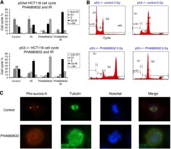
PHA-680632 exhibits 30- to 200-fold higher IC50 values for FLT3, LCK, PLK1, STLK2, VEGFR2, and VEGFR3 in comparison to Aurora A. Strong antiproliferative action against a variety of cell types is exhibited by PHA-680632. The following have different IC50 values: 0.32, 0.41, 0.06, 1.17, 0.56, 0.62, 0.29, 0.11, 1.56, 0.62, 0.07 for C33A, HeLa, HCT116, HT29, LOVO, A549, MCF7, A2780, U2OS, DU145, U937, HL60, and NHDF. ,0.13,0.41 micrometers. Tumor cells become polyploid due to PHA-680632. Treatment with PHA-680632 cells produces a phenotype resembling Aurora A or B depletion [1]. In certain cancer cell lines, PHA680632 induces polyploidy and inhibits colony formation. Cancer cells respond better to radiation when PHA680632 inhibits Aurora-A, particularly p53-deficient cells [2].
Antiproliferative activity against human cancer cell lines [1]: PHA-680632 showed potent antiproliferative effects across Aurora A-overexpressing cancer cell lines, with IC₅₀ values ranging from 22 nM to 35 nM. Specific examples include: - HCT116 (colorectal cancer, p53 wild-type): IC₅₀ = 25 nM - MCF-7 (breast cancer, p53 wild-type): IC₅₀ = 30 nM - H1299 (lung cancer, p53-deficient): IC₅₀ = 22 nM - SK-OV-3 (ovarian cancer, p53-mutant): IC₅₀ = 35 nM - Induction of G2/M cell cycle arrest [1]: Treatment of HCT116 cells with PHA-680632 (20 nM) for 24 hours resulted in a 4.2-fold increase in G2/M phase cells—from 14% (vehicle control) to 59% (treated group)—as detected by propidium iodide (PI) staining and flow cytometry. This arrest was associated with abnormal mitotic spindle formation (observed in 65% of treated cells via α-tubulin immunofluorescence). - Induction of cancer cell apoptosis [1]: MCF-7 cells treated with PHA-680632 (30 nM) for 48 hours showed a 3.8-fold increase in annexin V-positive apoptotic cells (38% vs. 10% in control). Western blot analysis confirmed elevated levels of cleaved caspase-3 (3.2-fold) and cleaved PARP (2.9-fold) compared to control. - Radiation sensitization in p53-deficient cells [2]: In H1299 (p53-deficient) lung cancer cells, co-treatment with PHA-680632 (20 nM) and ionizing radiation (2 Gy) reduced clonogenic survival by 50% compared to radiation alone (survival fraction: 0.2 vs. 0.4). This sensitization was associated with increased DNA double-strand breaks (elevated γ-H2AX foci, 2.5-fold vs. radiation alone) and prolonged G2/M arrest. |
| ln Vivo |
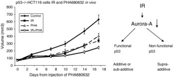
In animal studies, PHA-680632 reduces the growth of tumors. When PHA-680632 was administered at a dose of 45 mg/kg in the HL60 human acute myeloid leukemia xenograft model, 85% of TGIs occurred without any severe side effects. PHA-680632 therapy at 60 mg/kg iv for 5 days produced 78% of TGIs without any toxicities in the A2780 human ovarian cancer model [1]. PHA680632 does not function as a radiosensitizer, but it is linked to radiation-associated additive effects in cancer cells, especially in p53-deficient cells [2].
HCT116 colorectal cancer xenograft model [1]: Female nude mice (6–7 weeks old, n=8 per group) bearing HCT116 xenografts were treated with PHA-680632 at 50 mg/kg via oral gavage once daily for 14 days. This treatment resulted in 75% tumor growth inhibition (TGI) compared to vehicle control. At study end, tumor volume in the treated group was 190 ± 28 mm³, versus 760 ± 45 mm³ in the control group (p < 0.001). No significant body weight loss (<5%) was observed in the treated mice. - H1299 (p53-deficient) lung cancer xenograft model (combination with radiation) [2]: Nude mice bearing H1299 xenografts were randomized to four groups (n=6 per group): vehicle, PHA-680632 (30 mg/kg oral daily), radiation (4 Gy weekly for 3 weeks), or combination. The combination group achieved 85% TGI, significantly higher than single-agent effects (40% for PHA-680632 alone, 55% for radiation alone). Immunohistochemistry of tumors showed 70% reduction in phospho-TPX2 (Aurora A activity marker) and 2.3-fold increase in γ-H2AX (DNA damage marker) in the combination group. |
| Enzyme Assay |
Aurora A kinase activity assay (HTRF format) [1]:
Recombinant human Aurora A kinase (complexed with TPX2 to enhance catalytic activity) was incubated with PHA-680632 (serial concentrations: 0.01 nM to 500 nM), ATP (10 μM), and a biotinylated TPX2-derived peptide substrate (containing the Aurora A phosphorylation site at Ser466) in kinase buffer (50 mM Tris-HCl, 10 mM MgCl₂, 1 mM DTT, 0.01% BSA, pH 7.5) at 30°C for 60 minutes. The reaction was terminated by adding 50 mM EDTA. Phosphorylated substrate was detected using a streptavidin-conjugated europium cryptate (fluorescence donor) and a phospho-specific antibody labeled with XL665 (fluorescence acceptor). Fluorescence resonance energy transfer (FRET) signals were measured using a microplate reader, and IC₅₀ values were calculated by fitting dose-response curves to a four-parameter logistic model.
- Aurora B kinase selectivity assay [1]: To confirm selectivity, the above assay was repeated using recombinant human Aurora B kinase (complexed with INCENP) and a biotinylated histone H3 (Ser10) peptide substrate. PHA-680632 was tested at concentrations up to 1000 nM, and no significant inhibition (<10%) was observed, confirming its selectivity for Aurora A. |
| Cell Assay |
Antiproliferation assay (MTT method) [1]:
Cancer cells (e.g., HCT116, MCF-7, H1299) were seeded in 96-well plates at a density of 3×10³ cells/well and incubated overnight at 37°C (5% CO₂). PHA-680632 was added at serial concentrations (1 nM to 200 nM), and cells were cultured for 72 hours. MTT reagent (5 mg/mL) was added to each well, and the plates were incubated for 4 hours at 37°C. The formazan product was dissolved in DMSO, and absorbance was measured at 570 nm. IC₅₀ values were defined as the concentration of PHA-680632 that inhibited 50% of cell proliferation, calculated using GraphPad Prism software.
- Cell cycle analysis (PI staining) [1]: HCT116 cells were seeded in 6-well plates at 5×10⁵ cells/well and treated with PHA-680632 (20 nM) or vehicle for 24 hours. Cells were harvested by trypsinization, washed with cold PBS, and fixed in 70% ethanol at -20°C overnight. Fixed cells were washed again with PBS, resuspended in PI staining solution (50 μg/mL PI, 100 μg/mL RNase A, 0.1% Triton X-100 in PBS), and incubated at 37°C for 30 minutes. Cell cycle distribution (G0/G1, S, G2/M phases) was analyzed using a flow cytometer, and the percentage of cells in each phase was quantified using ModFit software. - Clonogenic survival assay (combination with radiation) [2]: H1299 cells were seeded in 6-well plates at 200 cells/well and incubated overnight. Cells were treated with PHA-680632 (20 nM) for 2 hours, then exposed to ionizing radiation (0–6 Gy) using a linear accelerator. After radiation, cells were cultured for 14 days to allow colony formation. Colonies were fixed with methanol, stained with crystal violet, and counted manually. Survival fractions were calculated as (number of colonies in treated group / number of colonies in control group) × plating efficiency, and radiation survival curves were generated. - Western blot for Aurora A substrates and DNA damage markers [1,2]: - [1] HCT116 cells treated with PHA-680632 (10–50 nM) for 6 hours were lysed in RIPA buffer (supplemented with protease and phosphatase inhibitors). Protein extracts (30 μg per lane) were separated by 10% SDS-PAGE, transferred to PVDF membranes, and probed with antibodies against phospho-TPX2 (Ser466), total TPX2, and β-actin (loading control). - [2] H1299 cells treated with PHA-680632 (20 nM) + radiation (2 Gy) were lysed 24 hours post-treatment, and membranes were probed with antibodies against γ-H2AX (DNA damage marker) and β-actin. Signals were detected using enhanced chemiluminescence (ECL) reagent, and band intensities were quantified using ImageJ software. |
| Animal Protocol |
Dissolved in 20% Tween-80 in 5% glucose solution; 40 mg/kg; i.p. injection BID Mice (female athymic nude) xenografts models of p53 / HCT116 cells
HCT116 colorectal cancer xenograft model [1]: Female nude mice (6–7 weeks old) were subcutaneously injected with 5×10⁶ HCT116 cells (suspended in a 1:1 mixture of PBS and Matrigel) into the right flank. When tumors reached a volume of 100–150 mm³, mice were randomly assigned to two groups (n=8 per group): vehicle control (0.5% carboxymethylcellulose sodium + 0.1% Tween 80 in distilled water) and PHA-680632 treatment. PHA-680632 was dissolved in the vehicle at a concentration of 10 mg/mL and administered via oral gavage at 50 mg/kg once daily for 14 days. Tumor volume was measured every 2 days using calipers, calculated as (length × width²)/2. Mouse body weight was also measured every 2 days to monitor toxicity. - H1299 lung cancer xenograft model (combination with radiation) [2]: Female nude mice were subcutaneously implanted with 1×10⁷ H1299 cells (mixed with Matrigel). When tumors reached ~120 mm³, mice were randomized to four groups (n=6 per group): 1. Vehicle: 0.5% CMC + 0.1% Tween 80, oral daily for 21 days; 2. PHA-680632: 30 mg/kg oral daily for 21 days; 3. Radiation: 4 Gy localized tumor radiation once weekly for 3 weeks; 4. Combination: PHA-680632 (30 mg/kg oral daily) + radiation (4 Gy weekly). Tumor volume and body weight were measured twice weekly. At study end (day 21), tumors were excised for immunohistochemical analysis. |
| ADME/Pharmacokinetics |
Oral bioavailability [1]: In male Sprague-Dawley rats, the oral bioavailability of PHA-680632 (20 mg/kg) was 30%. Plasma concentration-time curves showed that the peak plasma concentration (Cmax) of 1.0 μg/mL was reached 1.8 hours after administration, and the terminal half-life (t₁/₂) was 4.5 hours. Intravenous pharmacokinetics (rat) [1]: In rats, after intravenous injection of PHA-680632 (5 mg/kg), the clearance (CL) was 15 mL/min/kg, the steady-state volume of distribution (Vss) was 5.0 L/kg, and the t₁/₂ was 4.2 hours.
- Plasma protein binding rate [1]: PHA-680632 showed high plasma protein binding rates in human (95%), rat (94%), and mouse (93%) plasmas, as determined by equilibrium dialysis. Dialysis was performed at 37°C for 4 hours using a 10 kDa molecular weight cutoff membrane, and the plasma concentration of PHA-680632 was 1 μg/mL. - Metabolic stability [1]: The half-life of PHA-680632 in human liver microsomes was 3.8 hours (moderate metabolic stability); the half-life in rat liver microsomes was 4.3 hours. Liquid chromatography-tandem mass spectrometry (LC-MS/MS) identified the major metabolites as monohydroxylated derivatives (accounting for 55% of the total metabolites), mainly formed through CYP3A4-mediated oxidation. |
| Toxicity/Toxicokinetics |
Acute oral toxicity (mice) [1]: Female CD-1 mice were given a single oral dose of PHA-680632 up to 2000 mg/kg without death. At doses ≥1500 mg/kg, mice showed transient decreases in kinetic activity, but recovered within 24 hours. At doses ≤1000 mg/kg, no significant changes in body weight were observed. Chronic oral toxicity (rats) [1]: Male Sprague-Dawley rats were given PHA-680632 (50 mg/kg, once daily) for 28 consecutive days. Mild myelosuppression was observed: white blood cell count decreased by 18% compared to the solvent control group, while red blood cell count and platelet count remained within the normal range. Serum liver function indicators (ALT, AST) and kidney function indicators (BUN, creatinine) levels were not significantly different from the control group, and pathological examination of liver, kidney and heart tissues revealed no treatment-related lesions.
- Toxicity of combined radiotherapy[2]: In the H1299 xenograft model, combined therapy (PHA-680632 + radiotherapy) did not increase toxicity compared to monotherapy: no significant weight loss (<5%), skin irritation, or organ damage was observed in the treatment group mice. |
| References |
|
| Additional Infomation |
Chemical Classification and Design [1]: PHA-680632 is an indolecarbazole derivative optimized for efficient and selective inhibition of Aurora A kinase. It was designed to address the limitations of earlier non-selective Aurora inhibitors, which could lead to off-target toxicity (e.g., myelosuppression) due to inhibition of Aurora B. Mechanism of Action [1,2]: [1] PHA-680632 inhibits Aurora A kinase, a key regulator of spindle assembly and centrosome maturation. Inhibition disrupts mitotic processes, leading to G2/M phase cell cycle arrest, mitotic catastrophe, and apoptosis in Aurora A-overexpressing cancer cells.
- [2] In p53-deficient cells, PHA-680632 enhances radiosensitivity by prolonging G2/M phase arrest (providing more time for radiation-induced DNA damage) and inhibiting DNA repair pathways (by reducing Aurora A-mediated γ-H2AX dephosphorylation). - Clinical significance [2]: PHA-680632 has the potential to treat p53-deficient cancers (a common subtype that responds poorly to radiotherapy). Preclinical data show that it can overcome radioresistance in p53-deficient models, supporting its use in combination with radiotherapy. |
| Molecular Formula |
C28H35N7O2
|
|
|---|---|---|
| Molecular Weight |
501.62
|
|
| Exact Mass |
501.285
|
|
| CAS # |
398493-79-3
|
|
| Related CAS # |
|
|
| PubChem CID |
11249084
|
|
| Appearance |
White to off-white solid powder
|
|
| Density |
1.3±0.1 g/cm3
|
|
| Boiling Point |
709.0±60.0 °C at 760 mmHg
|
|
| Flash Point |
382.6±32.9 °C
|
|
| Vapour Pressure |
0.0±2.3 mmHg at 25°C
|
|
| Index of Refraction |
1.676
|
|
| LogP |
3.04
|
|
| Hydrogen Bond Donor Count |
3
|
|
| Hydrogen Bond Acceptor Count |
5
|
|
| Rotatable Bond Count |
6
|
|
| Heavy Atom Count |
37
|
|
| Complexity |
769
|
|
| Defined Atom Stereocenter Count |
0
|
|
| SMILES |
O=C(N1CC2=C(NN=C2NC(C3=CC=C(N4CCN(C)CC4)C=C3)=O)C1)NC5=C(C=CC=C5CC)CC
|
|
| InChi Key |
OBWNXGOQPLDDPS-UHFFFAOYSA-N
|
|
| InChi Code |
InChI=1S/C28H35N7O2/c1-4-19-7-6-8-20(5-2)25(19)29-28(37)35-17-23-24(18-35)31-32-26(23)30-27(36)21-9-11-22(12-10-21)34-15-13-33(3)14-16-34/h6-12H,4-5,13-18H2,1-3H3,(H,29,37)(H2,30,31,32,36)
|
|
| Chemical Name |
N-(2,6-diethylphenyl)-3-(4-(4-methylpiperazin-1-yl)benzamido)pyrrolo[3,4-c]pyrazole-5(1H,4H,6H)-carboxamide
|
|
| Synonyms |
PHA 680632; PHA-680632; PHA 680632.
|
|
| HS Tariff Code |
2934.99.9001
|
|
| Storage |
Powder -20°C 3 years 4°C 2 years In solvent -80°C 6 months -20°C 1 month |
|
| Shipping Condition |
Room temperature (This product is stable at ambient temperature for a few days during ordinary shipping and time spent in Customs)
|
| Solubility (In Vitro) |
DMSO: 100 mg/mL (199.4 mM)
Water: 1 mg/mL (Insoluble) Ethanol: 1 mg/mL |
|---|---|
| Solubility (In Vivo) |
Solubility in Formulation 1: ≥ 2.08 mg/mL (4.15 mM) (saturation unknown) in 10% DMSO + 40% PEG300 + 5% Tween80 + 45% Saline (add these co-solvents sequentially from left to right, and one by one), clear solution.
For example, if 1 mL of working solution is to be prepared, you can add 100 μL of 20.8 mg/mL clear DMSO stock solution to 400 μL PEG300 and mix evenly; then add 50 μL Tween-80 to the above solution and mix evenly; then add 450 μL normal saline to adjust the volume to 1 mL. Preparation of saline: Dissolve 0.9 g of sodium chloride in 100 mL ddH₂ O to obtain a clear solution. Solubility in Formulation 2: ≥ 2.08 mg/mL (4.15 mM) (saturation unknown) in 10% DMSO + 90% (20% SBE-β-CD in Saline) (add these co-solvents sequentially from left to right, and one by one), clear solution. For example, if 1 mL of working solution is to be prepared, you can add 100 μL of 20.8 mg/mL clear DMSO stock solution to 900 μL of 20% SBE-β-CD physiological saline solution and mix evenly. Preparation of 20% SBE-β-CD in Saline (4°C,1 week): Dissolve 2 g SBE-β-CD in 10 mL saline to obtain a clear solution. View More
Solubility in Formulation 3: ≥ 2.08 mg/mL (4.15 mM) (saturation unknown) in 10% DMSO + 90% Corn Oil (add these co-solvents sequentially from left to right, and one by one), clear solution. Solubility in Formulation 4: 10% Tween 80: 30 mg/mL |
| Preparing Stock Solutions | 1 mg | 5 mg | 10 mg | |
| 1 mM | 1.9935 mL | 9.9677 mL | 19.9354 mL | |
| 5 mM | 0.3987 mL | 1.9935 mL | 3.9871 mL | |
| 10 mM | 0.1994 mL | 0.9968 mL | 1.9935 mL |
*Note: Please select an appropriate solvent for the preparation of stock solution based on your experiment needs. For most products, DMSO can be used for preparing stock solutions (e.g. 5 mM, 10 mM, or 20 mM concentration); some products with high aqueous solubility may be dissolved in water directly. Solubility information is available at the above Solubility Data section. Once the stock solution is prepared, aliquot it to routine usage volumes and store at -20°C or -80°C. Avoid repeated freeze and thaw cycles.
Calculation results
Working concentration: mg/mL;
Method for preparing DMSO stock solution: mg drug pre-dissolved in μL DMSO (stock solution concentration mg/mL). Please contact us first if the concentration exceeds the DMSO solubility of the batch of drug.
Method for preparing in vivo formulation::Take μL DMSO stock solution, next add μL PEG300, mix and clarify, next addμL Tween 80, mix and clarify, next add μL ddH2O,mix and clarify.
(1) Please be sure that the solution is clear before the addition of next solvent. Dissolution methods like vortex, ultrasound or warming and heat may be used to aid dissolving.
(2) Be sure to add the solvent(s) in order.
Influence of PHA680632 on cell cycle in p53wt vs p53−/− HCT116 cells. Br J Cancer. 2007 Dec 17; 97(12): 1664–1672. |
In vivo tumour growth delay after PHA680632 and irradiation. Br J Cancer, 2007, 97(12), 1664-1672. |
Products are for research use only; We do not sell to patients
Copyright 2020 InvivoChem LLC | All Rights Reserved


 COA
COA