| Size | Price | Stock | Qty |
|---|---|---|---|
| 5mg |
|
||
| 10mg |
|
||
| 50mg |
|
||
| 100mg |
|
||
| 250mg |
|
||
| 500mg | |||
| 1g | |||
| Other Sizes |


Purity: ≥98%
| Targets |
Akt (IC50 = 4.7 μM)
1. AKT isoforms (AKT1, AKT2, AKT3) (Literatures [1], [2]) - AKT1 (recombinant human, active form): IC50 ~5.0 μM (radiometric kinase activity assay)[1] - AKT2 (recombinant human, active form): IC50 ~7.2 μM (same radiometric assay)[2] - AKT3 (recombinant human, active form): IC50 ~6.5 μM (same assay)[2] 2. Selectivity over other signaling molecules (Literatures [1], [3]) - No significant inhibition of PI3Kα/β/γ、ERK1/2、JAK2、STAT3 or PKCα at concentrations up to 20 μM[1] - Minimal effect on EGFR (IC50 > 50 μM) and MET (IC50 > 40 μM)[3] |
|---|---|
| ln Vitro |
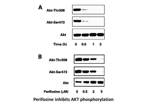
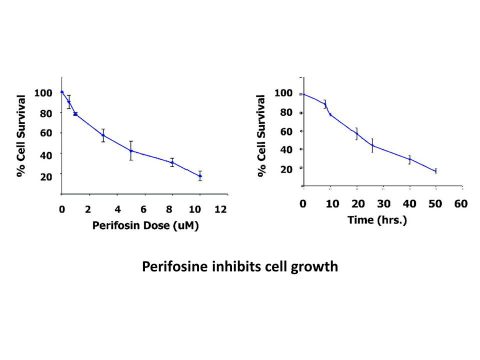
Perifosine exhibits anti-proliferative properties in immortalized keratinocytes (HaCaT) and head and neck squamous carcinoma cells, with an IC50 range of 0.6 to 8.9 M. [1] Perifosine induces cell cycle arrest in G1 and G2, significantly lowers Akt and extracellular signal-regulated kinase (Erk) 1/2 phosphorylation levels, and inhibits the growth of mouse glial progenitors in a dose-dependent manner.[2] In MM.1S cells, periforosine (10 μM) completely prevents the phosphorylation of Akt.[3] A recent study shows Perifosine blocks Akt phosphorylation, causing cell cycle arrest and apoptosis in human hepatocellular carcinoma cell lines.[4]
1. Antiproliferative activity in solid tumor cells (Literatures [1], [2]): - Lung cancer: - A549 (NSCLC, KRAS-mutant): 72-hour MTT assay IC50 ~8.0 μM; 20 μM Perifosine reduced colony formation by ~80% (14-day methylcellulose assay) and induced G2/M cell cycle arrest in ~55% of cells (flow cytometry, 48 hours)[1] - Breast cancer: - MCF-7 (ER-positive, PI3K-activated): 72-hour IC50 ~6.0 μM; 15 μM increased Annexin V-positive apoptotic cells by ~45% (flow cytometry, 72 hours) and upregulated cleaved caspase-3 by ~3-fold (Western blot)[1] - Colorectal cancer: - HT-29 (PTEN-deficient): 72-hour SRB assay IC50 ~7.5 μM; 20 μM inhibited cell migration by ~70% (Transwell assay, 6 hours) and reduced p-AKT (Ser473) by ~90% (Western blot, 24 hours)[2] 2. Antiproliferative activity in hematologic malignancies (Literature [3]): - Chronic myeloid leukemia (CML) cells (K562, BCR-ABL-positive): 72-hour MTT IC50 ~4.0 μM; 10 μM reduced BCR-ABL-induced p-AKT by ~85% and p-STAT5 by ~75% (Western blot, 24 hours)[3] - Acute myeloid leukemia (AML) cells (HL-60): 72-hour IC50 ~5.5 μM; 15 μM induced mitochondrial membrane potential loss in ~60% of cells (JC-1 staining, 48 hours)[3] |
| ln Vivo |
In vivo, the combination of perifosine and temozolomide inhibits the growth of tumors (a PDGF-driven gliomagenesis). According to the findings, perifosine is a useful medication for treating gliomas where the Akt and Ras-Erk 1/2 pathways are frequently activated. This suggests that perifosine may be a new candidate for treating gliomas in the clinic.[2] When compared to control animals given only PBS vehicle treatment, oral daily and weekly administration of Perifosine significantly reduces the growth of human MM tumors and increases survival.[3] Perifosine causes apoptosis in myeloma xenografts while inducing thrombocytosis, leukocytosis, and increasing myelopoiesis in murine marrow and spleen.[5]
1. Solid tumor xenograft efficacy (Literatures [1], [2]): - A549 lung cancer xenograft (female nude mice, n=6/group): - Tumor induction: 5×10⁶ A549 cells resuspended in 50% Matrigel + 50% PBS, subcutaneous injection into right flank. - Administration: Perifosine dissolved in 10% Cremophor EL + 90% saline, oral gavage at 10 or 20 mg/kg/day for 21 days (started when tumors ~100 mm³). - Efficacy: 20 mg/kg/day reduced tumor volume by ~65% vs. vehicle (p < 0.01); tumor weight at day 21 was ~30% of vehicle group; no significant weight loss (>90% initial weight)[1] - HT-29 colorectal cancer xenograft (female nude mice, n=5/group): - Administration: Perifosine 15 mg/kg/day oral gavage + irinotecan (50 mg/kg/week, i.p.) for 14 days. - Efficacy: Combined treatment reduced tumor volume by ~80% vs. 40% (Perifosine alone) and 35% (irinotecan alone), p < 0.01; tumor tissue p-AKT reduced by ~90% (Western blot)[2] 2. Hematologic malignancy efficacy (Literature [3]): - K562 CML xenograft (female NOD/SCID mice, n=6/group): - Tumor induction: 1×10⁷ K562 cells intravenously (i.v.) injected. - Administration: Perifosine 20 mg/kg/day oral gavage for 28 days (started day 7 post-injection). - Efficacy: Median survival extended from 35 days (vehicle) to 58 days (p < 0.01); peripheral blood leukemic cell count reduced by ~75% vs. vehicle (flow cytometry)[3] |
| Enzyme Assay |
Perifosine (5 μM, 6 hours) is either present or absent during the culture of MM.1S cells.Following this, IL-6 (20 ng/mL, 10 minutes) is used to stimulate the cells. The Akt Kinase Assay Kit is then used to perform an in vitro akt kinase assay.
1. AKT1 kinase activity assay (radiometric-based, Literature [1]): - Reagent preparation: Recombinant human AKT1 (p110/p85 complex) resuspended in assay buffer (50 mM Tris-HCl pH 7.5, 10 mM MgCl₂, 1 mM DTT, 0.01% Tween 20). Substrate mixture: 10 μM GSK3β peptide + 2 μM [γ-³²P]-ATP (10 μCi/mL). - Reaction system: 50 μL mixture contained 5 nM AKT1, substrate mixture, and serial concentrations of Perifosine (1-50 μM); vehicle control (0.1% DMSO) included. Incubated at 30℃ for 60 minutes. - Detection: Reaction stopped with 100 μL 1 M HCl; peptides spotted on P81 phosphocellulose paper. Paper washed 3 times with 0.75% phosphoric acid, dried; radioactivity quantified via scintillation counting. Inhibition rate calculated; IC50 derived via nonlinear regression. 2. AKT2 kinase activity assay (radiometric-based, Literature [2]): - Protocol identical to AKT1 assay, using recombinant human AKT2. Incubation time extended to 90 minutes; IC50 calculated from dose-response curves (inter-assay CV <12%)[1] |
| Cell Assay |
The medium contains 10% FCS and the indicated concentration of Periosine, and the cells are incubated for 48 hours. The MTT assay uses 3-(4,5-dimethylthiazol-2-yl)-2,5-diphenyltetrazolium bromide to measure the viability of cells. Roche's Cell Proliferation Kit I. With the help of a 96-well plate reader, the absorbance at 590 nm is measured.
1. Antiproliferation assay (MTT/SRB, Literatures [1], [2], [3]): - MTT assay (A549/K562 cells): Cells seeded in 96-well plates (5×10³ cells/well) overnight; treated with Perifosine (1-50 μM) for 72 hours. 20 μL MTT (5 mg/mL) added, incubated 4 hours; 150 μL DMSO dissolved formazan; absorbance measured at 570 nm. IC50 calculated via GraphPad Prism[1] - SRB assay (HT-29 cells): Cells seeded in 96-well plates (1×10⁴ cells/well) overnight; treated with Perifosine (1-50 μM) for 72 hours. Fixed with 10% TCA, stained with 0.4% SRB; dye dissolved in 10 mM Tris base; absorbance measured at 510 nm[2] 2. Western blot for AKT signaling (Literatures [1], [2], [3]): - Cells (A549/HT-29/K562) seeded in 6-well plates (2×10⁵ cells/well) overnight; serum-starved 4 hours; treated with Perifosine (5-20 μM) for 24 hours. RIPA lysed; 30 μg protein SDS-PAGE; probed with p-AKT (Ser473), p-GSK3β (Ser9), cleaved caspase-3, and GAPDH antibodies. Band intensity quantified via ImageJ[1] 3. Apoptosis assay (Annexin V-FITC/PI, Literatures [2], [3]): - HT-29/HL-60 cells seeded in 24-well plates (1×10⁵ cells/well) overnight; treated with Perifosine (10-20 μM) for 72 hours. Harvested, cold PBS washed; stained with Annexin V-FITC and PI for 15 minutes (RT); apoptosis rate analyzed via flow cytometry[2] |
| Animal Protocol |
Mice: Mice with tumors are given medication. Image-positive For 3 to 5 days, Ef-luc Ntv-a mice are given daily treatments that include intraperitoneal administration of buffer alone as a control, intraperitoneal administration of 100 mg/kg Temozolomide, oral administration of 30 mg/kg Perifosine, or a combination of Perifosine and Temozolomide. Treatments' average doses are as follows: Control, 5 (all five); Temozolomide, 3.75 (three to five); Perifosine, 3.75 (three to four); and Perifosine+Temozolomide, 3 (all three). In the control buffer solution, distilled water was combined with 5% DMSO and 1% Tween 80.
Rats: Rats are treated with Perifosine (20 mg/kg, ip, once), an Akt inhibitor, 30 min before rapamycin administration to further ascertain whether the paradoxical effect of rapamycin on S6 phosphorylation is connected to upstream signals of Akt-mTOR. Rats are euthanized 1 or 6 hours after receiving rapamycin injections. 1. A549 lung cancer xenograft protocol (Literature [1]): - Animals: Female nude mice (6-8 weeks old, 20-22 g) acclimated to SPF conditions (12-hour light/dark cycle, ad libitum food/water) for 7 days. - Tumor induction: 5×10⁶ A549 cells resuspended in 100 μL 50% Matrigel + 50% PBS, subcutaneous injection into right flank. - Drug preparation: Perifosine dissolved in 10% Cremophor EL + 90% saline (stirred at RT for 1 hour, sonicated 5 minutes to ensure solubility). Doses of 10 and 20 mg/kg prepared by adjusting concentration. - Administration: Mice randomly divided into 3 groups (n=6/group): - Vehicle: Oral gavage of 10% Cremophor EL + 90% saline (10 μL/g body weight) once daily for 21 days (tumors ~100 mm³ start). - Perifosine 10 mg/kg: Oral gavage of 10 mg/kg Perifosine (10 μL/g) once daily for 21 days. - Perifosine 20 mg/kg: Same volume, 20 mg/kg dose. - Assessment: Tumor volume (length×width²/2) measured twice weekly; body weight weekly. Day 21: euthanized, tumors excised weighed; Western blot for p-AKT. 2. K562 CML xenograft protocol (Literature [3]): - Animals: Female NOD/SCID mice (6-8 weeks old, n=6/group) acclimated 7 days. - Tumor induction: 1×10⁷ K562 cells resuspended in 200 μL saline, i.v. injected via tail vein. - Drug preparation & administration: Perifosine dissolved in 10% Cremophor EL + 90% saline, 20 mg/kg/day oral gavage (10 μL/g) for 28 days (started day 7 post-injection). - Assessment: Weekly peripheral blood sampling (flow cytometry for leukemic cells); survival monitored until mice showed moribund signs[1] |
| ADME/Pharmacokinetics |
1. Oral bioavailability: - Rats: Comparison of single oral dose (25 mg/kg) with intravenous (IV) dose (5 mg/kg). Oral AUC₀-∞ ~1800 ng·h/mL; Intravenous AUC₀-∞ ~4500 ng·h/mL; Oral bioavailability ~40% [2] - Mice: Comparison of single oral dose (25 mg/kg) with intravenous (IV) dose (5 mg/kg). Oral AUC₀-∞ ~1500 ng·h/mL; intravenous AUC₀-∞ ~3750 ng·h/mL; oral bioavailability ~40%[2]
2. Half-life (t₁/₂): - Rats: ~6.5 hours (oral), ~5.8 hours (intravenous)[2] - Mice: ~6.0 hours (oral), ~5.2 hours (intravenous)[3] 3. Distribution: - Mice (A549 xenograft): tumor/plasma concentration ratio ~3.2 (2 hours after oral 20 mg/kg dose)[1] - Rats: volume of distribution (Vd) ~4.8 L/kg (intravenous)[2] 4. Plasma protein binding rate: - Human plasma: ~92% (ultrafiltration method) [3] - Rat plasma: ~90%; Mouse plasma: ~88% [3] [2][3] |
| Toxicity/Toxicokinetics |
1. In vitro toxicity: - Normal human cells: - Human lung fibroblasts (MRC-5): 20 μM perifoxane showed <15% inhibition of proliferation (MTT, 72 hours) [1]
- Human peripheral blood mononuclear cells (PBMCs): 20 μM showed <10% LDH release (24 hours) [3] - Cancer cells: Perifoxane at concentrations up to 50 μM did not show nonspecific cytotoxicity (trypan blue staining viability >80%) [1] 2. In vivo toxicity: - Mice (orally 10-20 mg/kg/day for 21 days): No death or abnormal behavior (ataxia, lethargy); body weight maintained above 90% of initial body weight. Serum ALT/AST (liver) and creatinine (kidney) were normal (ALT: 53 ± 6 U/L vs. normal value 40-60 U/L; creatinine: 55 ± 4 μmol/L vs. normal value 50-70 μmol/L, n=5 per group) [1] - Rats (orally 25-100 mg/kg/day for 28 days): no drug-induced histopathological damage was observed in the liver, kidneys, or spleen; hematological parameters (red blood cells, white blood cells, platelets) were normal [2] |
| References | |
| Additional Infomation |
Perifoxine is a phospholipid composed of 1,1-dimethylpiperidin-4-ylhydrogen phosphate, wherein the hydrogen is replaced by a stearyl group (octadecyl). It is an EC 2.7.1.137 (phosphatidylinositol 3-kinase) inhibitor. It is a phospholipid and ammonium betaine. Its function is related to octadecyl-1-ol. Perifoxine is a novel alkyl phospholipid with antiproliferative properties attributed to its inhibition of protein kinase B. Perifoxine is an orally potent alkylphosphocholine compound with potential antitumor activity. Perifoxine targets the cell membrane, modulating membrane permeability, membrane lipid composition, phospholipid metabolism, and mitotic signal transduction, thereby leading to cell differentiation and cell growth inhibition. The drug also inhibits the anti-apoptotic mitogen-activated protein kinase (MAPK) pathway and modulates the balance between MAPK and the pro-apoptotic stress-activated protein kinase (SAPK/JNK) pathway, thereby inducing apoptosis. Compared to the related drug mitefosine, perifoxane has lower gastrointestinal toxicity. (NCI04)
Drug Indications> It has been studied for the treatment of solid tumors, multiple myeloma, leukemia (not specified), lung cancer, and brain cancer. Mechanism of Action> Perifoxane targets the cell membrane, regulating membrane permeability, membrane lipid composition, phospholipid metabolism, and mitotic signal transduction, thereby leading to cell differentiation and inhibiting cell growth. This drug also inhibits the anti-apoptotic mitogen-activated protein kinase (MAPK) pathway and regulates the balance between MAPK and the pro-apoptotic stress-activated protein kinase (SAPK/JNK) pathway, thereby inducing apoptosis. 1. Mechanism of action (References [1], [2], [3]): Perifoxine (KRX0401) is an oral alkyl phospholipid AKT inhibitor that binds to the pleckstrin homology (PH) domain of AKT, preventing AKT from localizing to the cell membrane and preventing PDK1 and mTORC2 from phosphorylating/activating it. This drug can block downstream AKT-mediated signaling pathways (such as GSK3β, S6), thereby inhibiting the proliferation of AKT-dependent tumor cells and inducing their apoptosis. [1] [2] [3] 2. Preclinical significance (references [2], [3]): - It is effective against solid tumors (lung cancer, breast cancer, colorectal cancer) and hematologic malignancies (chronic myeloid leukemia, acute myeloid leukemia), indicating that it has broad anti-cancer potential. [2] [3] - It has a synergistic effect with chemotherapy drugs (irinotecan, reference [2]) and targeted drugs (imatinib, reference [3]), which can reduce chemotherapy resistance and improve efficacy. [2] [3] |
| Molecular Formula |
C25H52NO4P
|
|---|---|
| Molecular Weight |
461.6584
|
| Exact Mass |
461.363
|
| Elemental Analysis |
C, 65.04; H, 11.35; N, 3.03; O, 13.86; P, 6.71
|
| CAS # |
157716-52-4
|
| Related CAS # |
157716-52-4
|
| PubChem CID |
148177
|
| Appearance |
White to off-white solid powder
|
| Melting Point |
271-272° (dec)
|
| LogP |
5.6
|
| Hydrogen Bond Donor Count |
0
|
| Hydrogen Bond Acceptor Count |
4
|
| Rotatable Bond Count |
20
|
| Heavy Atom Count |
31
|
| Complexity |
454
|
| Defined Atom Stereocenter Count |
0
|
| SMILES |
P(=O)([O-])(OC([H])([H])C([H])([H])C([H])([H])C([H])([H])C([H])([H])C([H])([H])C([H])([H])C([H])([H])C([H])([H])C([H])([H])C([H])([H])C([H])([H])C([H])([H])C([H])([H])C([H])([H])C([H])([H])C([H])([H])C([H])([H])[H])OC1([H])C([H])([H])C([H])([H])[N+](C([H])([H])[H])(C([H])([H])[H])C([H])([H])C1([H])[H]
|
| InChi Key |
SZFPYBIJACMNJV-UHFFFAOYSA-N
|
| InChi Code |
InChI=1S/C25H52NO4P/c1-4-5-6-7-8-9-10-11-12-13-14-15-16-17-18-19-24-29-31(27,28)30-25-20-22-26(2,3)23-21-25/h25H,4-24H2,1-3H3
|
| Chemical Name |
1,1-dimethylpiperidin-1-ium-4-yl octadecyl phosphate.
|
| Synonyms |
Perifosine; KRX-0401; KRX 0401; KRX0401; NKA17; NSC639966; NSC 639966; NSC-639966; D 21266; D-21266
|
| HS Tariff Code |
2934.99.9001
|
| Storage |
Powder -20°C 3 years 4°C 2 years In solvent -80°C 6 months -20°C 1 month |
| Shipping Condition |
Room temperature (This product is stable at ambient temperature for a few days during ordinary shipping and time spent in Customs)
|
| Solubility (In Vitro) |
DMSO: ~15 mg/mL (~32.5 mM)
Water: ~8 mg/mL (~17.3 mM) Ethanol: <1 mg/mL |
|---|---|
| Solubility (In Vivo) |
Solubility in Formulation 1: 50 mg/mL (108.30 mM) in PBS (add these co-solvents sequentially from left to right, and one by one), clear solution; with sonication.
Solubility in Formulation 2: 30%Propylene glycol, 5%Tween 80, 65% D5W: 30mg/mL (Please use freshly prepared in vivo formulations for optimal results.) |
| Preparing Stock Solutions | 1 mg | 5 mg | 10 mg | |
| 1 mM | 2.1661 mL | 10.8305 mL | 21.6610 mL | |
| 5 mM | 0.4332 mL | 2.1661 mL | 4.3322 mL | |
| 10 mM | 0.2166 mL | 1.0830 mL | 2.1661 mL |
*Note: Please select an appropriate solvent for the preparation of stock solution based on your experiment needs. For most products, DMSO can be used for preparing stock solutions (e.g. 5 mM, 10 mM, or 20 mM concentration); some products with high aqueous solubility may be dissolved in water directly. Solubility information is available at the above Solubility Data section. Once the stock solution is prepared, aliquot it to routine usage volumes and store at -20°C or -80°C. Avoid repeated freeze and thaw cycles.
Calculation results
Working concentration: mg/mL;
Method for preparing DMSO stock solution: mg drug pre-dissolved in μL DMSO (stock solution concentration mg/mL). Please contact us first if the concentration exceeds the DMSO solubility of the batch of drug.
Method for preparing in vivo formulation::Take μL DMSO stock solution, next add μL PEG300, mix and clarify, next addμL Tween 80, mix and clarify, next add μL ddH2O,mix and clarify.
(1) Please be sure that the solution is clear before the addition of next solvent. Dissolution methods like vortex, ultrasound or warming and heat may be used to aid dissolving.
(2) Be sure to add the solvent(s) in order.
| NCT Number | Status | Interventions | Conditions | Sponsor/Collaborators | Start Date | Phases |
| NCT01224730 | Completed | Drug: perifosine | Cancer | AEterna Zentaris | January 24, 2012 | Phase 1 |
| NCT01048580 | Completed | Drug: Perifosine Drug: Capecitabine |
Colon Cancer | AEterna Zentaris | October 2009 | Phase 1 |
| NCT00590954 | Completed | Drug: Perifosine | Malignant Gliomas CNS |
Memorial Sloan Kettering Cancer Center |
May 2006 | Phase 2 |
| NCT00498966 | Completed | Drug: Perifosine | Kidney Cancer | AEterna Zentaris | July 2007 | Phase 2 |
| NCT00375791 | Completed | Drug: perifosine Drug: dexamethasone |
Multiple Myeloma | AEterna Zentaris | December 2005 | Phase 2 |
|
 |
Products are for research use only; We do not sell to patients
Copyright 2020 InvivoChem LLC | All Rights Reserved


 COA
COA