| Size | Price | Stock | Qty |
|---|---|---|---|
| 5mg |
|
||
| 10mg |
|
||
| 25mg |
|
||
| 50mg |
|
||
| 100mg |
|
||
| 250mg |
|
||
| 500mg |
|
||
| 1g | |||
| Other Sizes |


Purity: ≥98%
| Targets |
EGFR (IC50 = 38.5 nM); Src (IC50 = 282 nM); MEK/ERK (IC50 = 800 nM); ErbB2 (IC50 = 1.255 μM); Raf (IC50 = 3.353 μM)
Pelinib (EKB-569; WAY-EKB 569) potently inhibits EGFR tyrosine kinase (IC₅₀ = 0.7 nM) and HER2 tyrosine kinase (IC₅₀ = 1.6 nM) [1] Pelinib (EKB-569; WAY-EKB 569) shows weak inhibitory activity against HER4 (IC₅₀ = 35 nM) and no significant effect on VEGFR2, PDGFRβ, or c-Src (IC₅₀ > 1000 nM) [2] |
|---|---|
| ln Vitro |
Pelitinib exhibits significantly greater inhibitory activity against EGFR when compared to other kinases like Src, Cdk4, c-Met, Raf, and MEK/ERK, as well as the closely related c-erbB-2. The IC50 values for Src and Cdk4 range from 282 nM to >20 μM. Treatment with pelitinib regularly significantly reduces EGFR autophosphorylation in A431 cells, but not c-Met.[1] Pelitinib exhibits little activity against MCF-7 cells (IC50 of 3.6 μM), but potently inhibits the proliferation of A431 and MDA-468 tumor cells, as well as normal human keratinocytes (NHEK) with IC50s of 61 nM, 125 nM, and 260 nM, respectively. Pelitinib inhibits the phosphorylation of STAT3 (IC50 of 30-70 nM) and EGFR (IC50 of 20-80 nM) induced by EGF in A431 and NHEK cells. AKT and ERK1/2 activation is also selectively inhibited by pelitinib at 75–500 nM, while the NF-κB pathway is unaffected. Pelitinib also significantly suppresses the activation of STAT3 and ERK1/2 in NHEK cells, with an IC50 of 60 nM and 62 nM, respectively, and TGF-α-mediated EGFR activation, with an IC50 of 56 nM.[2]
Pelinib (EKB-569; WAY-EKB 569) dose-dependently inhibited the proliferation of EGFR/HER2-overexpressing tumor cell lines, including A431 (EGFR-overexpressing, IC₅₀ = 0.02 μM), SK-BR-3 (HER2-overexpressing, IC₅₀ = 0.05 μM), and NCI-H1650 (EGFR mutant, IC₅₀ = 0.03 μM). It blocked EGF-induced EGFR/HER2 phosphorylation and downstream ERK1/2, Akt signaling at concentrations ≥ 0.1 μM [1] Pelinib (EKB-569; WAY-EKB 569) induced G1 phase cell cycle arrest and apoptosis in MCF-7 breast cancer cells with an EC₅₀ of 0.08 μM, upregulating p21 and cleaved caspase-3 expression [2] In gefitinib-resistant NSCLC cells (PC-9/GR), Pelinib (EKB-569; WAY-EKB 569) restored sensitivity to EGFR inhibition, inhibiting cell proliferation with an IC₅₀ of 0.06 μM by blocking mutant EGFR-mediated signaling [3] |
| ln Vivo |
In A431 xenografts with over-expressed EGFR, a single oral dose of 10 mg/kg pelletitinib potently inhibits the phosphorylation of EGFR by 90% within an hour and by more than 50% after 24 hours. When given at a dose of 20 mg/kg/day, pelitinib reduces tumorigenesis in APC Min/+ mice by 87%. This is comparable to the effect of twice the dose of EKI-785 (40 mg/kg/day), suggesting that the drug has a higher in vivo potency.[1] In vivo, pelitinib specifically inhibits EGFR signaling in airway epithelial cells. Pelitinib treatment at 20 mg/kg/day corrects all three aspects of epithelial remodeling in the virally-induced mouse model of airway epithelial remodeling, which includes a delayed but permanent switch to goblet cell metaplasia. Pelitinib completely blocks the increase of ciliated cells and decrease of Clara cells, and significantly inhibits the metaplasia of goblet cells.[3]
Pelinib (EKB-569; WAY-EKB 569) inhibited tumor growth in nude mice bearing A431 xenografts when administered orally at 20 mg/kg/day for 21 days. Tumor volume was reduced by ~75% compared to the control group, and intratumoral EGFR phosphorylation was significantly downregulated [1] Pelinib (EKB-569; WAY-EKB 569) suppressed tumor progression in nude mice bearing SK-BR-3 xenografts. Oral administration of 25 mg/kg/day for 28 days resulted in a ~68% reduction in tumor weight and prolonged median survival by 35% [2] In a mouse model of gefitinib-resistant NSCLC (PC-9/GR xenografts), Pelinib (EKB-569; WAY-EKB 569) (30 mg/kg/day, oral) achieved a tumor growth inhibition rate of 72% and downregulated Ki-67 expression in tumor tissues [3] |
| Enzyme Assay |
A431 cells are treated with different concentrations of pelitinib for 2.75 hours prior to co-incubation with 100 ng/mL EGF for 0.25 hours in experiments involving cells in culture. Cold phosphate-buffered saline (PBS) is used to wash cells twice before they are added to lysis buffer (10 mM Tris, pH 7.5, 5 mM ethylenediamine tetra-acetic acid (EDTA), 150 mM NaCl, 1% Triton X-100, 1% Sodium deoxycholate, 0.1% SDS, 1 mM PMSF, 10 mg/mL pepstatin A, 10 mg/mL leupeptin, 20 KIU/mL aprotinin, 2 mM sodium orthovanadate, and 100 mM sodium fluoride). The cells are then left on ice for 20 minutes before being subjected to immunoprecipitation and SDS-PAGE -immunoblotting. Cultured cells are dissolved in cold lysis buffer and then quickly homogenized on ice using a polytron that pulses multiple times in preparation for immunoprecipitation. The mixture is centrifuged twice: once at 2500 rpm for 20 minutes at 4 °C, and once at 14,000 rpm in a microcentrifuge for 10 minutes at 4 °C. Supernatants (1000 μg protein) are incubated with 15 mL of EGFR polyclonal antibody for 2 hours at 4 °C. Following two hours, add 50 μL of protein G plus/protein A agarose beads and incubate at 4 °C for two hours while rotating constantly. Beads are boiled in Laemmli sample buffer for two minutes following washing with lysis buffer. The proteins are then separated using SDS-PAGE, put on an immobilon membrane, and probed using an anti-phosphotyrosine antibody that has been coupled to horseradish peroxidase (HRP) for an overnight period. The ECL reagent is used in the development of membranes. Membrane removal and re-probing with antibodies specific to the receptor yield the total amount of EGFR protein. Densitometry is used to quantify bands, and ImageQuant software along with a Molecular Dynamics laser transmittance scanner are used.
Recombinant EGFR and HER2 kinase domains were individually incubated with ATP and specific peptide substrates in the presence of serial dilutions of Pelinib (EKB-569; WAY-EKB 569). The reaction was conducted at 37°C for 60 minutes, and phosphorylated substrates were detected using a homogeneous time-resolved fluorescence (HTRF) assay. Inhibition rates were calculated by comparing fluorescence intensity with vehicle controls, and IC₅₀ values were derived from dose-response curves [1] Recombinant HER4, VEGFR2, and PDGFRβ kinase domains were tested using the same protocol to assess selectivity. Reaction conditions were identical, and IC₅₀ values were determined to confirm preferential targeting of EGFR and HER2 [2] |
| Cell Assay |
In 96-well plates, cells are seeded, and after two hours, pelletinib is added. The cells are then incubated for five days. Following incubation, each well's medium is taken out and replaced with 150 μL of fresh medium and 50 μL of a 1 mg/mL MTT solution. Following a 2-hour incubation period at 37 °C, 150 μL of DMSO is added to the medium, and the absorbance at 540 nm in every well is measured. The data are linearly regressed to determine the IC50.
A431, SK-BR-3, and NCI-H1650 cells were seeded in 96-well plates at 5×10³ cells/well and treated with Pelinib (EKB-569; WAY-EKB 569) (0.001-0.5 μM) for 72 hours. Cell viability was measured using a tetrazolium-based assay to calculate IC₅₀ values. For Western blot analysis, cells were treated with 0.05-0.2 μM drug and stimulated with EGF, then lysed and probed with antibodies against phosphorylated EGFR/HER2, ERK1/2, Akt, and GAPDH [1] MCF-7 cells were treated with Pelinib (EKB-569; WAY-EKB 569) (0.04-0.2 μM) for 24 hours. Cell cycle distribution was analyzed by flow cytometry after propidium iodide staining. Apoptosis was detected by Annexin V-FITC/PI staining, and p21/cleaved caspase-3 expression was assessed by Western blot [2] PC-9/GR cells were seeded in 96-well plates and treated with Pelinib (EKB-569; WAY-EKB 569) (0.01-0.1 μM) for 72 hours. Cell viability was assessed by MTT assay, and mutant EGFR signaling proteins were detected by Western blot [3] |
| Animal Protocol |
Mice: A431 tumor cells measuring 5x10 6 are subcutaneously inserted into athymic nu/nu female mice for in vivo experiments. Animals receiving treatment receive one gavage of 10 mg/kg EKB-569 in pH 2.0 water when tumor masses approach 200–300 mg. The tumors are removed and chopped into 1-mm pieces for analysis from both drug-treated and control animals[1].
Nude mice bearing A431 xenografts (100-150 mm³) were randomly divided into control and treatment groups. Pelinib (EKB-569; WAY-EKB 569) was suspended in 0.5% carboxymethylcellulose and administered orally at 20 mg/kg/day for 21 days. Tumor volume was measured every 3 days, and mice were euthanized to collect tumors for Western blot analysis of EGFR phosphorylation [1] Nude mice bearing SK-BR-3 xenografts were treated with Pelinib (EKB-569; WAY-EKB 569) orally at 25 mg/kg/day for 28 days. Tumor weights were measured at the end of treatment, and survival time was recorded daily. Tumor tissues were processed for immunohistochemical staining of Ki-67 [2] Nude mice bearing PC-9/GR xenografts were treated with Pelinib (EKB-569; WAY-EKB 569) orally at 30 mg/kg/day for 24 days. Tumor volume was recorded twice weekly, and mice were euthanized to collect tumors for Western blot detection of mutant EGFR signaling proteins [3] |
| ADME/Pharmacokinetics |
In mice, the bioavailability of a single oral dose of 20 mg/kg Pelinib (EKB-569; WAY-EKB 569) was approximately 62%. The plasma half-life was approximately 8.5 hours, and the maximum plasma concentration (Cmax) of 3.6 μg/mL was reached 2 hours after administration [1]. In rats, the 24-hour AUC₀ of 25 mg/kg Pelinib (EKB-569; WAY-EKB 569) was 42.8 μg·h/mL. The drug was widely distributed in tumor tissue, liver, and lungs, with a tumor/plasma concentration ratio of approximately 3.2 [2].
|
| Toxicity/Toxicokinetics |
Mice treated with Pelinib (EKB-569; WAY-EKB 569) at a dose of 20 mg/kg/day for 21 days showed mild weight loss (approximately 7%) and transient diarrhea (15% of animals), but no significant hepatotoxicity or nephrotoxicity was observed. Serum ALT, AST, and creatinine levels were all within the normal range [1]. The plasma protein binding rate of Pelinib (EKB-569; WAY-EKB 569) in human plasma was approximately 97%, as determined by balanced dialysis. In long-term toxicity studies (28 days, 25 mg/kg/day, orally), no serious hematologic or gastrointestinal toxicities were observed in rats [2].
|
| References | |
| Additional Infomation |
Peritinib is an aminoquinoline compound, belonging to the nitrile, monocarboxylic acid amide, and monochlorobenzene classes. It is a protein kinase inhibitor. Peritinib (EKB-569) is a potent, low-molecular-weight, selective, and irreversible epidermal growth factor receptor (EGFR) inhibitor currently under development as an anticancer drug. Peritinib is a 3-cyanoquinoline pan-ErbB tyrosine kinase inhibitor with potential antitumor activity. Peritinib irreversibly covalently binds to epidermal growth factor receptors (EGFR) ErbB-1, -2, and -4, thereby inhibiting receptor phosphorylation and signal transduction, leading to apoptosis and inhibition of proliferation in tumor cells overexpressing these receptors.
Drug Indications It has been investigated in the treatment of colorectal cancer and lung cancer. Pelinib (EKB-569; WAY-EKB 569) is an irreversible small molecule inhibitor that covalently binds to the ATP binding sites of EGFR and HER2, permanently blocking their tyrosine kinase activity and downstream signaling pathways involved in tumor proliferation and survival[1]. It has shown potential therapeutic effects in gefitinib-resistant non-small cell lung cancer (NSCLC) by targeting mutant EGFR, making it a candidate drug for treating patients resistant to first-generation EGFR inhibitors.[3] Pelinib (EKB-569; WAY-EKB 569) has been evaluated in a phase I clinical trial in advanced solid tumors, showing manageable toxicity and preliminary antitumor activity in EGFR/HER2-positive patients[2] |
| Molecular Formula |
C24H23CLFN5O2
|
|
|---|---|---|
| Molecular Weight |
467.92
|
|
| Exact Mass |
467.152
|
|
| Elemental Analysis |
C, 61.60; H, 4.95; Cl, 7.58; F, 4.06; N, 14.97; O, 6.84
|
|
| CAS # |
257933-82-7
|
|
| Related CAS # |
|
|
| PubChem CID |
6445562
|
|
| Appearance |
White to off white powder
|
|
| Density |
1.3±0.1 g/cm3
|
|
| Boiling Point |
655.5±55.0 °C at 760 mmHg
|
|
| Melting Point |
173-178ºC
|
|
| Flash Point |
350.2±31.5 °C
|
|
| Vapour Pressure |
0.0±2.0 mmHg at 25°C
|
|
| Index of Refraction |
1.645
|
|
| LogP |
5.95
|
|
| Hydrogen Bond Donor Count |
2
|
|
| Hydrogen Bond Acceptor Count |
7
|
|
| Rotatable Bond Count |
8
|
|
| Heavy Atom Count |
33
|
|
| Complexity |
729
|
|
| Defined Atom Stereocenter Count |
0
|
|
| SMILES |
ClC1=C(C([H])=C([H])C(=C1[H])N([H])C1=C(C#N)C([H])=NC2=C([H])C(=C(C([H])=C21)N([H])C(/C(/[H])=C(\[H])/C([H])([H])N(C([H])([H])[H])C([H])([H])[H])=O)OC([H])([H])C([H])([H])[H])F
|
|
| InChi Key |
WVUNYSQLFKLYNI-AATRIKPKSA-N
|
|
| InChi Code |
InChI=1S/C24H23ClFN5O2/c1-4-33-22-12-20-17(11-21(22)30-23(32)6-5-9-31(2)3)24(15(13-27)14-28-20)29-16-7-8-19(26)18(25)10-16/h5-8,10-12,14H,4,9H2,1-3H3,(H,28,29)(H,30,32)/b6-5+
|
|
| Chemical Name |
(E)-N-[4-(3-chloro-4-fluoroanilino)-3-cyano-7-ethoxyquinolin-6-yl]-4-(dimethylamino)but-2-enamide
|
|
| Synonyms |
Pelitinib; EKB569; WAY-172569; EKB-569; EKB 569; WAY 172569; WAY172569
|
|
| HS Tariff Code |
2934.99.9001
|
|
| Storage |
Powder -20°C 3 years 4°C 2 years In solvent -80°C 6 months -20°C 1 month |
|
| Shipping Condition |
Room temperature (This product is stable at ambient temperature for a few days during ordinary shipping and time spent in Customs)
|
| Solubility (In Vitro) |
DMSO: ~13 mg/mL (~27.8 mM)
Water: 1 mg/mL (Insoluble) Ethanol: 1 mg/mL |
|---|---|
| Solubility (In Vivo) |
Solubility in Formulation 1: ≥ 2.5 mg/mL (5.34 mM) (saturation unknown) in 10% DMSO + 40% PEG300 + 5% Tween80 + 45% Saline (add these co-solvents sequentially from left to right, and one by one), clear solution.
For example, if 1 mL of working solution is to be prepared, you can add 100 μL of 25.0 mg/mL clear DMSO stock solution to 400 μL PEG300 and mix evenly; then add 50 μL Tween-80 to the above solution and mix evenly; then add 450 μL normal saline to adjust the volume to 1 mL. Preparation of saline: Dissolve 0.9 g of sodium chloride in 100 mL ddH₂ O to obtain a clear solution. Solubility in Formulation 2: 2.5 mg/mL (5.34 mM) in 10% DMSO + 90% (20% SBE-β-CD in Saline) (add these co-solvents sequentially from left to right, and one by one), suspension solution; with ultrasonication. For example, if 1 mL of working solution is to be prepared, you can add 100 μL of 25.0 mg/mL clear DMSO stock solution to 900 μL of 20% SBE-β-CD physiological saline solution and mix evenly. Preparation of 20% SBE-β-CD in Saline (4°C,1 week): Dissolve 2 g SBE-β-CD in 10 mL saline to obtain a clear solution. View More
Solubility in Formulation 3: 30% PEG400+0.5% Tween80+5% propylene glycol: 30 mg/mL |
| Preparing Stock Solutions | 1 mg | 5 mg | 10 mg | |
| 1 mM | 2.1371 mL | 10.6856 mL | 21.3712 mL | |
| 5 mM | 0.4274 mL | 2.1371 mL | 4.2742 mL | |
| 10 mM | 0.2137 mL | 1.0686 mL | 2.1371 mL |
*Note: Please select an appropriate solvent for the preparation of stock solution based on your experiment needs. For most products, DMSO can be used for preparing stock solutions (e.g. 5 mM, 10 mM, or 20 mM concentration); some products with high aqueous solubility may be dissolved in water directly. Solubility information is available at the above Solubility Data section. Once the stock solution is prepared, aliquot it to routine usage volumes and store at -20°C or -80°C. Avoid repeated freeze and thaw cycles.
Calculation results
Working concentration: mg/mL;
Method for preparing DMSO stock solution: mg drug pre-dissolved in μL DMSO (stock solution concentration mg/mL). Please contact us first if the concentration exceeds the DMSO solubility of the batch of drug.
Method for preparing in vivo formulation::Take μL DMSO stock solution, next add μL PEG300, mix and clarify, next addμL Tween 80, mix and clarify, next add μL ddH2O,mix and clarify.
(1) Please be sure that the solution is clear before the addition of next solvent. Dissolution methods like vortex, ultrasound or warming and heat may be used to aid dissolving.
(2) Be sure to add the solvent(s) in order.
| NCT Number | Recruitment | interventions | Conditions | Sponsor/Collaborators | Start Date | Phases |
| NCT00098501 | Completed | Drug: pelitinib Drug: temsirolimus |
Unspecified Adult Solid Tumor, Protocol Specific |
National Cancer Institute (NCI) |
October 2004 | Phase 1 |
| NCT00072748 | Completed | Drug: EKB-569 | Colorectal Neoplasms Colonic Neoplasms |
Wyeth is now a wholly owned subsidiary of Pfizer |
October 2004 | Phase 2 |
| NCT00067548 | Completed | Drug: EKB-569 | Non-Small-Cell Lung Carcinoma Lung Neoplasms |
Wyeth is now a wholly owned subsidiary of Pfizer |
January 2005 | Phase 2 |
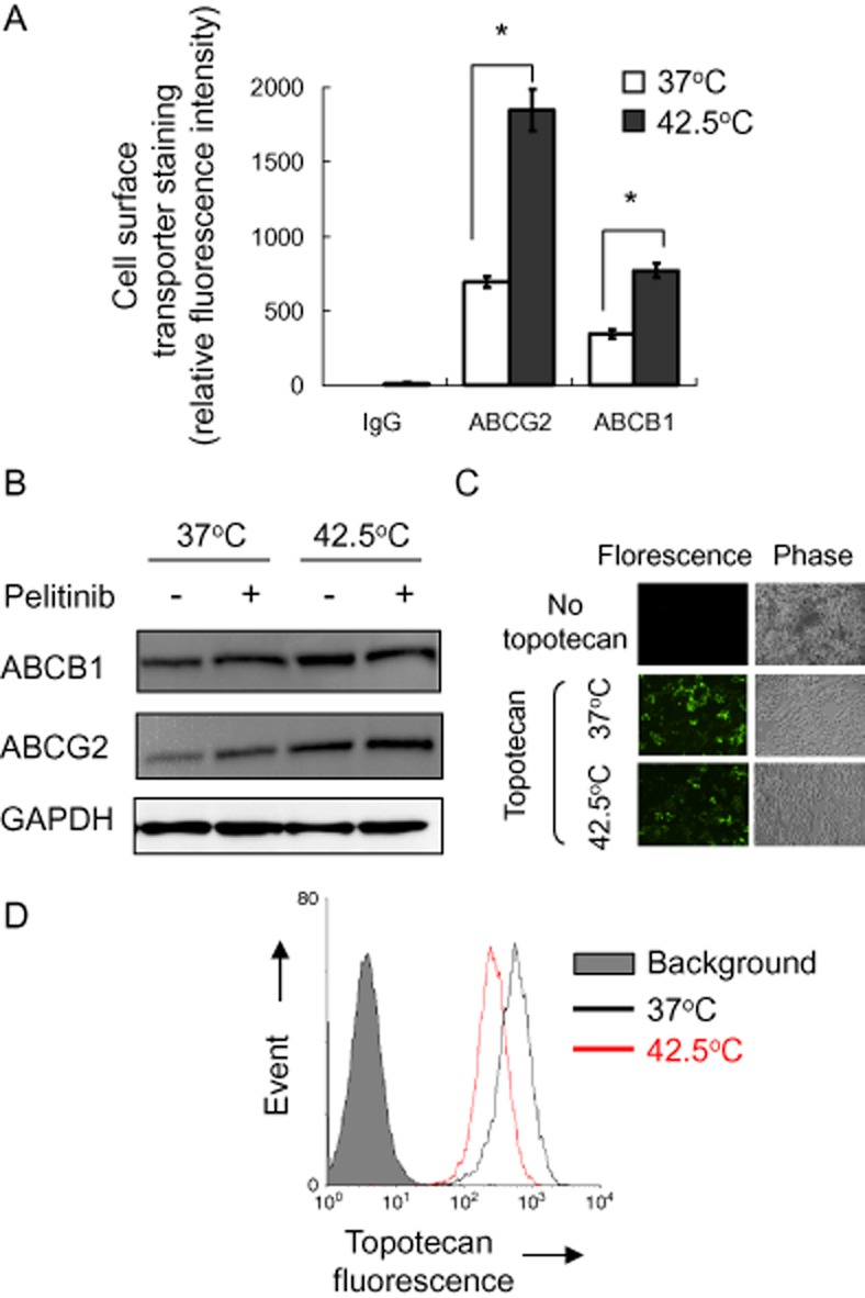
Cell surface expression and functional analysis of ABCG2 and ABCB1 in A549 cells with or without hyperthermia (42.5°C) treatment.Br J Pharmacol.2015 Aug;172(16):4089-106. |
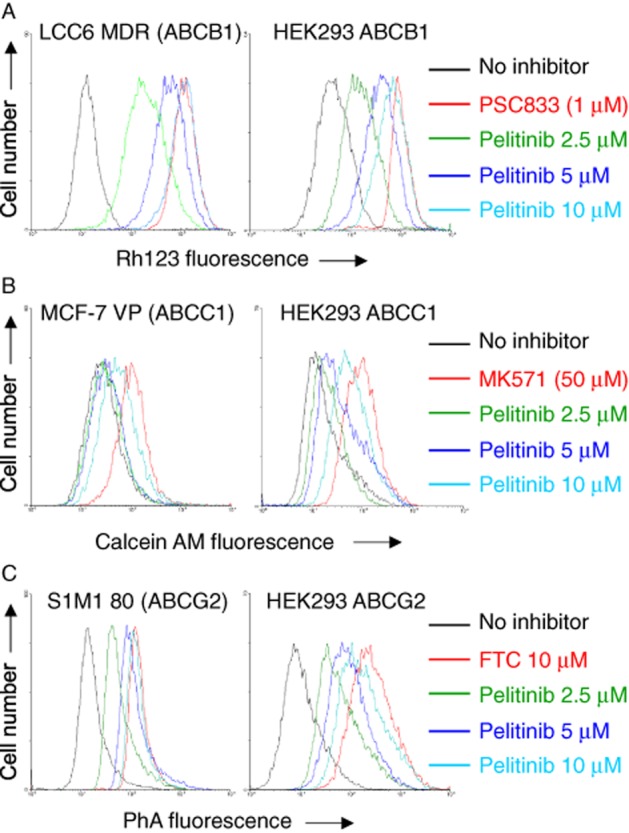
Inhibition of ABCB1-, ABCC1- or ABCG2-mediated efflux of fluorescent probe substrate bypelitinibin drug-resistant cells overexpressing the transporters (left panel) or HEK293 cells stably transfected with the three transporters (right panel).Br J Pharmacol.2015 Aug;172(16):4089-106. |
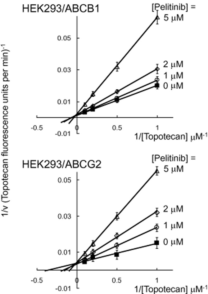
Inhibition kinetics of ABCB1- and ABCG2-mediated topotecan efflux bypelitinib.Br J Pharmacol.2015 Aug;172(16):4089-106. |
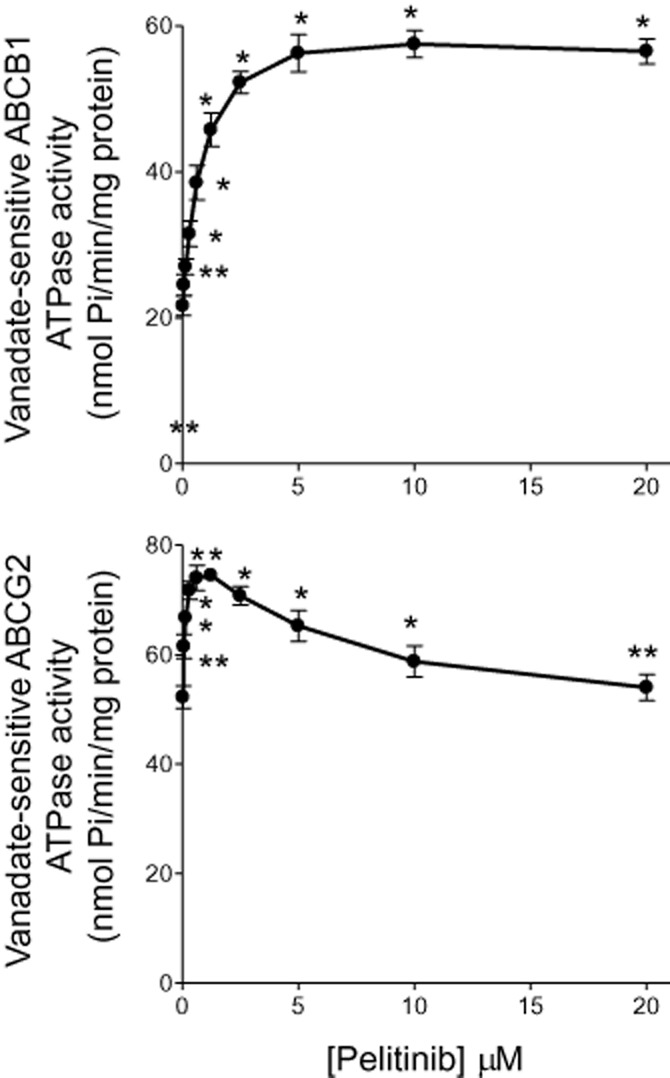 Effect ofpelitinibon the ATPase activity of ABCB1 (top panel) and ABCG2 (bottom panel).Br J Pharmacol.2015 Aug;172(16):4089-106. Effect ofpelitinibon the ATPase activity of ABCB1 (top panel) and ABCG2 (bottom panel).Br J Pharmacol.2015 Aug;172(16):4089-106. |
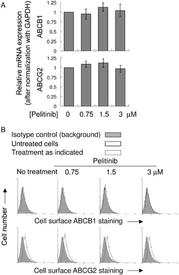
mRNA and cell surface expression of ABCB1 and ABCG2 in A549 after treatment withpelitinib.Br J Pharmacol.2015 Aug;172(16):4089-106. |
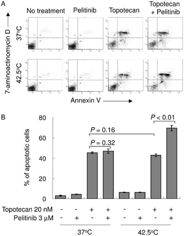
elitinibsensitized A549 cells to apoptosis specifically after exposure to hyperthermia.Br J Pharmacol.2015 Aug;172(16):4089-106. |
Products are for research use only; We do not sell to patients
Copyright 2020 InvivoChem LLC | All Rights Reserved


 COA
COA