| Size | Price | Stock | Qty |
|---|---|---|---|
| 1g |
|
||
| 2g |
|
||
| 5g |
|
||
| 10g |
|
||
| 25g | |||
| Other Sizes |
Paeonol (also known as NSC-401442; NSC401442; Peonol), a naturally occurring phenolic compound isolated from Paeonia suffruticosa (moutan cortex) and Cynanchum paniculatum which are used in traditional Chinese herbs/medicines. Paeonol has a range of pharmacologic functions such as antiaggregatory,anti-inflammatory and anti-ischemia reperfusion damage and anti-lipid peroxidation effects. It acts as a MAO inhibitor with an IC50 of about 50 μM.
| Targets |
Inhibitor of monoamine oxidase A (MAO-A) (IC50 = 53.4 μM) and monoamine oxidase B (MAO-B) (IC50 = 62.7 μM), showing moderate inhibitory activity against both MAO isoforms with slightly higher selectivity for MAO-A [1]
|
|---|---|
| ln Vitro |
It is discovered that pedonol inhibits MAO A in a dose-dependent manner, with an IC50 value of 54.6 μM. With an IC50 of 42.5 μM, eonol is demonstrated to inhibit MAO-B in a dose-dependent way. The calculated Ki for Paeonol is 51.1 μM. With a Ki value of 38.2 μM, Paeonol's inhibition of MAO B is of the competitive type[1].
In vitro MAO enzyme inhibition assays using rat brain mitochondria showed that Paeonol exhibited concentration-dependent inhibitory effects on both MAO-A and MAO-B: - At a concentration of 10 μM, Paeonol inhibited MAO-A activity by 12% and MAO-B activity by 9%; - At 50 μM, the inhibition rates of MAO-A and MAO-B increased to 38% and 31%, respectively; - At 100 μM, the inhibition rates reached 45% (MAO-A) and 38% (MAO-B); - The half-maximal inhibitory concentrations (IC50) were determined as 53.4 μM for MAO-A and 62.7 μM for MAO-B via curve fitting [1] |
| ln Vivo |
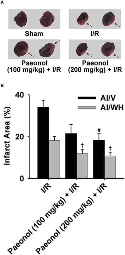
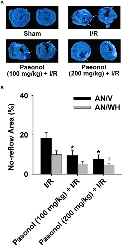
When compared to the I/R group [AN/V (%): 18.2±2.9], the 200 mg/kg Paeonol+I/R group [AN/V (%): 7.6±2.2, p<0.01] and the 100 mg/kg Paeonol+I/R group [AN/V (%): 9.4±2.8, p<0.05] both exhibit reduced amounts of no-reflow area in the ventricles. Specifically, compared to the I/R group [AN/WH (%): 10.0±1.9][2], the 200 mg/kg Paeonol + I/R group showed significantly lessened no-reflow in the whole heart [AN/WH (%): 4.6±1, p<0.05].
In a rat model of myocardial ischemia-reperfusion (I/R)-induced no-reflow (established by 30 minutes of left anterior descending coronary artery (LAD) ligation followed by 120 minutes of reperfusion), intravenous administration of Paeonol at doses of 5 mg/kg and 10 mg/kg (administered at the start of reperfusion) significantly improved cardiac function and alleviated no-reflow: - Regional myocardial blood flow (RMBF) in the risk area: Compared with the I/R model group (RMBF = 0.21 ± 0.03 mL/min/g), RMBF increased to 0.38 ± 0.04 mL/min/g (5 mg/kg group, +81%) and 0.52 ± 0.05 mL/min/g (10 mg/kg group, +148%); - No-reflow area: The percentage of no-reflow area relative to the risk area was 68.3 ± 4.2% in the model group, which decreased to 45.1 ± 3.8% (5 mg/kg group, -34%) and 32.6 ± 3.1% (10 mg/kg group, -52%) after Paeonol treatment; - Myocardial infarction area: The infarction area/risk area ratio was 52.7 ± 3.9% in the model group, and was reduced to 36.2 ± 3.5% (5 mg/kg group, -31%) and 25.8 ± 2.8% (10 mg/kg group, -51%) in Paeonol-treated groups; - Hemodynamic parameters: Paeonol (10 mg/kg) increased left ventricular systolic pressure (LVSP) by 28% and left ventricular ejection fraction (LVEF) by 32%, while decreasing left ventricular end-diastolic pressure (LVEDP) by 41% compared to the model group [2] |
| Enzyme Assay |
MAO-A activity assay: Mitochondria were isolated from rat whole brain by differential centrifugation (10,000×g for 20 minutes, followed by 120,000×g for 70 minutes) to obtain the MAO enzyme source. The reaction system (total volume 1 mL) contained 50 mM phosphate buffer (pH 7.5), 0.15 mM 5-hydroxytryptamine (5-HT, specific substrate for MAO-A), 0.3 mg/mL mitochondrial protein, and Paeonol at different concentrations (10 μM–200 μM). The mixture was incubated at 37°C for 30 minutes, and the reaction was terminated by adding 0.2 mL of 1 M perchloric acid. After centrifugation (12,000×g for 10 minutes), the supernatant was collected, and the concentration of the reaction product (5-hydroxyindoleacetic acid, 5-HIAA) was measured by high-performance liquid chromatography (HPLC) with ultraviolet detection (λ = 280 nm). The inhibition rate of MAO-A was calculated by comparing the 5-HIAA level with that of the control group (without Paeonol), and the IC50 value was obtained by fitting the concentration-inhibition curve using GraphPad Prism software [1]
- MAO-B activity assay: The enzyme source was the same rat brain mitochondria as used in MAO-A assay. The reaction system was identical to that of MAO-A assay except that the substrate was replaced with 0.15 mM phenylethylamine (specific substrate for MAO-B). After incubation and termination, the supernatant was analyzed by HPLC (ultraviolet detection at λ = 254 nm) to quantify the reaction product (phenylacetic acid). The MAO-B inhibition rate was calculated, and the IC50 value was determined via curve fitting [1] |
| Animal Protocol |
100, 200 mg/kg; oral
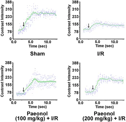
Male Wistar rats Rat model of myocardial I/R-induced no-reflow establishment and drug administration: Male Sprague-Dawley rats (250–300 g) were randomly divided into 4 groups (n=8 per group): sham-operated group, I/R model group, Paeonol 5 mg/kg group, and Paeonol 10 mg/kg group. Rats were anesthetized with sodium pentobarbital (intraperitoneal injection), tracheotomized, and mechanically ventilated. A left thoracotomy was performed to expose the heart, and a 6-0 silk suture was placed around the LAD. For the I/R model and Paeonol groups, the LAD was ligated with a slipknot for 30 minutes (confirmed by myocardial blanching and ST-segment elevation on electrocardiogram), followed by 120 minutes of reperfusion (slipknot released). Paeonol was dissolved in normal saline containing 5% dimethyl sulfoxide (DMSO), and administered via the tail vein at a volume of 1 mL/kg immediately after the start of reperfusion; the model group received the same volume of 5% DMSO-normal saline, and the sham group underwent thoracotomy without LAD ligation [2] - Detection of myocardial no-reflow and infarction area: At the end of reperfusion, 0.5 mL of fluorescent microspheres (2 μm diameter) was injected into the left ventricle to label perfused myocardium. The heart was excised, and the left ventricle was sliced into 5 transverse sections (1–2 mm thick). The sections were stained with 2,3,5-triphenyltetrazolium chloride (TTC, 1% w/v) at 37°C for 15 minutes (viable myocardium appeared red, infarcted myocardium appeared pale). The no-reflow area (non-fluorescent region) and infarction area (pale region) were imaged using a fluorescence microscope and quantified with Image-Pro Plus software. Regional myocardial blood flow (RMBF) was measured using a radioactive microsphere technique (141Ce-labeled microspheres) before ischemia and at the end of reperfusion [2] |
| ADME/Pharmacokinetics |
Metabolism / Metabolites
Paeoniflorin's known metabolites include (2-acetyl-5-methoxyphenyl) hydrogen sulfate. |
| References | |
| Additional Infomation |
Paeonol belongs to the phenol and methoxybenzene class of compounds and is a metabolite. Paeoniflorin has been reported to exist in Clausena dunniana, Ficus benghalensis and other organisms with relevant data. See also: Paeonia lactiflora root (part); Paeonia X suffruticosa root (part). Paeoniflorin (2'-hydroxy-4'-methoxyacetophenone) is a natural phenolic compound that has been isolated mainly from the roots of Paeonia suffruticosa Andr. and other plants. The moderate inhibitory activity of paeoniflorin against MAO-A and MAO-B suggests its potential application in the treatment of neurological diseases associated with monoamine metabolism disorders, such as depression and Parkinson's disease [1]. Paeoniflorin’s protective effect against myocardial no-reflow is thought to involve multiple mechanisms: (1) dilating coronary microvessels to improve microcirculation perfusion; (2) inhibiting neutrophil activation and adhesion to reduce microvascular obstruction; and (3) scavenging reactive oxygen species (ROS) to alleviate microvascular damage caused by oxidative stress. The dose-dependent effect of paeoniflorin (10 mg/kg is more effective than 5 mg/kg) supports its potential as a treatment for myocardial ischemia/reperfusion injury [2].
|
| Molecular Formula |
C9H10O3
|
|
|---|---|---|
| Molecular Weight |
166.17
|
|
| Exact Mass |
166.062
|
|
| CAS # |
552-41-0
|
|
| Related CAS # |
Paeonol-d3;55712-78-2
|
|
| PubChem CID |
11092
|
|
| Appearance |
White to off-white solid powder
|
|
| Density |
1.2±0.1 g/cm3
|
|
| Boiling Point |
301.9±22.0 °C at 760 mmHg
|
|
| Melting Point |
48-50 °C(lit.)
|
|
| Flash Point |
122.3±15.8 °C
|
|
| Vapour Pressure |
0.0±0.7 mmHg at 25°C
|
|
| Index of Refraction |
1.538
|
|
| LogP |
2.16
|
|
| Hydrogen Bond Donor Count |
1
|
|
| Hydrogen Bond Acceptor Count |
3
|
|
| Rotatable Bond Count |
2
|
|
| Heavy Atom Count |
12
|
|
| Complexity |
167
|
|
| Defined Atom Stereocenter Count |
0
|
|
| InChi Key |
UILPJVPSNHJFIK-UHFFFAOYSA-N
|
|
| InChi Code |
InChI=1S/C9H10O3/c1-6(10)8-4-3-7(12-2)5-9(8)11/h3-5,11H,1-2H3
|
|
| Chemical Name |
1-(2-Hydroxy-4-methoxyphenyl)ethanone
|
|
| Synonyms |
|
|
| HS Tariff Code |
2934.99.9001
|
|
| Storage |
Powder -20°C 3 years 4°C 2 years In solvent -80°C 6 months -20°C 1 month |
|
| Shipping Condition |
Room temperature (This product is stable at ambient temperature for a few days during ordinary shipping and time spent in Customs)
|
| Solubility (In Vitro) |
|
|||
|---|---|---|---|---|
| Solubility (In Vivo) |
Solubility in Formulation 1: ≥ 2.5 mg/mL (15.04 mM) (saturation unknown) in 10% DMSO + 40% PEG300 + 5% Tween80 + 45% Saline (add these co-solvents sequentially from left to right, and one by one), suspension solution.
For example, if 1 mL of working solution is to be prepared, you can add 100 μL of 25.0 mg/mL clear DMSO stock solution to 400 μL PEG300 and mix evenly; then add 50 μL Tween-80 to the above solution and mix evenly; then add 450 μL normal saline to adjust the volume to 1 mL. Preparation of saline: Dissolve 0.9 g of sodium chloride in 100 mL ddH₂ O to obtain a clear solution. Solubility in Formulation 2: ≥ 2.5 mg/mL (15.04 mM) (saturation unknown) in 10% DMSO + 90% (20% SBE-β-CD in Saline) (add these co-solvents sequentially from left to right, and one by one), clear solution. For example, if 1 mL of working solution is to be prepared, you can add 100 μL of 25.0 mg/mL clear DMSO stock solution to 900 μL of 20% SBE-β-CD physiological saline solution and mix evenly. Preparation of 20% SBE-β-CD in Saline (4°C,1 week): Dissolve 2 g SBE-β-CD in 10 mL saline to obtain a clear solution. View More
Solubility in Formulation 3: ≥ 2.5 mg/mL (15.04 mM) (saturation unknown) in 10% DMSO + 90% Corn Oil (add these co-solvents sequentially from left to right, and one by one), clear solution. Solubility in Formulation 4: ≥ 2.5 mg/mL (15.04 mM) (saturation unknown) in 10% EtOH + 90% (20% SBE-β-CD in Saline) (add these co-solvents sequentially from left to right, and one by one), clear solution. For example, if 1 mL of working solution is to be prepared, you can add 100 μL of 25.0 mg/mL clear EtOH stock solution to 900 μL of 20% SBE-β-CD physiological saline solution and mix well. Preparation of 20% SBE-β-CD in Saline (4°C,1 week): Dissolve 2 g SBE-β-CD in 10 mL saline to obtain a clear solution. Solubility in Formulation 5: 10% EtOH + 90% Corn Oil |
| Preparing Stock Solutions | 1 mg | 5 mg | 10 mg | |
| 1 mM | 6.0179 mL | 30.0897 mL | 60.1793 mL | |
| 5 mM | 1.2036 mL | 6.0179 mL | 12.0359 mL | |
| 10 mM | 0.6018 mL | 3.0090 mL | 6.0179 mL |
*Note: Please select an appropriate solvent for the preparation of stock solution based on your experiment needs. For most products, DMSO can be used for preparing stock solutions (e.g. 5 mM, 10 mM, or 20 mM concentration); some products with high aqueous solubility may be dissolved in water directly. Solubility information is available at the above Solubility Data section. Once the stock solution is prepared, aliquot it to routine usage volumes and store at -20°C or -80°C. Avoid repeated freeze and thaw cycles.
Calculation results
Working concentration: mg/mL;
Method for preparing DMSO stock solution: mg drug pre-dissolved in μL DMSO (stock solution concentration mg/mL). Please contact us first if the concentration exceeds the DMSO solubility of the batch of drug.
Method for preparing in vivo formulation::Take μL DMSO stock solution, next add μL PEG300, mix and clarify, next addμL Tween 80, mix and clarify, next add μL ddH2O,mix and clarify.
(1) Please be sure that the solution is clear before the addition of next solvent. Dissolution methods like vortex, ultrasound or warming and heat may be used to aid dissolving.
(2) Be sure to add the solvent(s) in order.
| NCT Number | Recruitment | interventions | Conditions | Sponsor/Collaborators | Start Date | Phases |
| NCT04657926 | Completed | Drug: APPA Drug: Placebo |
Osteoarthritis | AKL Research and Development | September 9, 2020 | Phase 2 |
|
|
|
Products are for research use only; We do not sell to patients
Copyright 2020 InvivoChem LLC | All Rights Reserved