| Size | Price | Stock | Qty |
|---|---|---|---|
| 25mg |
|
||
| 50mg |
|
||
| 100mg |
|
||
| 250mg |
|
||
| 500mg |
|
||
| 1g |
|
||
| Other Sizes |


Purity: ≥98%
| Targets |
HER2 (IC50 = 59 nM); EGFR (IC50 = 92 nM); KDR (IC50 = 800 nM); Src (IC50 = 1.4 μM)
Neratinib (HKI-272; PB272; Nerlynx) potently inhibits HER2 (IC₅₀ = 59 nM) and EGFR (IC₅₀ = 92 nM) tyrosine kinases. It weakly inhibits KDR (IC₅₀ = 0.8 μM) and Src (IC₅₀ = 1.4 μM), and shows no significant inhibitory activity against Akt, CDK1/2/4, IKK-2, MK-2, PDK1, c-Raf, and c-Met [1] |
|---|---|
| ln Vitro |
Neratinib shows no action against tyrosine kinase c-Met[1], Akt, cyclin D1/cdk4, cyclin E/cdk2, cyclin B1/cdk1, IKK-2, MK-2, PDK1, c-Raf, and Tpl-2, among other serine-threonine kinases[1].
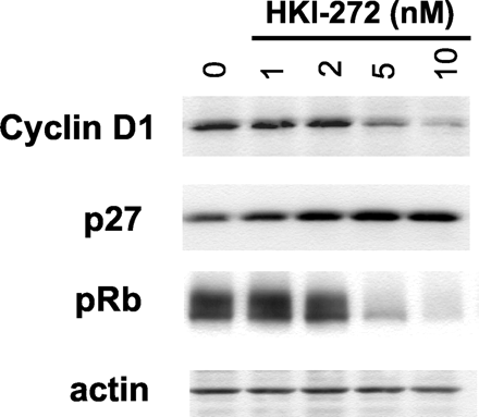
Neratinib is much less active in cell lines that express neither EGFR nor HER-2 (3T3, MDA-MB-435, and SW620), and it inhibits the proliferation of cell lines that exhibit high levels of HER-2 (3T3/neu, SK-Br-3, and BT474)[1]. Neratinib (0-2 nM, 12-16 h) arrests BT474 cell cycle at G1-S phase[1]. Neratinib causes cyclin D1 levels to be down-regulated, Akt and MAPK phosphorylation to be inhibited, and p27 to be induced[1]. Neratinib (HKI-272; PB272; Nerlynx) selectively inhibits the proliferation of HER2-transfected 3T3 cells (3T3/neu) and HER2-overexpressing breast cancer cell lines SK-Br-3 and BT474 with IC₅₀ values of 2-3 nM, which is more than 230 times more potent than its effect on untransfected 3T3 cells and EGFR/HER2-negative cell lines MDA-MB-435 and SW620 [1] In osteosarcoma cell lines U2-OS and 143B, Neratinib (HKI-272; PB272; Nerlynx) exhibits time-dependent antiproliferative activity. It also reduces cell migration and invasion by downregulating the expression of phosphorylated HER2 (p-HER2), PI3K, and phosphorylated Akt (p-Akt) [1] The drug blocks HER2 and EGFR downstream signaling pathways, leading to decreased phosphorylation of key signaling molecules and subsequent inhibition of tumor cell proliferation and survival [1] |
| ln Vivo |
Neratinib (HKI-272) exhibits anticancer properties against cancer cells that express high levels of EGFR or HER-2 (0-80 mg/kg/day; i.e., 42 days)[1].
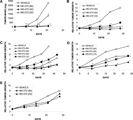
In vivo, HKI-272 is active in HER-2- and EGFR-dependent tumor xenograft models when dosed orally on a once daily schedule. On the basis of its favorable preclinical pharmacological profile, HKI-272 has been selected as a candidate for additional development as an antitumor agent in breast and other HER-2-dependent cancers.[1] Oral administration of Neratinib (HKI-272; PB272; Nerlynx) significantly inhibits the growth of HER2-overexpressing tumor xenografts in nude mice. It reduces tumor volume and weight by suppressing intratumoral HER2 signaling and proliferation [1] In combination with standard chemotherapy regimens, Neratinib (HKI-272; PB272; Nerlynx) enhances the antitumor efficacy in xenograft models, showing synergistic effects on tumor growth inhibition [1] |
| Enzyme Assay |
Prepared as 10 mg/mL stocks in DMSO, neratinib is diluted in 25 mM HEPES (pH 7.5; 0.002 ng/mL–20 μg/mL). In 96-well ELISA plates, purified recombinant COOH-terminal fragments of HER2 (amino acids 676-1255) or epidermal growth factor receptor (EGFR) (amino acids 645-1186) are diluted in 100 mM HEPES (pH 7.5) and 50% glycerol. The mixture is then incubated with increasing concentrations of Neratinib in 4 mM HEPES (pH 7.5), 0.4 mM MnCl2, 20 μM sodium vanadate, and 0.2 mM DTT for 15 minutes at room temperature. 40 μM ATP and 20 mM MgCl2 are added to start the kinase reaction, which is then left to run at room temperature for an hour. Wash the plates, then use anti-phospho-tyrosine antibodies (15 ng/well) labeled with europium to detect phosphorylation. Using a Victor2 fluorescence reader (excitation wavelength of 340 nm and emission wavelength of 615 nm), the signal is detected following the washing and enhancement stages. An inhibition curve is used to determine the concentration of Neratinib (IC50) at which receptor phosphorylation is inhibited by 50%.
Activity of HER-2 and EGFR cytoplasmic domains was measured by an autophosphorylation assay using time-resolved fluorometry. Compounds were prepared as 10 mg/ml stocks in DMSO and diluted in 25 mm HEPES (pH 7.5; 0.002 ng/ml–20 μg/ml). Enzyme [diluted in 100 mm HEPES (pH 7.5) and 50% glycerol] was incubated with inhibitor in 4 mm HEPES (pH 7.5), 0.4 mm MnCl2, 20 μm sodium vanadate, and 0.2 mm DTT for 15 min at room temperature in 96-well ELISA plates. The kinase reaction was initiated by the addition of 40 μm ATP and 20 mm MgCl2 and allowed to proceed for 1 h at room temperature. Plates were washed, and phosphorylation was detected using Europium-labeled anti-phospho-tyrosine antibodies (15 ng/well; Wallac). After washing and enhancement steps according to the manufacturer’s recommendations, signal was detected using a Victor2 fluorescence reader (excitation wavelength 340 nm, emission wavelength 615 nm). The concentration of compound that inhibited receptor phosphorylation by 50% (IC50) was calculated from inhibition curves.[1] Assays for other kinases were performed using recombinant enzymes expressed in bacterial, insect, or human cell lines. All enzymes used were serine-threonine kinases, except c-Met, KDR, src (tyrosine kinases), and MEK1 (dual specificity). Substrates used were peptides (Akt, IKK-2, MK2, PDK1, src, and Tpl2), proteins (cyclin D1/CDK4, cyclin E/CDK2, cyclin B1/CDK1, and c-Raf), poly(glutamic acid4-tyrosine) (KDR), or the kinase itself (autophosphorylation; c-met). Phosphorylation was measured using TMB peroxidase substrate for cyclin/cyclin-dependent kinase (cdk), LabChip for MK-2, or DELPHIA/LANCE for all others.[1] Recombinant HER2 and EGFR kinase domains were incubated with ATP and specific peptide substrates in the presence of serial dilutions of Neratinib (HKI-272; PB272; Nerlynx). The reaction mixtures were maintained at a constant temperature for a set period, and phosphorylated substrates were detected using a spectrophotometric method. Inhibition rates were calculated by comparing the signal intensity with vehicle control groups, and IC₅₀ values were derived from dose-response curves [1] Kinase activity assays for KDR, Src, and other non-target kinases were performed using the same protocol to evaluate the selectivity of the drug [1] |
| Cell Assay |
Different concentrations of Neratinib are applied to cells (3T3, 3T3/neu, A431, BT474, SK-Br-3, MDA-MB-435, and SW480) for a period of either two or six days. A protein-binding dye called sulforhodamine B is used to measure cell proliferation. In short, cells are thoroughly cleaned with water after being fixed with 10% trichloroacetic acid. After staining the cells with 0.1% sulforhodamine B, they are rinsed in 5% acetic acid. After solubilizing the protein-associated dye in 10 mM Tris, the absorbance is calculated at 450 nM. Inhibition curves are used to calculate the concentration of neratinib (IC50) that inhibits cell proliferation by 50%.
HER2-overexpressing SK-Br-3, BT474 cells, and HER2-transfected 3T3 cells were seeded in 96-well plates at a density of 5×10³ cells/well and treated with Neratinib (HKI-272; PB272; Nerlynx) at concentrations ranging from 0.001 to 100 nM for 72 hours. Cell viability was measured using a colorimetric assay to determine IC₅₀ values [1] Osteosarcoma cells U2-OS and 143B were treated with different concentrations of Neratinib (HKI-272; PB272; Nerlynx) for 24, 48, and 72 hours. Wound healing and Transwell assays were used to assess cell migration and invasion, respectively. Western blot analysis was performed to detect the expression levels of p-HER2, PI3K, Akt, and p-Akt [1] For signaling pathway analysis, tumor cells were treated with the drug for 24 hours, lysed, and probed with antibodies against phosphorylated and total forms of downstream signaling molecules to evaluate the inhibition of HER2/EGFR pathways [1] |
| Animal Protocol |
Female athymic (nude) mice, tumor xenograft[1]
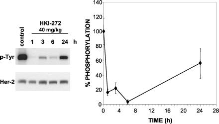
10, 20, 40, 60 or 80 mg/kg/day Gavage, 42 days Tumor Xenograft Studies.[1] Tumor cells (maintained in tissue culture) or tumor fragments were implanted s.c. in the flanks of female athymic (nude) mice. For estrogen-dependent cell lines (BT474, MCF-7, and SK-OV-3), animals were implanted with hormone pellets (0.72 mg of 17-β estradiol, 60-day release) 1 week before implantation of tumors. Additionally, SK-OV-3 cells were suspended in Matrigel basement membrane matrix for implantation. Treatment was initiated after tumors had reached a size of 90–200 mg, following random assignment of the animals to different treatment groups (staging, day 0). For 3T3/neu xenografts, treatment was initiated the day after tumor implantation (day 0). HKI-272 was formulated in 0.5% methocellulose-0.4% polysorbate-80 (Tween 80) and administered daily, p.o., by gavage. Tumor mass [(length × width2)/2] was determined every 7 days. Tumor outgrowth in all xenograft studies, except 3T3/neu, was expressed as relative tumor growth: the ratio of the mean tumor mass to the mean tumor mass on day 0. Inhibition of tumor growth was calculated relative to vehicle-treated controls. Statistical significance of inhibition was demonstrated using one-tailed Student’s t test (equal variance) after log transformation of the data.[1] HER-2 Phosphorylation in Xenografts.[1] Athymic female nude mice (5 animals/group) were implanted s.c. with BT474 tumor fragments (∼30 mm3). When tumors reached 200–300 mg, animals were given a single oral dose (40 mg/kg) of HKI-272 in pH 2.0 water. Tumors from control and treated animals were excised at 1, 3, 6, and 24 h and minced. Tumor fragments were suspended in 10 mm Tris (pH 7.5), 5 mm EDTA, 150 mm NaCl, 1% Triton X-100, 1% sodium deoxycholate, 0.1% SDS, 1 mm phenylmethylsulfonyl fluoride, 10 μg/ml pepstatin, 10 μg/ml leupeptin, 10 μg/ml aprotinin, 2 mm sodium vanadate, and 100 mm sodium fluoride and lysed by homogenization on ice with a polytron. After clarification by centrifugation, protein concentration in lysates was estimated using the Bio-Rad DC protein assay. Sixty μg of lysate pooled from each group were analyzed by SDS-PAGE and immunoblotting with phospho-tyrosine-specific antibodies. Pooled extracts were also immunoprecipitated using 4 μg of anti-HER-2 antibodies for 1 h at 4°C. Immune complexes were collected on protein A-agarose, washed, and analyzed by immunoblotting using phospho-tyrosine-specific antibodies. Extracts from individual tumors were analyzed to determine variability between animals. Nude mice bearing HER2-overexpressing SK-Br-3 or BT474 xenografts (tumor volume ~100 mm³) were randomly divided into control and treatment groups. Neratinib (HKI-272; PB272; Nerlynx) was suspended in 0.5% carboxymethylcellulose and administered orally at doses of 10-40 mg/kg/day for 21 consecutive days. Tumor volume was measured every 3 days, and mice were euthanized at the end of treatment to collect tumor tissues for immunohistochemical analysis of proliferation markers [1] In combination therapy experiments, nude mice with tumor xenografts were treated with Neratinib (HKI-272; PB272; Nerlynx) (20 mg/kg/day orally) plus standard chemotherapy agents. The drug was administered 1 hour before chemotherapy, and the treatment cycle was repeated every 7 days for 3 cycles. Tumor weight and survival time of mice were recorded [1] |
| ADME/Pharmacokinetics |
Absorption, Distribution and Excretion
The time to peak concentration (Tmax) of neratinib and its main active metabolites M3, M6, and M7 is 2–8 hours. Co-administration with a high-fat meal increases Cmax by 1.7-fold and total exposure by 2.2-fold. Co-administration with a standard meal increases Cmax by 1.2-fold and total exposure by 1.1-fold. Co-administration with proton pump inhibitors or other gastric acid inhibitors reduces Cmax by 71% and total exposure by 65%. 97.1% of the total dose is excreted in feces, and 1.13% in urine. The steady-state apparent volume of distribution is 6433 L. After multiple dosings, the total clearance is 216 L/h (after initial dosing) and 281 L/h (steady-state). Metabolism/Metabolites Neratinib is primarily metabolized via CYP3A4. It is also metabolized in small amounts by flavin-containing monooxygenases. The systemic exposures of the active metabolites M3, M6, M7, and M11 of neratinib are 15%, 33%, 22%, and 4%, respectively. Biological Half-Life After a single dose, the mean elimination half-life is 7–17 hours. After multiple doses, the mean plasma half-life of neratinib is 14.6 hours, M3 is 21.6 hours, M6 is 13.8 hours, and M7 is 10.4 hours. Neratinib (HKI-272; PB272; Nerlynx) has oral bioavailability, reaching peak plasma concentrations 2–8 hours after oral administration. The drug has a plasma half-life of 7-17 hours in the human body, supporting once-daily administration [1]. The drug is widely distributed in tumor tissues, and its tumor-to-plasma concentration ratio is beneficial for effectively targeting HER2-overexpressing tumors [1]. |
| Toxicity/Toxicokinetics |
Hepatotoxicity
Elevated serum transaminase levels are not uncommon during neratinib treatment, occurring in up to 10% of patients, but only 1% to 2% of patients experience levels exceeding five times the upper limit of normal. In premarketing studies, no clinically significant neratinib-related liver injury cases were observed; serum enzyme elevations were typically mild and self-limiting, without symptoms or jaundice. Hepatotoxicity may be a class effect of HER2 protein kinase inhibitors, although the frequency and severity vary among different drugs. Specific details of neratinib-related liver injury, such as latency, serum enzyme profiles, clinical features, and disease course, have not been published. Other tyrosine kinase receptor inhibitors typically cause liver injury within days or weeks of treatment initiation, manifesting as a sudden increase in hepatocyte enzymes, with a moderate to severe course. Immune hypersensitivity and autoimmune features are uncommon. Patients with a history of cirrhosis or liver dysfunction due to hepatic tumor burden have an increased risk of clinically significant liver injury and liver failure. However, there is currently no conclusive evidence linking neratinib to clinically visible cases of liver injury. Probability Score: E (Unproven but suspected cause of clinically visible liver damage). Use during pregnancy and lactation> ◉ Overview of use during lactation There is currently no information regarding the clinical use of neratinib during lactation. Because neratinib and its metabolites bind to plasma proteins at a rate exceeding 99%, its levels in breast milk may be very low. The manufacturer recommends discontinuing breastfeeding during neratinib treatment. ◉ Effects on breastfed infants No published information found as of the revision date. ◉ Effects on lactation and breast milk No published information found as of the revision date. Protein binding> Neratinib binds to human plasma proteins at a rate exceeding 99%. It binds to human serum albumin and α1 acid glycoprotein. In preclinical studies, neratinib (HKI-272; PB272; Nerlynx) showed mild to moderate toxicity at therapeutic doses, including transient diarrhea and slight weight loss in mice. No significant hepatotoxicity or nephrotoxicity was observed, and serum ALT, AST, and creatinine levels were normal[1]. Neratinib (HKI-272; PB272; Nerlynx) has approximately 95% plasma protein binding in human plasma, which may affect its distribution and potential drug interactions[1]. |
| References | |
| Additional Infomation |
Neratinib is a quinoline compound with a cyano group at position 3, a 3-chloro-4-(2-pyridinemethoxy)aniline group at position 4, a 4-dimethylamino-trans-but-2-enamid group at position 6, and an ethoxy group at position 7. It is a tyrosine kinase inhibitor and an antitumor drug. It belongs to the quinoline and nitrile class of compounds. Neratinib was approved in July 2017 for extended adjuvant therapy in human epidermal growth factor receptor 2 (HER2)-positive breast cancer. Puma Biotechnology Inc. obtained approval for the drug under the brand name Nerlynx. Currently, neratinib is being investigated for the treatment of several other types of cancer. Neratinib is a kinase inhibitor. Its mechanism of action is as a kinase inhibitor and a P-glycoprotein inhibitor. Neratinib is an oral tyrosine kinase receptor inhibitor used for extended adjuvant therapy in early-stage breast cancer. The incidence of transient elevations in serum transaminase levels during neratinib treatment is low, but there is no conclusive evidence that it is associated with clinically significant liver injury with jaundice. Neratinib is an orally administered 6,7-disubstituted-4-aniline-quinoline-3-nitrile compound and an irreversible inhibitor of HER-2 receptor tyrosine kinase, possessing potential antitumor activity. Neratinib binds irreversibly to the HER-2 receptor, thereby reducing intracellular autophosphorylation; its mechanism of action may be targeting cysteine residues in the receptor's ATP-binding pocket. Treatment of cells with this drug leads to inhibition of downstream signal transduction events and cell cycle regulation pathways; cell cycle arrest at the G1-S (Gap 1/DNA synthesis) phase transition; ultimately resulting in reduced cell proliferation. Neratinib also inhibits epidermal growth factor receptor (EGFR) kinase and the proliferation of EGFR-dependent cells. See also: neratinib maleate (its active ingredient).
Drug Indications Neratinib is indicated for adult patients with early-stage HER2-overexpressing/amplified breast cancer as extended adjuvant therapy following trastuzumab adjuvant therapy. FDA Label Neratinib is indicated for adult patients with early-stage hormone receptor-positive, HER2-overexpressing/amplified breast cancer who have completed less than one year of prior trastuzumab adjuvant therapy. Breast Cancer Treatment Mechanism of Action Neratinib binds to and irreversibly inhibits EGFR, HER2, and HER4. This prevents autophosphorylation of tyrosine residues on the receptors and reduces oncogenic signaling via mitogen-activated protein kinases and the Akt pathway. Pharmacodynamics Neratinib is a tyrosine kinase inhibitor with antitumor activity against epidermal growth factor receptor (EGFR), HER2, and human epidermal growth factor receptor 4 (HER4)-positive cancers. Nerlatinib (HKI-272; PB272; Nerlynx) is an irreversible pan-HER tyrosine kinase inhibitor that exerts its antitumor effect by covalently binding to the kinase domains of HER2, EGFR, and HER4, thereby permanently blocking downstream signaling pathways involved in tumor cell proliferation, migration, and survival[1]. It is mainly used to treat HER2-positive breast cancer, especially for patients who have previously received trastuzumab treatment. This drug can overcome trastuzumab resistance caused by HER2 extracellular domain truncation or EGFR co-activation[1]. |
| Molecular Formula |
C30H29CLN6O3
|
|---|---|
| Molecular Weight |
557.04
|
| Exact Mass |
556.198
|
| Elemental Analysis |
C, 64.68; H, 5.25; Cl, 6.36; N, 15.09; O, 8.62
|
| CAS # |
698387-09-6
|
| Related CAS # |
Neratinib maleate;915942-22-2;Neratinib-d6;1259519-18-0
|
| PubChem CID |
9915743
|
| Appearance |
White to light brown solid powder
|
| Density |
1.3±0.1 g/cm3
|
| Boiling Point |
757.0±60.0 °C at 760 mmHg
|
| Melting Point |
184 °C
|
| Flash Point |
411.6±32.9 °C
|
| Vapour Pressure |
0.0±2.5 mmHg at 25°C
|
| Index of Refraction |
1.667
|
| LogP |
5.46
|
| Hydrogen Bond Donor Count |
2
|
| Hydrogen Bond Acceptor Count |
8
|
| Rotatable Bond Count |
11
|
| Heavy Atom Count |
40
|
| Complexity |
881
|
| Defined Atom Stereocenter Count |
0
|
| SMILES |
ClC1=C(C([H])=C([H])C(=C1[H])N([H])C1=C(C#N)C([H])=NC2=C([H])C(=C(C([H])=C12)N([H])C(/C(/[H])=C(\[H])/C([H])([H])N(C([H])([H])[H])C([H])([H])[H])=O)OC([H])([H])C([H])([H])[H])OC([H])([H])C1=C([H])C([H])=C([H])C([H])=N1
|
| InChi Key |
JWNPDZNEKVCWMY-VQHVLOKHSA-N
|
| InChi Code |
InChI=1S/C30H29ClN6O3/c1-4-39-28-16-25-23(15-26(28)36-29(38)9-7-13-37(2)3)30(20(17-32)18-34-25)35-21-10-11-27(24(31)14-21)40-19-22-8-5-6-12-33-22/h5-12,14-16,18H,4,13,19H2,1-3H3,(H,34,35)(H,36,38)/b9-7+
|
| Chemical Name |
(E)-N-[4-[3-chloro-4-(pyridin-2-ylmethoxy)anilino]-3-cyano-7-ethoxyquinolin-6-yl]-4-(dimethylamino)but-2-enamide
|
| Synonyms |
Neratinib; HKI-272; PB272; HKI272; PB 272; HKI 272; 698387-09-6; HKI-272; Neratinib (HKI-272); Nerlynx; HKI 272; UNII-JJH94R3PWB; JJH94R3PWB; PB-272; trade name: Nerlynx
|
| HS Tariff Code |
2934.99.9001
|
| Storage |
Powder -20°C 3 years 4°C 2 years In solvent -80°C 6 months -20°C 1 month |
| Shipping Condition |
Room temperature (This product is stable at ambient temperature for a few days during ordinary shipping and time spent in Customs)
|
| Solubility (In Vitro) |
DMSO: 3~12 mg/mL (vary from batch to batch)
Water: 1 mg/mL (Insoluble) Ethanol: 1 mg/mL |
|---|---|
| Solubility (In Vivo) |
Solubility in Formulation 1: 2 mg/mL (3.59 mM) in 10% DMSO + 90% (20% SBE-β-CD in Saline) (add these co-solvents sequentially from left to right, and one by one), suspension solution; with sonication.
For example, if 1 mL of working solution is to be prepared, you can add 100 μL of 20.0 mg/mL clear DMSO stock solution to 900 μL of 20% SBE-β-CD physiological saline solution and mix evenly. Preparation of 20% SBE-β-CD in Saline (4°C,1 week): Dissolve 2 g SBE-β-CD in 10 mL saline to obtain a clear solution. Solubility in Formulation 2: ≥ 0.83 mg/mL (1.49 mM) (saturation unknown) in 10% DMSO + 40% PEG300 + 5% Tween80 + 45% Saline (add these co-solvents sequentially from left to right, and one by one), clear solution. For example, if 1 mL of working solution is to be prepared, you can add 100 μL of 8.3 mg/mL clear DMSO stock solution to 400 μL of PEG300 and mix evenly; then add 50 μL of Tween-80 to the above solution and mix evenly; then add 450 μL of normal saline to adjust the volume to 1 mL. Preparation of saline: Dissolve 0.9 g of sodium chloride in 100 mL ddH₂ O to obtain a clear solution. View More
Solubility in Formulation 3: 30% PEG400+0.5% Tween80+5% propylene glycol: 5 mg/mL Solubility in Formulation 4: 3.33 mg/mL (5.98 mM) in 0.5% MC 0.5% Tween-80 (add these co-solvents sequentially from left to right, and one by one), suspension solution; with ultrasonication. |
| Preparing Stock Solutions | 1 mg | 5 mg | 10 mg | |
| 1 mM | 1.7952 mL | 8.9760 mL | 17.9520 mL | |
| 5 mM | 0.3590 mL | 1.7952 mL | 3.5904 mL | |
| 10 mM | 0.1795 mL | 0.8976 mL | 1.7952 mL |
*Note: Please select an appropriate solvent for the preparation of stock solution based on your experiment needs. For most products, DMSO can be used for preparing stock solutions (e.g. 5 mM, 10 mM, or 20 mM concentration); some products with high aqueous solubility may be dissolved in water directly. Solubility information is available at the above Solubility Data section. Once the stock solution is prepared, aliquot it to routine usage volumes and store at -20°C or -80°C. Avoid repeated freeze and thaw cycles.
Calculation results
Working concentration: mg/mL;
Method for preparing DMSO stock solution: mg drug pre-dissolved in μL DMSO (stock solution concentration mg/mL). Please contact us first if the concentration exceeds the DMSO solubility of the batch of drug.
Method for preparing in vivo formulation::Take μL DMSO stock solution, next add μL PEG300, mix and clarify, next addμL Tween 80, mix and clarify, next add μL ddH2O,mix and clarify.
(1) Please be sure that the solution is clear before the addition of next solvent. Dissolution methods like vortex, ultrasound or warming and heat may be used to aid dissolving.
(2) Be sure to add the solvent(s) in order.
Innovative Trial for Understanding the Impact of Targeted Therapies in NF2-Related Schwannomatosis (INTUITT-NF2)
CTID: NCT04374305
Phase: Phase 2 Status: Active, not recruiting
Date: 2024-11-18
Effect of HKI-272 on cell cycle regulatory proteins. BT474 cells were incubated with HKI-272 for 12–16 h at 37°C. Protein extracts were analyzed by immunoblotting using cyclin D1, p27, or retinoblastoma (Rb)-specific antibodies. Actin antibodies were used as control. Cancer Res. 2004 Jun 1;64(11):3958-65. |
In vivo activity of HKI-272. A, groups of 10 mice were implanted with 2 × 10⁶ 3T3/neu cells. Animals were treated with the vehicle or HKI-272 on days 1–10 (p.o.), beginning the day after implantation. B–E, mice were implanted with BT474 tumor fragments (30 mm3; B), 5 × 10⁶ SK-OV-3 cells (C), 5 × 10⁶ A431 cells (D), or MCF-7 tumor fragments (30 mm3; E). Cancer Res. 2004 Jun 1;64(11):3958-65. |
Inhibition of HER-2 phosphorylation in xenografts. BT474 tumor-bearing mice (5 animals/group) were treated with vehicle or a single dose of HKI-272. Tumors from control and treated mice were dissected at various times after compound administration. Cancer Res. 2004 Jun 1;64(11):3958-65. |
Products are for research use only; We do not sell to patients
Copyright 2020 InvivoChem LLC | All Rights Reserved


 COA
COA