| Size | Price | Stock | Qty |
|---|---|---|---|
| 100mg |
|
||
| 500mg |
|
||
| 1g |
|
||
| 2g |
|
||
| 5g |
|
||
| 10g | |||
| Other Sizes | |||
| 10 mM * 1 mL in DMSO |
|


Purity: ≥98%
Mevastatin (CS500; ML236B; Mevastatin; Compactin; ML-236B), an approved anti-hyperliperdemic drug of the statin class, is a potent and competitive inhibitor of HMG-Coenzyme A (HMG-CoA) reductase with anti-hyperlipidemic effects. It has a binding affinity of around 10,000-fold higher than the native ubstrate HMG-CoA. Mevastatin is a naturally occuring compound isolated from the mold Penicillium citrinum by Akira Endo in the 1970s, and is generally considered to be the first statin drug but it never made to the market due to undesired adverse effects.
| Targets |
Selective inhibitor of 3-hydroxy-3-methylglutaryl-coenzyme A (HMG-CoA) reductase (the rate-limiting enzyme in cholesterol biosynthesis), the core target of Mevastatin. Additionally, its non-lipid effects (e.g., neurite outgrowth) involve activation of epidermal growth factor receptor (EGFR) [4]
|
|---|---|
| ln Vitro |
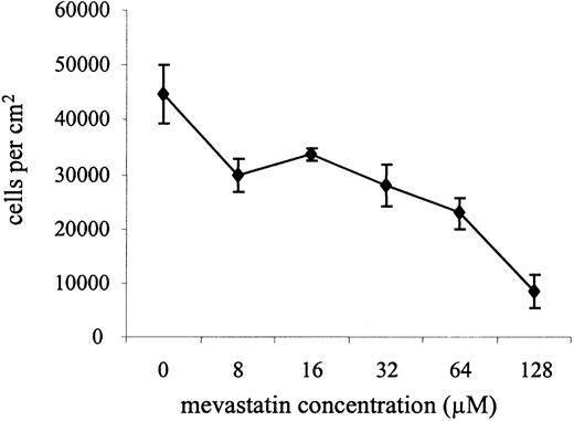
Treatment with levofloxacin (0–128 μM; 5 days; Caco-2 cells) reduces cell number in a dose-dependent manner [1]. Treatment with mevastatin (32-128 μM; 24-72 hours; Caco-2 cells) causes cell cycle arrest in two stages: early G0/G1 and late G2/M [1]. Cyclin-dependent kinase (cdk) 4 and 6 as well as cyclin D1 were downregulated after 72 hours of mevastatin (32-128 μM) treatment in Caco-2 cells; in contrast, the levels of cdk 2 and cdk E protein stayed unaltered. Mevastatin dramatically increases p21 and p27, two cell cycle inhibitors [1]. Treatment with mevastatin (16-256 μM; Caco-2 cells) dose-dependently promotes apoptosis [1]. Mevastatin treatment of Neuro2a cells for 24 hours resulted in neurite outgrowth and increased expression of the neuronal marker protein NeuN. The important kinases ERK1/2, Akt/protein kinase B, and epidermal growth factor receptor (EGFR) are phosphorylated in response to mevastatin. Mevastatin-induced axonal development is inhibited by PI3K, EGFR, and mitogen-activated protein kinase cascade inhibition [4].
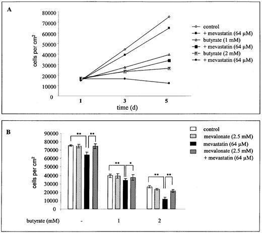
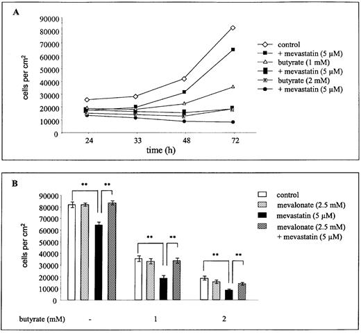
Synergistic inhibition of colorectal carcinoma cell proliferation with butyrate: - In human colorectal carcinoma Caco-2 cells, co-treatment with Mevastatin (0.1 μM, 1 μM, 10 μM) and sodium butyrate (5 mM) for 72 hours showed synergistic antiproliferative effects: - 1 μM Mevastatin alone reduced cell viability by 15%, while co-treatment with 5 mM butyrate reduced viability by 60% (MTT assay) [1] - Mechanism: 1 μM Mevastatin + 5 mM butyrate upregulated p21WAF1/CIP1 protein by 3.2-fold (Western blot), arresting cells in the G1 phase (cell cycle analysis via flow cytometry, G1 phase ratio increased from 55% to 75%) [1] - Induction of neurite outgrowth in neuroblastoma cells via EGFR activation: - In human neuroblastoma SH-SY5Y cells, Mevastatin (0.1 μM, 1 μM, 5 μM) treatment for 48 hours concentration-dependently promoted neurite outgrowth: - 5 μM Mevastatin increased the percentage of cells with neurites (length > 2× cell body diameter) from 10% to 65% (phase-contrast microscopy counting) [4] - EGFR activation: 5 μM Mevastatin increased EGFR phosphorylation (Tyr1173) by 2.8-fold and downstream ERK1/2 phosphorylation by 2.5-fold (Western blot); EGFR inhibitor (AG1478, 1 μM) abolished neurite outgrowth, confirming EGFR dependence [4] |
| ln Vivo |
In wild-type 129-SV/eVTAcBr male mice and eNOS-deficient male mice, mevastatin (2–20 mg/kg; administered daily via ALZET micro-osmotic pump) increases endothelial nitric oxide levels synthase (eNOS) mRNA and protein, reduces infarct size, and improves neurological deficits in a dose- and time-dependent manner [2]. Mevastatin (2.5 pmol/hour) administered locally promotes bone morphogenetic protein-2 (BMP-2) mRNA and receptor activator of NF-κB ligand (RANKL) mRNA expression in addition to boosting bone turnover. The transplanted bone mass in the MRL/MpJ mice [3].
Reduction of stroke damage and upregulation of eNOS in mice: 1. Animals: Male C57BL/6 mice (8–10 weeks old, 25–30 g) were randomized into 3 groups (n=10/group): Sham, Stroke + Vehicle, Stroke + Mevastatin [2] 2. Stroke model: Transient middle cerebral artery occlusion (MCAO) for 90 minutes, followed by reperfusion [2] 3. Treatment: Mevastatin (2 mg/kg/day, dissolved in 0.9% saline) was administered via intraperitoneal injection, starting 24 hours before MCAO and continuing for 3 days post-reperfusion [2] 4. Results: - Cerebral infarction volume: Reduced by 40% vs. Stroke + Vehicle (2,3,5-triphenyltetrazolium chloride [TTC] staining) [2] - Neurological function: Neurological deficit score (0–5 scale) decreased from 3.8 (Vehicle) to 1.9 (Mevastatin) [2] - Endothelial nitric oxide synthase (eNOS): Cerebral cortex eNOS protein increased by 2.3-fold (Western blot) [2] - Enhancement of grafted bone healing in MRL/MpJ mice: 1. Animals: Female MRL/MpJ mice (8 weeks old, 20–22 g) were randomized into 2 groups (n=8/group): Bone graft + Vehicle, Bone graft + Mevastatin [3] 2. Bone graft model: Syngeneic femoral bone grafts (5 mm length) were implanted into the dorsal subcutaneous pocket [3] 3. Treatment: Mevastatin (1 mg/kg/day, suspended in 0.5% CMC-Na) was administered via oral gavage for 4 weeks post-implantation [3] 4. Results: - Bone mineral density (BMD): Grafted bone BMD increased by 35% vs. Vehicle (dual-energy X-ray absorptiometry [DXA]) [3] - Bone formation: Histological analysis showed increased osteoblast number (by 40%) and mineralized tissue area (by 30%) [3] |
| Cell Assay |
Cell Viability Assay[2]
Cell Types: Caco-2 cells Tested Concentrations: 0 µM, 8 µM, 16 µM, 32 µM, 64 µM, 128 µM Incubation Duration: 5 days Experimental Results: Caused a dose-dependent decrease in cell number. Cell Cycle Analysis[2] Cell Types: Caco-2 cells Tested Concentrations: 32 µM, 64 µM, 128 µM Incubation Duration: 24 hrs (hours), 48 hrs (hours), 72 hrs (hours) Experimental Results: Caused a dose-dependent increase of cells in G0/G1 and G2/ M phases of the cell cycle. Western Blot Analysis[2] Cell Types: Caco-2 cells Tested Concentrations: 32 µM, 64 µM, 128 µM Incubation Duration: 72 hrs (hours) Experimental Results: Resulted in a down-regulation of cyclin-dependent kinases (cdk ) 4 and cdk 6 as well as cyclin D1. Caco-2 cell proliferation and cell cycle assay : 1. Cell culture: Caco-2 cells were seeded in 96-well plates (5×10³ cells/well) or 6-well plates (2×10⁵ cells/well) in DMEM medium supplemented with 10% FBS, 100 U/mL penicillin, and 100 μg/mL streptomycin. Cells were cultured at 37°C, 5% CO2 for 24 hours to attach [1] 2. Drug treatment: Mevastatin (0.1 μM, 1 μM, 10 μM) was added alone or with sodium butyrate (5 mM). Vehicle group received 0.1% DMSO. Cells were incubated for 72 hours [1] 3. Proliferation assay: MTT solution (5 mg/mL) was added to 96-well plates for 4 hours. Formazan crystals were dissolved with DMSO, and absorbance at 570 nm was measured to calculate viability [1] 4. Cell cycle analysis: Cells in 6-well plates were harvested, fixed with 70% ethanol, stained with propidium iodide (PI), and analyzed via flow cytometry to determine G1, S, and G2/M phase distributions [1] 5. Western blot: Cells were lysed with RIPA buffer (含protease inhibitors). 30 μg protein was separated by 10% SDS-PAGE, transferred to PVDF membranes, and probed with anti-p21WAF1/CIP1 and anti-β-actin antibodies [1] - SH-SY5Y cell neurite outgrowth assay : 1. Cell culture: SH-SY5Y cells were seeded in 24-well plates (1×10⁴ cells/well) pre-coated with poly-L-lysine, in RPMI 1640 medium (10% FBS) at 37°C, 5% CO2 [4] 2. Drug treatment: Mevastatin (0.1 μM, 1 μM, 5 μM) was added; for EGFR inhibition experiments, cells were pre-treated with AG1478 (1 μM) for 1 hour. Cells were incubated for 48 hours [4] 3. Neurite outgrowth quantification: Images were captured via phase-contrast microscopy. Cells with neurites longer than twice the cell body diameter were counted; neurite length was measured using ImageJ software [4] 4. Western blot: Lysates were probed with antibodies against phospho-EGFR (Tyr1173), total EGFR, phospho-ERK1/2, total ERK1/2, and β-actin [4] |
| Animal Protocol |
Animal/Disease Models: Wild-type 129-SV/eVTAcBr male mice and eNOS-deficient male mice (18-22 g) with the filament model[2]
Doses: 2 mg/kg or 20 mg/kg Route of Administration: Delivered via 7- or 14-day ALZET miniosmotic pumps implanted subcutaneously (sc); daily; for 7, 14, or 28 days Experimental Results: Increased levels of endothelial nitric oxide synthase (eNOS) mRNA and protein, decreased infarct size, and improved neurological deficits in a dose- and time- dependent manner. Mouse transient MCAO stroke model : 1. Animal preparation: Male C57BL/6 mice were anesthetized with isoflurane (3% induction, 1.5% maintenance). Body temperature was maintained at 37±0.5°C via a heating pad [2] 2. MCAO induction: A 6-0 nylon suture with a silicone-coated tip was inserted into the external carotid artery and advanced to occlude the middle cerebral artery (MCA) for 90 minutes. Sham group received the same surgery without suture insertion [2] 3. Grouping and treatment: Mice were randomized into 3 groups: - Sham: No MCAO + 0.9% saline (intraperitoneal injection); - Stroke + Vehicle: MCAO + 0.9% saline; - Stroke + Mevastatin: MCAO + Mevastatin 2 mg/kg/day (intraperitoneal injection, once daily, starting 24 hours pre-MCAO for 3 days post-reperfusion) [2] 4. Sample collection and detection: - Infarction volume: 3 days post-reperfusion, brains were removed, sectioned into 2 mm slices, stained with TTC, and infarction area was quantified via ImageJ [2] - Neurological scoring: Evaluated at 24 and 72 hours post-reperfusion using a 5-point scale (0=normal, 5=moribund) [2] - Western blot: Cerebral cortex tissue was lysed to detect eNOS protein [2] - MRL/MpJ mouse bone graft model : 1. Animal anesthesia: Female MRL/MpJ mice were anesthetized with ketamine (80 mg/kg) and xylazine (10 mg/kg) via intraperitoneal injection [3] 2. Bone graft implantation: Syngeneic femoral bones were harvested from donor mice, cut into 5 mm segments, and implanted into the dorsal subcutaneous pocket of recipient mice [3] 3. Grouping and treatment: Recipient mice were randomized into 2 groups: - Bone graft + Vehicle: 0.5% CMC-Na (oral gavage, once daily for 4 weeks post-implantation); - Bone graft + Mevastatin: Mevastatin 1 mg/kg/day (suspended in 0.5% CMC-Na, oral gavage, once daily for 4 weeks) [3] 4. Sample collection and detection: - BMD measurement: Grafted bones were harvested 4 weeks post-implantation, and BMD was measured via DXA [3] - Histology: Bones were fixed in 4% paraformaldehyde, decalcified, embedded in paraffin, sectioned, and stained with hematoxylin-eosin (H&E) to count osteoblasts and measure mineralized area [3] |
| Toxicity/Toxicokinetics |
In vitro cytotoxicity: - Caco-2 cells: Mevastatin alone (maximum concentration 10 μM, treatment for 72 hours) showed low cytotoxicity (cell viability > 80%, MTT assay); no significant toxicity was observed when treated with butyrate [1] - SH-SY5Y cells: Mevastatin (maximum concentration 5 μM, treatment for 48 hours) had no adverse effect on cell viability (cell viability > 90%) [4] - In vivo safety: - Stroke mice (2 mg/kg/day, 4 days): No significant changes in serum ALT, AST, BUN, or creatinine levels were observed compared to the sham-operated group; no clinical symptoms of toxicity (drowsiness, weight loss) were observed [2] - Bone transplant mice (1 mg/kg/day, 4 weeks): Weight gain was comparable to the vector group; no histological abnormalities were observed in the liver or kidneys [3]
|
| References |
[1]. Wächtershäuser A, et al. HMG-CoA reductase inhibitor mevastatin enhances the growth inhibitory effect of butyrate in the colorectal carcinoma cell line Caco-2. Carcinogenesis. 2001 Jul;22(7):1061-7.
[2]. Amin-Hanjani S, Stagliano NE, Yamada M, et al. Mevastatin, an HMG-CoA reductase inhibitor, reduces stroke damage and upregulates endothelial nitric oxide synthase in mice. Stroke. 2001 Apr;32(4):980-6. [3]. Sugazaki M, Hirotani H, Echigo S, et al. Effects of mevastatin on grafted bone in MRL/MpJ mice. Connect Tissue Res. 2010 Apr;51(2):105-12. [4]. Evangelopoulos ME, Weis J, Krüttgen A. Mevastatin-induced neurite outgrowth of neuroblastoma cells via activation of EGFR. J Neurosci Res. 2009 Jul;87(9):2138-44. |
| Additional Infomation |
Mevastatin is a carboxylic acid ester, a structural derivative of pravastatin lacking an allyl hydroxyl group. It is a hydroxymethylglutaryl-CoA reductase inhibitor (statin), originally isolated from Penicillium citrinum and Penicillium breve. Its clinical use as a lipid-lowering drug has been discontinued due to animal toxicity reports. Mevastatin has multiple functions, including as a fungal metabolite, an EC 3.4.24.83 (anthrax lethal factor endopeptidase) inhibitor, an antifungal agent, a Penicillium metabolite, and an apoptosis inducer. It is a carboxylic acid ester, a statin (naturally occurring), a hexahydronaphthalene compound, a 2-pyranone compound, and a polyketide compound. Mevastatin, or compatine, is a cholesterol-lowering drug isolated from Penicillium citrinum. It was the first statin cholesterol-lowering drug to be discovered. In 1971, Akira Endo of Sankyo Co., Ltd. in Japan discovered a class of compounds that appeared to lower plasma cholesterol levels while searching for antibiotic compounds produced by fungi. Two years later, the research team isolated a compound with a structure similar to hydroxymethylglutaric acid (HMG), which could inhibit the incorporation of acetic acid. Researchers hypothesized that this compound could bind to reductase and named it campatin. Mevastatin is a competitive inhibitor of HMG-CoA reductase, with a binding affinity 10,000 times higher than the HMG-CoA substrate itself. Mevastatin is a prodrug that requires hydrolysis of the lactone ring in vivo to be activated. It was once one of the lead compounds in the development of synthetic compounds. Mevastatin has been reported to exist in Penicillium cyclopium, Morus lhou, and other organisms with relevant data. Mevastatin is an HMG-CoA reductase inhibitor, initially isolated from Pythium ultimum. Mevastatin was the first statin drug to enter clinical trials. Due to its numerous side effects, it is currently not used for clinical treatment. Mechanism of Action Mevastatin's structure is similar to HMG, a substituent for the endogenous substrate of HMG-CoA reductase. Mevastatin is a prodrug activated in vivo via the hydrolysis of its lactone ring. The hydrolyzed lactone ring mimics the tetrahedral intermediate produced by the reductase, giving the drug a 10,000-fold higher affinity for its substrate than its natural substrate. The bicyclic moiety of mevastatin binds to the coenzyme A moiety at its active site. Pharmacodynamics The primary cause of cardiovascular disease is the formation of atherosclerotic plaques. Mevastatin reduces the risk of cardiovascular disease by lowering hepatic cholesterol production. Mevastatin competitively inhibits HMG-CoA reductase. This inhibition blocks the rate-limiting step in cholesterol synthesis. Lowered hepatic cholesterol levels lead to increased uptake of low-density lipoprotein (LDL) cholesterol, thereby reducing circulating cholesterol levels.
Background and Classification: Mevastatin (also known as Compatin) is a naturally occurring statin that was first isolated from Penicillium citrinum in 1976. It is the prototype of HMG-CoA reductase inhibitors and laid the foundation for the development of synthetic statins (such as lovastatin and atorvastatin)[1][4] - Core and Pleiotropic Mechanisms: - Lipid-lowering Mechanism: Inhibits HMG-CoA reductase to block the synthesis of mevalonate, thereby reducing the production of cholesterol in the liver (although not directly measured in the selected literature, this is its recognized core function)[1][2] - Pleiotropic Effects: - Anticancer Effects: Synergistically works with butyrate to inhibit the proliferation of colorectal cancer cells by upregulating p21[1] - Neuroprotective Effects: Reduces stroke-induced brain damage by upregulating eNOS (improving vascular function). [2] - Neurotrophic effect: Promotes neurite growth of neuroblastoma cells through the EGFR-ERK signaling pathway. [4] - Osteoprotective effect: Enhances bone graft healing by increasing osteoblast activity and mineralization. [3] - Clinical status: Mevastatin itself has not been approved for clinical use (due to its lower potency and solubility compared to later statins), but it is an important research tool for studying the pharmacology of statins and developing treatment strategies for cancer, neurological diseases, and bone diseases. [1][2][3][4] |
| Molecular Formula |
C23H34O5
|
|
|---|---|---|
| Molecular Weight |
390.51
|
|
| Exact Mass |
390.24
|
|
| CAS # |
73573-88-3
|
|
| Related CAS # |
|
|
| PubChem CID |
64715
|
|
| Appearance |
White to off-white solid powder
|
|
| Density |
1.1±0.1 g/cm3
|
|
| Boiling Point |
555.0±50.0 °C at 760 mmHg
|
|
| Melting Point |
151-153 °C
|
|
| Flash Point |
186.5±23.6 °C
|
|
| Vapour Pressure |
0.0±3.4 mmHg at 25°C
|
|
| Index of Refraction |
1.535
|
|
| LogP |
3.57
|
|
| Hydrogen Bond Donor Count |
1
|
|
| Hydrogen Bond Acceptor Count |
5
|
|
| Rotatable Bond Count |
7
|
|
| Heavy Atom Count |
28
|
|
| Complexity |
637
|
|
| Defined Atom Stereocenter Count |
7
|
|
| SMILES |
O(C([C@@]([H])(C([H])([H])[H])C([H])([H])C([H])([H])[H])=O)[C@@]1([H])C([H])([H])C([H])([H])C([H])=C2C([H])=C([H])[C@]([H])(C([H])([H])[H])[C@]([H])(C([H])([H])C([H])([H])[C@]3([H])C([H])([H])[C@]([H])(C([H])([H])C(=O)O3)O[H])[C@@]12[H]
|
|
| InChi Key |
AJLFOPYRIVGYMJ-INTXDZFKSA-N
|
|
| InChi Code |
InChI=1S/C23H34O5/c1-4-14(2)23(26)28-20-7-5-6-16-9-8-15(3)19(22(16)20)11-10-18-12-17(24)13-21(25)27-18/h6,8-9,14-15,17-20,22,24H,4-5,7,10-13H2,1-3H3/t14-,15-,17+,18+,19-,20-,22-/m0/s1
|
|
| Chemical Name |
[(1S,7S,8S,8aR)-8-[2-[(2R,4R)-4-hydroxy-6-oxooxan-2-yl]ethyl]-7-methyl-1,2,3,7,8,8a-hexahydronaphthalen-1-yl] (2S)-2-methylbutanoate
|
|
| Synonyms |
|
|
| HS Tariff Code |
2934.99.9001
|
|
| Storage |
Powder -20°C 3 years 4°C 2 years In solvent -80°C 6 months -20°C 1 month Note: This product requires protection from light (avoid light exposure) during transportation and storage. |
|
| Shipping Condition |
Room temperature (This product is stable at ambient temperature for a few days during ordinary shipping and time spent in Customs)
|
| Solubility (In Vitro) |
|
|||
|---|---|---|---|---|
| Solubility (In Vivo) |
Solubility in Formulation 1: ≥ 2.5 mg/mL (6.40 mM) (saturation unknown) in 10% DMSO + 40% PEG300 + 5% Tween80 + 45% Saline (add these co-solvents sequentially from left to right, and one by one), clear solution.
For example, if 1 mL of working solution is to be prepared, you can add 100 μL of 25.0 mg/mL clear DMSO stock solution to 400 μL PEG300 and mix evenly; then add 50 μL Tween-80 to the above solution and mix evenly; then add 450 μL normal saline to adjust the volume to 1 mL. Preparation of saline: Dissolve 0.9 g of sodium chloride in 100 mL ddH₂ O to obtain a clear solution. Solubility in Formulation 2: ≥ 2.5 mg/mL (6.40 mM) (saturation unknown) in 10% DMSO + 90% (20% SBE-β-CD in Saline) (add these co-solvents sequentially from left to right, and one by one), clear solution. For example, if 1 mL of working solution is to be prepared, you can add 100 μL of 25.0 mg/mL clear DMSO stock solution to 900 μL of 20% SBE-β-CD physiological saline solution and mix evenly. Preparation of 20% SBE-β-CD in Saline (4°C,1 week): Dissolve 2 g SBE-β-CD in 10 mL saline to obtain a clear solution. View More
Solubility in Formulation 3: ≥ 2.5 mg/mL (6.40 mM) (saturation unknown) in 10% DMSO + 90% Corn Oil (add these co-solvents sequentially from left to right, and one by one), clear solution. |
| Preparing Stock Solutions | 1 mg | 5 mg | 10 mg | |
| 1 mM | 2.5608 mL | 12.8038 mL | 25.6075 mL | |
| 5 mM | 0.5122 mL | 2.5608 mL | 5.1215 mL | |
| 10 mM | 0.2561 mL | 1.2804 mL | 2.5608 mL |
*Note: Please select an appropriate solvent for the preparation of stock solution based on your experiment needs. For most products, DMSO can be used for preparing stock solutions (e.g. 5 mM, 10 mM, or 20 mM concentration); some products with high aqueous solubility may be dissolved in water directly. Solubility information is available at the above Solubility Data section. Once the stock solution is prepared, aliquot it to routine usage volumes and store at -20°C or -80°C. Avoid repeated freeze and thaw cycles.
Calculation results
Working concentration: mg/mL;
Method for preparing DMSO stock solution: mg drug pre-dissolved in μL DMSO (stock solution concentration mg/mL). Please contact us first if the concentration exceeds the DMSO solubility of the batch of drug.
Method for preparing in vivo formulation::Take μL DMSO stock solution, next add μL PEG300, mix and clarify, next addμL Tween 80, mix and clarify, next add μL ddH2O,mix and clarify.
(1) Please be sure that the solution is clear before the addition of next solvent. Dissolution methods like vortex, ultrasound or warming and heat may be used to aid dissolving.
(2) Be sure to add the solvent(s) in order.
| NCT Number | Recruitment | interventions | Conditions | Sponsor/Collaborators | Start Date | Phases |
| NCT02441400 | Terminated | Device: EndoStim LES Stimulation System |
GERD | EndoStim Inc. | May 2013 |
|
|
|
Products are for research use only; We do not sell to patients
Copyright 2020 InvivoChem LLC | All Rights Reserved


 NMR
NMR