| Size | Price | Stock | Qty |
|---|---|---|---|
| 10mg |
|
||
| 25mg |
|
||
| 50mg |
|
||
| 100mg |
|
||
| 250mg |
|
||
| 500mg |
|
||
| Other Sizes |


Purity: ≥98%
Lomeguatrib (formerly PaTrin-2) is a novel, potent and selective inhibitor of MGMT (O6-alkylguanine-DNA-alkyltransferase) with anticancer activity. It inhibits MGMT with IC50s of 9 nM and ∼6 nM in cell-free assay and MCF-7 cells, respectively.
| Targets |
O6-methylguanine-DNA methyltransferase (MGMT) (IC50 = 6-9 nM
|
||
|---|---|---|---|
| ln Vitro |
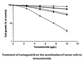
Lomeguatrib (Compound 10) is an O6-methylguanine methyltransferase (MGMT) inhibitor, with an IC50 of 9 nM in cell-free assay[1] and ∼6 nM in MCF-7 cells. Lomeguatrib (10 μM) considerably improves the growth inhibitory effects of temozolomide in MCF-7 cells (D60=10 μM with Lomeguatrib versus 400 μM without)[2].
Effect of Lomeguatrib (PaTrin-2) on MGMT activity and temozolomide sensitivity in MCF-7 cells [2] MCF-7 cells expressed high levels of MGMT (∼1540 fmoles mg−1 total protein). Exposure to Lomeguatrib (PaTrin-2) for 2 h resulted in extensive inactivation of MGMT in MCF-7 cells: the concentration required to inactivate 50% of the MGMT was around 6 nM (Figure 1). The sensitivity of the MCF-7 cells to the growth inhibitory effects of temozolomide was substantially increased by Lomeguatrib (PaTrin-2) . Growth amounting to 60% of control was seen after 400 μM temozolomide alone but following preincubation with 10 μM PaTrin-2, 60% growth occurred at 10 μM temozolomide (Figure 2), indicating a 40-fold increase in sensitivity. PaTrin-2 itself had no growth inhibitory effect. |
||
| ln Vivo |
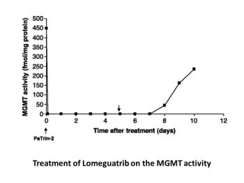
In MCF-7 xenografts, memeguatrib (20 mg/kg ip) totally inactivates MGMT in less than two hours, but has no discernible effect on tumor growth[2].
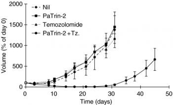
Effect of Lomeguatrib (PaTrin-2) on MGMT activity in host tissues and tumour [2] Extensive depletion of MGMT activity was seen in all host tissues measured after a single i.p. dose of 20 mg kg−1 PaTrin-2 (Figure 3). Depletion to below the limits of detection occurred in the kidney, while it was to ∼20, ∼35 and ∼40% of pretreatment values in liver, lung and bone marrow, respectively. The nadir was generally between 2 and 8 h and substantial activity (to over 50% of pretreatment levels) had returned by 24 h after dosing. In the MCF-7 xenografts, complete MGMT inactivation was seen between 2 and 8 h, and recovery of levels was only to ∼20% of pretreatment levels by 24 h after dosing. Slower recovery in the xenograft might reflect the relative strength of the human MGMT promoter or that the human protein is more extensively inactivated by PaTrin-2 or its putative metabolites. Effect of Lomeguatrib (PaTrin-2) and temozolomide on MCF-7 tumour growth [2] The median MCF-7 tqt in the vehicle control and PaTrin-2 only groups were ∼21 and ∼17 days, respectively. Neither temozolomide (tqt∼17 days) nor PaTrin-2 alone had any significant effect on xenograft growth. However, the combination of PaTrin-2 and temozolomide resulted in a median tumour quintupling time of ∼43 days representing an increase of ∼22 days). Toxicity, as measured by weight loss, was essentially unaffected by the addition of PaTrin-2 to the temozolomide treatment regimen. At the end of the treatment period, weight loss was ∼5% in both the temozolomide alone and combination groups. |
||
| Enzyme Assay |
MGMT Assay. [1]
The MGMT assay has been described previously. 45 Briefly, 200 μg of extracted cellular protein from HeLaS3 cells in 200 μL of 70 mM HEPES buffer (with 1 mM dithiothreitol (DTT), 5 mM EDTA, pH 7.8) was incubated at 37 °C with a defined concentration of MGMT inhibitor (added as a DMSO solution). After 30 min an excess of [3H]-methylated DNA (120 000 cpm) was added, and the incubation was continued for an additional 90 min. The reaction was stopped by the addition of 400 μL TCA (13%), and the DNA was hydrolyzed by heating the reaction mixture for 30 min at 98 °C. The precipitated protein was washed three times with 400-μL portions of 5% TCA, solubilized in 0.1 N NaOH, and analyzed by liquid scintillation counting using the cocktail Rotiszint eco plus and a TRI-CARB 2100 TR liquid scintillation analyzer. Enzyme activity was expressed as fmol of [3H]methyl transferred to TCA-insoluble protein material per mg of total cellular protein. Percent inhibition was calculated relative to untreated control samples. Each assay was repeated three times, and IC50 values were determined graphically from plots of percent inhibition vs inhibitor concentration. |
||
| Cell Assay |
MCF-7 cells (a human breast adenocarcinoma cell line) were grown as a monolayer in RPMI medium containing 10% foetal bovine serum, at 37°C in a humidified atmosphere of 5% CO2/95% air. [2]
To determine MGMT inactivation, cells (5 × 10~6) were incubated in the presence of increasing concentrations of Lomeguatrib (PaTrin-2) at 37°C, 5% CO2. After 2 h, cells were pelleted and resuspended in 10 ml PBS. This was repeated three times in order to remove any residual Lomeguatrib (PaTrin-2) . Finally, cells were pelleted and assayed for MGMT activity as previously described. Activity remaining, based on at least three points on the linear part of the protein-dependence curve, was calculated as a percentage of the activity in untreated cells. [2] To determine toxicity, the MTT [3′ (4,5-dimethylthiazol-2-yl)-2,5-diphenyl tetrazolium bromide] growth inhibition assay, based on the method of Carmichael et al (1987) was employed. Cells (1000 per well) were plated into a 96-well plate and following a 24 h attachment period, Lomeguatrib (PaTrin-2) was added to the cells. After 2 h incubation with Lomeguatrib (PaTrin-2) (10 μM) at 37°C, 5% CO2, increasing doses of temozolomide or vehicle were added and the cells were incubated for a further 4–5 days. At the end of the exposure period, 150 μg MTT was added to each well and plates were incubated for 3 h at 37°C, 5% CO2. The media were removed and the formazan crystals formed in the viable cells were solubilised in 200 μl DMSO. The absorbances at 540 and 690 nm were determined using a Titertek Multiscan ELISA plate reader and growth inhibition calculated as a percentage of the A540–A690 of untreated wells. [2] |
||
| Animal Protocol |
|
||
| References |
|
||
| Additional Infomation |
We synthesized a series of potential inhibitors of the human DNA repair protein O(6)-methylguanine-DNA methyltransferase (MGMT), characterized them in detail by nuclear magnetic resonance (NMR), and tested their ability to inhibit MGMT activity in vitro. These new compounds are ω-[O(6)-R-guanine-9-yl]-(CH(2))(n)-β-D-glucoside, where R is benzyl or 4-bromothiophene, and ω=n=2,4,12. We compared these new compounds with known inhibitors O(6)-benzylguanine (O(6)-BG), 8-aza-O(6)-benzylguanine (8-aza-BG), and O(6)-(4-bromothiophene)guanine (4-BTG). In vitro experiments showed that the IC50 values of O(6)-BG, 8-aza-O(6)-benzylguanine (8-aza-BG), and 4-bromothienylguanine (4-BTG) were 0.62, 0.038, and 0.009 μM, respectively. The potential advantage of glycosides lies in their increased water solubility and selective uptake by tumor cells. 4-BTG glycosides with n = 2, 4, and 6 exhibited moderate inhibitory activity, with IC50 values of approximately 0.5 μM, while the inhibitory activity of glycosides derived from BG and 8-aza-BG was significantly reduced compared to the parent compounds. 4-BTG glycosides with n = 8, 10, and 12 were potent inhibitors, with IC50 values of approximately 0.03 μM. To understand this phenomenon, we conducted extensive molecular modeling studies using the published MGMT crystal structure (PDB accession number: ). The inhibitor molecule was docked to the BG binding pocket and molecular dynamics simulations involving explicit water molecules were performed. Stabilization energies for the interaction of specific regions of the inhibitor with individual amino acid residues were calculated. The alkyl spacer group is located in a cleft of the MGMT helix 6. As the length of the spacer region increases, its interaction with multiple amino acid residues is enhanced, which play an important role in the nucleotide flipping mechanism required for DNA repair. [1] Tumors develop resistance to methylated chemotherapeutic agents such as dacarbazine and temozolomide, which is associated with the expression of the DNA repair protein O(6)-alkylguanine-DNA alkyltransferase (MGMT). There has been great interest in improving the efficacy of such O(6)-alkylated chemotherapeutic agents by pre-inactivating MGMT. We investigated the effects of modified guanine base O(6)-(4-bromophenyl)guanine (PaTrin-2, Patrin, Lomeguatrib) on MGMT activity and the effect of temozolomide on the inhibition of cell or xenograft growth in the human breast cancer sarcoma cell line MCF-7. PaTrin-2 effectively inactivated MGMT in MCF-7 cells (IC50 approximately 6 nM). In xenograft tumors, MGMT was completely inactivated within 2 hours of administration (20 mg kg⁻¹, intraperitoneal injection), with only slight recovery after 24 hours. In various mouse host tissues, MGMT inactivation ranged from complete inactivation to approximately 60%, with significant recovery after 24 hours in all cases. PaTrin-2 (10 μM) significantly enhanced the growth-inhibiting effect of temozolomide on MCF-7 cells (D₆₀ = 10 μM in the PaTrin-2 treatment group, 400 μM in the untreated group). In MCF-7 xenograft tumors, neither temozolomide (100 mg kg⁻¹, once daily for 5 days) nor PaTrin-2 (20 mg kg⁻¹, once daily for 5 days) had a significant effect on tumor growth. In contrast, the combination of PaTrin-2 and temozolomide significantly delayed tumor growth: the median time to five-fold tumor growth was prolonged by 22 days (P<0.005), and no significant increase in toxicity was observed based on animal body weight assessment. Therefore, the combination of PaTrin-2 and temozolomide may be beneficial for the treatment of human breast cancer. [2]
|
| Molecular Formula |
C10H8BRN5OS
|
|
|---|---|---|
| Molecular Weight |
326.17
|
|
| Exact Mass |
324.963
|
|
| Elemental Analysis |
C, 36.82; H, 2.47; Br, 24.50; N, 21.47; O, 4.91; S, 9.83
|
|
| CAS # |
192441-08-0
|
|
| Related CAS # |
|
|
| PubChem CID |
3025944
|
|
| Appearance |
Light yellow to yellow solid powder
|
|
| Density |
1.9±0.1 g/cm3
|
|
| Boiling Point |
683.8±65.0 °C at 760 mmHg
|
|
| Flash Point |
367.3±34.3 °C
|
|
| Vapour Pressure |
0.0±2.1 mmHg at 25°C
|
|
| Index of Refraction |
1.797
|
|
| LogP |
2.36
|
|
| Hydrogen Bond Donor Count |
2
|
|
| Hydrogen Bond Acceptor Count |
6
|
|
| Rotatable Bond Count |
3
|
|
| Heavy Atom Count |
18
|
|
| Complexity |
299
|
|
| Defined Atom Stereocenter Count |
0
|
|
| SMILES |
BrC1=CSC(COC2=C3N=CNC3=NC(N)=N2)=C1
|
|
| InChi Key |
JUJPKFNFCWJBCX-UHFFFAOYSA-N
|
|
| InChi Code |
InChI=1S/C10H8BrN5OS/c11-5-1-6(18-3-5)2-17-9-7-8(14-4-13-7)15-10(12)16-9/h1,3-4H,2H2,(H3,12,13,14,15,16)
|
|
| Chemical Name |
6-[(4-bromothiophen-2-yl)methoxy]-7H-purin-2-amine
|
|
| Synonyms |
PaTrin-2; Lomeguatrib; PaTrin 2; 192441-08-0; PaTrin-2; PaTrin 2; Lomeguatrib [INN]; Lomeguatrib [INN:BAN]; C10H8BrN5OS; 6-[(4-bromothiophen-2-yl)methoxy]-7H-purin-2-amine; PaTrin2; 2-Amino-6-[(4-bromo-2-thienyl)methoxy]-9H-purine
|
|
| HS Tariff Code |
2934.99.9001
|
|
| Storage |
Powder -20°C 3 years 4°C 2 years In solvent -80°C 6 months -20°C 1 month |
|
| Shipping Condition |
Room temperature (This product is stable at ambient temperature for a few days during ordinary shipping and time spent in Customs)
|
| Solubility (In Vitro) |
DMSO: 65 mg/mL (199.3 mM)
Water: 1 mg/mL (Insoluble) Ethanol: 1 mg/mL |
|---|---|
| Solubility (In Vivo) |
Solubility in Formulation 1: ≥ 2.08 mg/mL (6.38 mM) (saturation unknown) in 10% DMSO + 40% PEG300 + 5% Tween80 + 45% Saline (add these co-solvents sequentially from left to right, and one by one), clear solution.
For example, if 1 mL of working solution is to be prepared, you can add 100 μL of 20.8 mg/mL clear DMSO stock solution to 400 μL PEG300 and mix evenly; then add 50 μL Tween-80 to the above solution and mix evenly; then add 450 μL normal saline to adjust the volume to 1 mL. Preparation of saline: Dissolve 0.9 g of sodium chloride in 100 mL ddH₂ O to obtain a clear solution. Solubility in Formulation 2: ≥ 2.08 mg/mL (6.38 mM) (saturation unknown) in 10% DMSO + 90% (20% SBE-β-CD in Saline) (add these co-solvents sequentially from left to right, and one by one), clear solution. For example, if 1 mL of working solution is to be prepared, you can add 100 μL of 20.8 mg/mL clear DMSO stock solution to 900 μL of 20% SBE-β-CD physiological saline solution and mix evenly. Preparation of 20% SBE-β-CD in Saline (4°C,1 week): Dissolve 2 g SBE-β-CD in 10 mL saline to obtain a clear solution. View More
Solubility in Formulation 3: ≥ 2.08 mg/mL (6.38 mM) (saturation unknown) in 10% DMSO + 90% Corn Oil (add these co-solvents sequentially from left to right, and one by one), clear solution. |
| Preparing Stock Solutions | 1 mg | 5 mg | 10 mg | |
| 1 mM | 3.0659 mL | 15.3294 mL | 30.6589 mL | |
| 5 mM | 0.6132 mL | 3.0659 mL | 6.1318 mL | |
| 10 mM | 0.3066 mL | 1.5329 mL | 3.0659 mL |
*Note: Please select an appropriate solvent for the preparation of stock solution based on your experiment needs. For most products, DMSO can be used for preparing stock solutions (e.g. 5 mM, 10 mM, or 20 mM concentration); some products with high aqueous solubility may be dissolved in water directly. Solubility information is available at the above Solubility Data section. Once the stock solution is prepared, aliquot it to routine usage volumes and store at -20°C or -80°C. Avoid repeated freeze and thaw cycles.
Calculation results
Working concentration: mg/mL;
Method for preparing DMSO stock solution: mg drug pre-dissolved in μL DMSO (stock solution concentration mg/mL). Please contact us first if the concentration exceeds the DMSO solubility of the batch of drug.
Method for preparing in vivo formulation::Take μL DMSO stock solution, next add μL PEG300, mix and clarify, next addμL Tween 80, mix and clarify, next add μL ddH2O,mix and clarify.
(1) Please be sure that the solution is clear before the addition of next solvent. Dissolution methods like vortex, ultrasound or warming and heat may be used to aid dissolving.
(2) Be sure to add the solvent(s) in order.
|
Products are for research use only; We do not sell to patients
Copyright 2020 InvivoChem LLC | All Rights Reserved


 COA
COA