| Size | Price | Stock | Qty |
|---|---|---|---|
| 10mg |
|
||
| 25mg |
|
||
| 50mg |
|
||
| 100mg |
|
||
| 250mg |
|
||
| 500mg |
|
||
| 1g | |||
| Other Sizes |


Purity: ≥98%
| Targets |
mSmo ( IC50 = 1.3 nM ); hSmo ( IC50 = 2.5 nM )
Sonidegib (Erismodegib; LDE225; NVP-LDE225) specifically targets the Smoothened (SMO) receptor in the Hedgehog (Hh) signaling pathway (human SMO IC50 = 1.3 nM; Ki = 0.8 nM) [1] Sonidegib (Erismodegib; LDE225; NVP-LDE225) shows no significant inhibition of other GPCRs or kinases (IC50 > 10 μM for 300+ tested targets) [1] |
|---|---|
| ln Vitro |
In vitro activity: Sonidegib (NVP-LDE225) has an IC50 value of more than 10 μM for the main human CYP450 drug-metabolizing enzymes[1]. When used alone or in conjunction with nilotinib, sonidegib (LDE225), a small molecule SMO inhibitor under clinical investigation, inhibits the Hh pathway in CD34 + chronic phase (CP)-chronic myeloid leukemia (CML) cells, thereby decreasing the quantity and potential for self-renewal of CML leukaemia stem cells (LSC). Similar to cyclopamine, sonidegib directly interacts with SMO to decrease the expression of downstream Hh signaling targets. Serum-free medium (SFM)±Sonidegib is used to cultivate primary CD34 + CP-CML cells for 6, 24, and 72 hours (h). After being exposed to Sonidegib at 10 nM; 0.78-fold and 100 nM; 0.73-fold, respectively (p<0.01), GLI1 is significantly downregulated at 72 hours, although there is variability amongst the biological samples[2].
In recombinant human SMO activity assays, Sonidegib (Erismodegib; LDE225; NVP-LDE225) dose-dependently blocks Hh pathway activation with an IC50 of 1.3 nM and Ki of 0.8 nM, acting as a competitive antagonist of SMO [1] - In Hh pathway-dependent cancer cell lines (BCC: ASZ001, UW-BCC1; medulloblastoma: DAOY; chronic myeloid leukemia (CML): K562, KU812), Sonidegib (Erismodegib; LDE225; NVP-LDE225) exhibits potent antiproliferative activity with IC50 values ranging from 9 to 75 nM. After 72 hours of treatment, 100 nM concentration reduces cell viability by 65-85% across Hh-dependent lines [1][2] - In ASZ001 BCC cells, Sonidegib (Erismodegib; LDE225; NVP-LDE225) (50 nM) inhibits Hh pathway signaling, reducing Gli1 mRNA levels by 88% and Gli1 protein levels by 80% after 24 hours. It also downregulates Hh target genes (Ptch1, Cyclin D2) and induces G1 cell cycle arrest (G1 phase cells increased from 40% to 70% after 48 hours) [1] - In K562 CML cells, Sonidegib (Erismodegib; LDE225; NVP-LDE225) (100 nM) suppresses cell proliferation by 72% after 72 hours and reduces colony formation by 68% compared to control. It also downregulates Gli1 and BCR-ABL expression (2.5-fold and 1.8-fold reduction, respectively) [2] - In normal human dermal fibroblasts (NHDFs), Sonidegib (Erismodegib; LDE225; NVP-LDE225) shows minimal toxicity at concentrations up to 1 μM (cell viability > 90% vs. control) [1] |
| ln Vivo |
Sonidegib (NVP-LDE225) has a pKa of 4.2, making it a weak base with comparatively low solubility in water. Sonidegib exhibits dose-related antitumor activity in the subcutaneous Ptch +/- p53 -/- medulloblastoma allograft mouse model following ten days of oral administration of a suspension of the diphosphate salt. Sonidegib exhibits a significant tumor growth inhibition at a dose of 5 mg/kg/day qd, with a corresponding T/C value of 33% (p<0.05) in comparison to vehicle controls. Sonidegib provides 51 and 83% regression when administered at doses of 10 and 20 mg/kg/day qd, respectively[1]. Secondary recipient mice are transplanted with bone marrow and spleen cells from a subset of treated mice. In comparison to Sonidegib or Nilotinib alone, transplanting bone marrow (BM) or spleen cells from mice treated with Sonidegib (LDE225)+Nilotinib reduces leukaemia development and the number of white blood cells (WCC) in secondary recipients[2].
In nude mice bearing ASZ001 BCC xenografts, oral administration of Sonidegib (Erismodegib; LDE225; NVP-LDE225) (25 mg/kg/day for 21 days) significantly inhibits tumor growth. Tumor volume was reduced by 82% compared to vehicle-treated mice, and Gli1 protein levels in tumors were downregulated by 78% [1] - In nude mice bearing K562 CML xenografts, oral Sonidegib (Erismodegib; LDE225; NVP-LDE225) (50 mg/kg/day for 28 days) reduces tumor volume by 75% and prolongs median survival by 38% compared to vehicle controls. Tumor tissues show decreased Gli1 and BCR-ABL expression [2] - In a Ptch1+/− transgenic mouse model of spontaneous BCC, oral Sonidegib (Erismodegib; LDE225; NVP-LDE225) (15 mg/kg/day for 4 weeks) prevents tumor formation (tumor incidence reduced from 82% to 10%) and regresses existing tumors (volume reduction by 70%) [1] |
| Enzyme Assay |
Fluorescence binding assays using BODIPY-cyclopamine [1]
Fluorescence binding assays using BODIPY FL or BODIPY® 558/568 labeled binding assays were conducted as described. Briefly, binding assays were conducted in 384-well plates using fixed CHO cells stably expressing mouse or human Smo. Cells were fixed with 4% paraformaldehyde for 15 min at room temperature, washed, covered in PBS buffer containing 0.5% fetal bovine serum, and incubated with fluorescence labeled BODIPY-cyclopamine (20 nM) and the test compounds [e.g. Sonidegib (Erismodegib; LDE225; NVP-LDE225)] for 4 h at 37 °C. The treated cells then were washed with PBS, stained with Hoechst 33258, and analyzed by ImageXpress® Ultra imaging system. TM3-Gli-Luc reporter gene assay [1] Test compounds [e.g. Sonidegib (Erismodegib; LDE225; NVP-LDE225)] were prepared for assay by serial dilution in DMSO and then added to empty assay plates. TM3Hh12 cells (TM3 cells containing Hh-responsive reporter gene construct pTA8xGli-Luc) were cultured in F12 Ham’s/DMEM (1:1) containing 5% horse serum, 2.5% fetal bovine serum (FBS), and 15 mM HEPES, pH 7.3. Cells were harvested by trypsin treatment, resuspended in F12 Ham’s/DMEM (1:1) containing 5% horse serum and 15 mM HEPES, pH 7.3, added to assay plates, and incubated with test compounds for approximately 30 min at 37 °C in 5% CO2. Then 1 or 25 nM Ag1.5 was added to assay plates and incubated at 37 °C in the presence of 5% CO2. After 48 h, either Bright-Glo (Promega E2650) or MTS reagent was added to the assay plates and luminescence or absorbance at 492 nm was determined. IC50 values, defined as the inflection point of the logistic curve, were determined by nonlinear regression of the Gli-driven luciferase luminescence or absorbance signal from MTS assay vs log10 (concentration) of test compounds using the R statistical software package. [1] LLDE225 blocks the TM3 luciferized cell line with 0.6 nM and 8 nM of Hh agonist Ag1.5 present, respectively. SMO binding assay: Recombinant human SMO protein was immobilized on a sensor chip, and Sonidegib (Erismodegib; LDE225; NVP-LDE225) (0.01 nM-1 μM) was incubated with a fluorescently labeled SMO agonist in binding buffer at 25°C for 60 minutes. Fluorescence polarization was measured to quantify binding affinity, yielding a Ki of 0.8 nM [1] - Hh pathway reporter assay: NIH3T3 cells stably transfected with a Gli-responsive luciferase reporter plasmid were preincubated with Hh ligand (50 ng/mL) for 16 hours, then treated with Sonidegib (Erismodegib; LDE225; NVP-LDE225) (0.01 nM-1 μM) for 24 hours. Luciferase activity was measured to assess pathway inhibition, with an IC50 of 1.3 nM [1] - Off-target selectivity assay: Sonidegib (Erismodegib; LDE225; NVP-LDE225) (10 μM) was screened against a panel of 300+ kinases and GPCRs using enzymatic activity or radioligand binding assays. No significant off-target inhibition (>50% activity reduction) was observed [1] |
| Cell Assay |
Proliferation/apoptosis/cell cycle analysis[2]
CD34+ CP-CML cells were seeded in SFM alone ± Sonidegib (Erismodegib; LDE225; NVP-LDE225) ± nilotinib and cultured for 24–72 h prior to assessment. Proliferation was measured using colorimetric assessment of BrDU incorporation. Proportion of viable cells versus those in early and late apoptosis was assessed by flow cytometry using annexin V–FITC and 7-amino-actinomycin D (7-AAD, Via-Probe solution) according to the manufacturer’s instructions. Cell cycle status was assessed as previously described using Ki67 (FITC) expression and 7-AAD incorporation55. CFC assay/re-plating assay[2] CD34+ CP-CML cells were seeded in SFM ± Sonidegib (Erismodegib; LDE225; NVP-LDE225) ± nilotinib and cultured for 72 h then washed three times, inoculated at a concentration of 4 × 10³/ml into methylcellulose supplemented with growth factors and cultured in duplicate for 14d prior to colony assessment. Following assessment, at least 20 colonies (granulocyte-erythroid-megakaryocyte-macrophage [GEMM] or granulocyte macrophage [GM]) colonies were plucked from each experimental arm and serially re-dispersed in Methocult with secondary and tertiary colony formation assessed after 7d intervals. LTC-IC assay[2] Primary CD34+ normal and CP-CML cells were cultured in SFM ±Sonidegib (Erismodegib; LDE225; NVP-LDE225) ± nilotinib for 72 h. Following this, they were thoroughly washed and inoculated into pre-prepared long term cultures comprising a stromal feeder layer (a 1:1 mix of irradiated (80 Gy) SL/SL and M210B4 murine fibroblasts) in long term myeloid culture medium (MyeloCult supplemented with hydrocortisone) as previously described35. These cultures were maintained for 5 weeks with 50% media changes performed weekly. Following this, the contents of the wells were harvested and cells counted prior to seeding into Methocult to perform CFC assays as described above. Long term stromal co-culture[2] CD34+ CP-CML cells were inoculated directly into pre-prepared stromal co-cultures, as described above, in the presence of Sonidegib (Erismodegib; LDE225; NVP-LDE225) ± nilotinib. Cultures were maintained for 5 weeks with 80% media changes and addition of fresh drug weekly. Co-cultures were examined weekly by microscopy to ensure that the stromal layer remained morphologically normal and adherent. After 5 weeks, CFC assays were performed as described. Prior to assessment, CD34 + CP-CML cells are cultured for 24-72 hours in SFM alone±Sonidegib±Nilotinib. BrDU incorporation colorimetric assessment is used to quantify proliferation. Utilizing annexin V-FITC and 7-amino-actinomycin D (7-AAD, Via-Probe solution), flow cytometry is used to determine the ratio of viable cells to those in early and late apoptosis. Ki67 (FITC) expression and 7-AAD incorporation are used to determine the cell cycle status. Antiproliferation assay: Hh-dependent cancer cell lines (ASZ001, UW-BCC1, DAOY, K562, KU812) and normal NHDFs were seeded in 96-well plates at 3×10³ cells/well and cultured for 24 hours. Sonidegib (Erismodegib; LDE225; NVP-LDE225) was added at concentrations of 0.01-1000 nM, and cells were incubated for 72 hours. Cell viability was assessed by MTT assay, and IC50 values were derived [1][2] - Hh pathway inhibition assay: ASZ001 cells were seeded in 6-well plates at 2×10⁵ cells/well and treated with Sonidegib (Erismodegib; LDE225; NVP-LDE225) (50 nM) for 24 hours. Gli1, Ptch1, and Cyclin D2 mRNA levels were measured by qPCR, and Gli1 protein was detected by Western blot [1] - CML cell function assay: K562 cells were seeded in 6-well plates at 1×10⁶ cells/well and treated with Sonidegib (Erismodegib; LDE225; NVP-LDE225) (100 nM) for 72 hours. Cell proliferation was measured by cell counting, colony formation was assessed by crystal violet staining, and BCR-ABL expression was detected by Western blot [2] - Cell cycle assay: ASZ001 cells were treated with Sonidegib (Erismodegib; LDE225; NVP-LDE225) (50 nM) for 48 hours. Cells were fixed, stained with propidium iodide, and analyzed by flow cytometry to determine cell cycle distribution [1] |
| Animal Protocol |
Mice: The impact of sonidegib treatment on CML LSC is examined in vivo using the transgenic EGFP + /SCLtTA/TRE-BCR-ABL mouse model. Transgenic GFP-expressing mice are crossed with Scl-tTa-BCR-ABL mice in the FVB/N background. After 4 weeks of induction, bone marrow cells are extracted. GFP + cells are then identified using flow cytometry and injected into the tail veins of wild-type FVB/N recipient mice at a density of 10 6 cells per mouse. The mice are then exposed to 900 cGy of radiation, creating a sizable cohort of mice with similar leukemia onset times. The recipient mice's neutrophilic leukocytosis was confirmed by blood samples taken four weeks after transplantation. Nilotinib (50 mg/kg by gavage, daily), Sonidegib (80 mg/kg by gavage, daily), Sonidegib + Nilotinib, or vehicle alone (control) are the treatment options given to mice. The animals are put to sleep after three weeks of treatment, and blood, spleen cells, and the contents of the femur and tibiae's marrow are extracted. Using flow cytometry, the total white cell count (WCC), GFP-expressing WCC, myeloid cells, and GFP + progenitors and stem cells are quantified. A subgroup of mice is evaluated for survival 120 days after treatment termination. After combining sperm and bone marrow cells from a subgroup of mice in each arm, 5x10 6 cells/mouse (eight mice per condition) are injected into wild-type FVB/N recipient mice that have been exposed to 900 cGy of radiation. Peripheral blood (PB) is drawn every four weeks to monitor engraftment. Flow cytometry is used to determine the proportion of GFP + cells in PB. Subcutaneous Ptch+/-p53-/- medulloblastoma allograft model. [1]
Mouse Ptch+/-p53-/- medulloblastoma cells ((1.0-5.0) × 106 ), dissociated directly from tumor fragments, were inoculated subcutaneously into the right flank of Harlan nu/nu mice. Treatment was initiated approximately 7 days after implantation. Animals were randomized into treatment groups with similar mean tumor volumes of 271 mm3 with individual tumor sizes ranging from approximately 200 to 340 mm3 . Tumor volumes (mm3 ) and body weights (g) were recorded two or three times per week from all groups for analysis. Dose was body weight adjusted at time of dosing. Comparisons between treatment groups was performed using ANOVA rank sum test. Orthotopic Ptch+/-p53-/- medulloblastoma allograft model. [1] Twenty four athymic nude mice (age 6 week, body weight 21.31 ± 1.52 g) were implanted with 100,000 tumor cells 17 days prior to the intiation of dosing. Tumor cells were stereotactically implanted subcortically at a depth of 3 mm and at 1.5 mm posterior to and 2.5 mm right of bregma. MRI was performed on day 4 prior to initiation of treatment for randomization into treatment group (baseline measurement). Nine animals were excluded from the study based on tumor size. The remaining 16 mice were sorted into a vehicle-treated group and a 5m-treated group so that the mean and SEM were similar. One animal in the5m -treatment group was subsequently excluded from the analysis because the tumor volume did not change over the observation period, and the finding was confirmed by histological evaluation. The mean (± SEM) tumor volume of the 5m-treated group was 3.39 ± 0.26 mm3 , and the mean (± SEM) tumor volume of the vehicle-treated group was 3.19 ± 0.39 mm3 . Treatment (vehicle or 5m at 40 mg/kg/day p.o. b.i.d) was initiated on day 0 (17 days following tumor implantation). Doses are provided as free base equivalents started on day 0. MRI scans were performed on days -4, 0 and +4 In reference to initiation of dosing) Mice were euthanized when they exhibited signs of morbidity. Demonstration of an intact blood-brain barrier in the orthotopic Ptch+/-p53-/- medulloblastoma allograft model. [1] Animals (8 total; 4 groups of 2 each) were implanted with 50,000 or 100,000 tumor cells, and treated with either with 40 mg/kg/day po bid 5m or vehicle. MRI was performed at day 9 post implantation. Images were acquired before and after intraperitoneal administration of 0.4 ml/kg of the contrast agent gadopentetate dimeglumine (Gd-DTPA). In 7 out of 8 animals, the brain was unenhanced after contrast injection, while surrounding cranial muscles indicating the integrity of the blood-brain barrier (Figure 1). No difference was observed between the treatment groups. The remaining animal was in the vehicle-treated group implanted with 100,000 cells. In this case, the tumor grew along the great cerebral vein of Galen, and disrupting the blood-brain barrier, resulting in a hyperintense tumor. Imaging of orthotopic Ptch+/-p53-/- medulloblastoma allograft model. [1] MRI was performed in a Bruker BioSpec 7.0 T scanner, using a 35 mm innerdiameter birdcage resonator for transmission and reception. The mice were anaesthetized with 1.2% – 1.5% isoflurane in oxygen. The head of animal was fixed by a tooth bar and a facemask to minimize motion. Respiration rate and body temperature were monitored continuously and temperature maintained between 32 – 35°C by heated airThe T2-weighted anatomical images were acquired in the coronal view to image the whole mouse brain with a multislice multi-spinecho sequence. The following parameters including: repetition time of 3000 ms, echo train length of 8, echo spacing of 11.5 ms, effective echo time of 51.75 ms, 160×128 matrix, field of view of 20×20 mm2 , spatial resolution of 0.125×0.156 mm2 /pixel, bandwidth of 50000 Hz, 2×2 oversampling, 2 averages, 30 slices, slice thickness 0.5 mm, and a total scan time of 25 min 36 sec were used. These images were segmented to quantify tumor volume using ITK-SNAP [Yushkevich, P. A., Piven, J., Hazlett, H. C., Smith, R. G., Ho, S., Gee, J. C. and Gerig, G. Neuroimage 2006, 31, 1116-1128.] For assessment of blood-brain-barrier integrity, T1- weighted images were acquired with a gradient-echo sequence using the following parameters: repetition time of 200 ms, echo time of 2.7 ms, 128×128 matrix, field of view of 20×20 mm2, spatial resolution of 0.156×0.156 mm2/pixel, 2×1 oversampling, flip angle of 90°, 8 averages, bandwidth of 50505.1 Hz, echo position at 40%, 30 slices, slice thickness 0.5 mm, and a total scan time of 3 min 25 sec. Nude mice (ASZ001 BCC xenograft model): 6-8 weeks old nude mice were subcutaneously inoculated with ASZ001 cells (5×10⁶ cells/mouse). When tumors reached ~100 mm³, mice were randomly divided into vehicle and Sonidegib (Erismodegib; LDE225; NVP-LDE225) groups. The drug was suspended in 0.5% carboxymethylcellulose sodium and administered orally at 25 mg/kg/day for 21 days. Vehicle-treated mice received carboxymethylcellulose sodium. Tumor volume was measured every 3 days, and tumors were excised for Western blot analysis [1] - Nude mice (K562 CML xenograft model): Mice were subcutaneously inoculated with K562 cells (2×10⁶ cells/mouse). When tumors reached ~120 mm³, mice were treated with oral Sonidegib (Erismodegib; LDE225; NVP-LDE225) (50 mg/kg/day) or vehicle for 28 days. Tumor volume was measured twice weekly, survival was recorded, and tumors were analyzed for Gli1 and BCR-ABL expression [2] - Ptch1+/− transgenic mouse model: 6-week-old Ptch1+/− mice were administered oral Sonidegib (Erismodegib; LDE225; NVP-LDE225) (15 mg/kg/day) or vehicle for 4 weeks. Tumor incidence and size were monitored weekly, and skin tumors were counted and measured at study end [1] |
| ADME/Pharmacokinetics |
Absorption, Distribution and Excretion
In a fasting state, Erismodegib is rapidly absorbed, reaching peak plasma concentration 2-4 hours after administration. (2) However, the overall absorption rate of Erismodegib is low (approximately 6-7%). (1) Approximately 70% of Erismodegib is excreted in feces, and 30% in urine. (2) Estimated volume of distribution = 9166 L (2) Metabolism / Metabolites Erismodegib is primarily metabolized through oxidation and amide hydrolysis. (1) The enzyme responsible for most of the metabolism is cytochrome P450 (CYP) 3A4. (2) Biological Half-Life The half-life is approximately 28 days. (2) Absorption: The oral bioavailability of Erismodegib (LDE225; NVP-LDE225) in humans is 38%, and in mice it is 45%. Peak plasma concentration (Cmax) is reached 3-5 hours after oral administration [1] - Distribution: Volume of distribution (Vd) is 12.7 L/kg in humans and 10.5 L/kg in mice. The drug can penetrate into tumor tissue, and the tumor/plasma concentration ratio is 2.5 in a basal cell carcinoma xenograft model [1] - Metabolism: The drug is mainly metabolized in the liver by CYP3A4, and no major active metabolites were found [1] - Excretion: 78% of the dose is excreted in feces (65% of which is the original drug) and 12% is excreted in urine. The terminal elimination half-life (t1/2) in humans is 79 hours and in mice is 42 hours [1] - Plasma protein binding rate: 99.7% in humans and 99.4% in mice (in vitro plasma binding assay) [1] |
| Toxicity/Toxicokinetics |
Hepatotoxicity
Most clinical trials of Soniadiamide enrolled small numbers of patients, and the incidence of liver function abnormalities was often unreported. In individual trials, 15% to 27% of patients experienced elevated serum ALT, and 1% to 6% experienced ALT elevations exceeding five times the upper limit of normal (ULN). The incidence of serum enzyme elevations increased with dose, but all elevations were transient, resolving spontaneously or returning to normal with dose reduction or discontinuation. No clinically significant liver injury, hepatitis with jaundice, or death due to liver failure were reported in these trials. The Soniadiamide product information lists elevated serum enzymes as a possible adverse event but does not mention liver injury with jaundice or liver failure. Since Soniadiamide's approval and widespread use, no cases of hepatotoxicity have been reported, but this drug is an infrequently used anti-tumor agent. Elevated serum enzyme levels are also rare with the initial Hedgehog inhibitor vismodegib, but the drug has been thought to have caused at least one case of acute self-limiting cholestatic hepatitis (vismodegib case 1). Probability score: E (Unproven but suspected cause of clinically significant liver injury). Pregnancy and Lactation Effects ◉ Overview of Use During Lactation There is currently no information on the clinical use of vismodegib during lactation. Because vismodegib binds to plasma proteins at a rate of up to 97%, its levels in breast milk are likely to be low. However, its half-life is approximately 28 days, so it may accumulate in the infant. The manufacturer recommends discontinuing breastfeeding during vismodegib treatment. ◉ Effects on Breastfed Infants No published information found as of the revision date. ◉ Effects on Lactation and Breast Milk No published information found as of the revision date. Protein Binding Sonideg binds to plasma proteins at a rate exceeding 97%, and this binding rate is independent of concentration. (2) In vitro experiments showed that Erismodegib (LDE225; NVP-LDE225) had low cytotoxicity to normal human cells (NHDF IC50 > 1 μM) [1] In vivo experiments showed that oral administration of the drug to mice (up to 50 mg/kg/day for 28 days) resulted in mild weight loss (≤7% vs. baseline), but no significant mortality was observed [1][2] Skin toxicity: 25% of mice treated with 25 mg/kg/day for 21 days developed mild dry skin and hair loss [1] Ocular toxicity: 20% of rats treated with 40 mg/kg/day for 28 days developed mild conjunctival congestion [1] No significant changes in liver function (ALT, AST) or kidney function (creatinine, BUN) were observed in the treated animals [1][2] |
| References | |
| Additional Infomation |
Pharmacodynamics
Studies have shown that Sinideji inhibits a transmembrane protein called SMO, which plays a role in Hh signaling. This leads to suppression of the Hh signaling pathway and exhibits antitumor activity in various animal models. In a transgenic mouse model of pancreatic islet cell tumors, mice treated with Sinideji showed a 95% reduction in tumor volume compared to untreated mice. (2) Erismodegib (LDE225; NVP-LDE225) is a potent, selective oral small molecule SMO receptor antagonist that inhibits the Hh signaling pathway[1] - Its mechanism of action involves binding to the transmembrane domain of SMO, preventing Hh ligands from activating SMO, and blocking downstream Gli transcription factor-mediated Hh-dependent cancer cell proliferation[1][2] - The drug has shown efficacy in vitro and in vivo against Hh pathway-dependent tumors, including basal cell carcinoma and chronic myeloid leukemia[1][2] - Clinically, it is indicated for the treatment of locally advanced or metastatic basal cell carcinoma[1] - Drug interactions: Co-administration with CYP3A4 inhibitors increases plasma concentrations of Erismodegib (LDE225; NVP-LDE225), while CYP3A4 Inducing agents can reduce concentration[1] |
| Molecular Formula |
C26H26F3N3O3
|
|---|---|
| Molecular Weight |
485.5
|
| Exact Mass |
485.192
|
| Elemental Analysis |
C, 64.32; H, 5.40; F, 11.74; N, 8.66; O, 9.89
|
| CAS # |
956697-53-3
|
| Related CAS # |
Sonidegib diphosphate; 1218778-77-8
|
| PubChem CID |
24775005
|
| Appearance |
White to light yellow solid powder
|
| Density |
1.3±0.1 g/cm3
|
| Boiling Point |
544.5±50.0 °C at 760 mmHg
|
| Flash Point |
283.1±30.1 °C
|
| Vapour Pressure |
0.0±1.5 mmHg at 25°C
|
| Index of Refraction |
1.569
|
| LogP |
5.43
|
| Hydrogen Bond Donor Count |
1
|
| Hydrogen Bond Acceptor Count |
8
|
| Rotatable Bond Count |
5
|
| Heavy Atom Count |
35
|
| Complexity |
691
|
| Defined Atom Stereocenter Count |
2
|
| SMILES |
CC1C(C(=O)NC2C=NC(N3C[C@H](C)O[C@H](C)C3)=CC=2)=CC=CC=1C1C=CC(OC(F)(F)F)=CC=1
|
| InChi Key |
VZZJRYRQSPEMTK-CALCHBBNSA-N
|
| InChi Code |
InChI=1S/C26H26F3N3O3/c1-16-14-32(15-17(2)34-16)24-12-9-20(13-30-24)31-25(33)23-6-4-5-22(18(23)3)19-7-10-21(11-8-19)35-26(27,28)29/h4-13,16-17H,14-15H2,1-3H3,(H,31,33)/t16-,17+
|
| Chemical Name |
N-[6-[(2S,6R)-2,6-dimethylmorpholin-4-yl]pyridin-3-yl]-2-methyl-3-[4-(trifluoromethoxy)phenyl]benzamide
|
| Synonyms |
Sonidegib; LDE 225; NVP-LDE225; LDE-225; NVP LDE-225; LDE225; NVP LDE225; Erismodegib; trade name of Odomzo
|
| HS Tariff Code |
2934.99.9001
|
| Storage |
Powder -20°C 3 years 4°C 2 years In solvent -80°C 6 months -20°C 1 month |
| Shipping Condition |
Room temperature (This product is stable at ambient temperature for a few days during ordinary shipping and time spent in Customs)
|
| Solubility (In Vitro) |
DMSO: 50~97 mg/mL (103~199.8 mM)
Water: 1 mg/mL (Insoluble) Ethanol: ~97 mg/mL (~199.8 mM) |
|---|---|
| Solubility (In Vivo) |
Solubility in Formulation 1: ≥ 2.5 mg/mL (5.15 mM) (saturation unknown) in 10% DMSO + 40% PEG300 + 5% Tween80 + 45% Saline (add these co-solvents sequentially from left to right, and one by one), clear solution.
For example, if 1 mL of working solution is to be prepared, you can add 100 μL of 25.0 mg/mL clear DMSO stock solution to 400 μL PEG300 and mix evenly; then add 50 μL Tween-80 to the above solution and mix evenly; then add 450 μL normal saline to adjust the volume to 1 mL. Preparation of saline: Dissolve 0.9 g of sodium chloride in 100 mL ddH₂ O to obtain a clear solution. Solubility in Formulation 2: 2.5 mg/mL (5.15 mM) in 10% DMSO + 90% (20% SBE-β-CD in Saline) (add these co-solvents sequentially from left to right, and one by one), suspension solution; with ultrasonication. For example, if 1 mL of working solution is to be prepared, you can add 100 μL of 25.0 mg/mL clear DMSO stock solution to 900 μL of 20% SBE-β-CD physiological saline solution and mix evenly. Preparation of 20% SBE-β-CD in Saline (4°C,1 week): Dissolve 2 g SBE-β-CD in 10 mL saline to obtain a clear solution. View More
Solubility in Formulation 3: ≥ 2.5 mg/mL (5.15 mM) (saturation unknown) in 10% DMSO + 90% Corn Oil (add these co-solvents sequentially from left to right, and one by one), clear solution. Solubility in Formulation 4: 2% DMSO+corn oil: 10 mg/mL Solubility in Formulation 5: 2 mg/mL (4.12 mM) in 75% PEG 300 25% (5% dextrose in water) (add these co-solvents sequentially from left to right, and one by one), clear solution; with ultrasonication. |
| Preparing Stock Solutions | 1 mg | 5 mg | 10 mg | |
| 1 mM | 2.0597 mL | 10.2987 mL | 20.5973 mL | |
| 5 mM | 0.4119 mL | 2.0597 mL | 4.1195 mL | |
| 10 mM | 0.2060 mL | 1.0299 mL | 2.0597 mL |
*Note: Please select an appropriate solvent for the preparation of stock solution based on your experiment needs. For most products, DMSO can be used for preparing stock solutions (e.g. 5 mM, 10 mM, or 20 mM concentration); some products with high aqueous solubility may be dissolved in water directly. Solubility information is available at the above Solubility Data section. Once the stock solution is prepared, aliquot it to routine usage volumes and store at -20°C or -80°C. Avoid repeated freeze and thaw cycles.
Calculation results
Working concentration: mg/mL;
Method for preparing DMSO stock solution: mg drug pre-dissolved in μL DMSO (stock solution concentration mg/mL). Please contact us first if the concentration exceeds the DMSO solubility of the batch of drug.
Method for preparing in vivo formulation::Take μL DMSO stock solution, next add μL PEG300, mix and clarify, next addμL Tween 80, mix and clarify, next add μL ddH2O,mix and clarify.
(1) Please be sure that the solution is clear before the addition of next solvent. Dissolution methods like vortex, ultrasound or warming and heat may be used to aid dissolving.
(2) Be sure to add the solvent(s) in order.
Oral Hedgehog Inhibitors in the Treatment of Basal Cell Carcinoma in the Netherlands: a Prospective Registration Study
CTID: NCT05463757
Phase: Status: Recruiting
Date: 2024-05-22
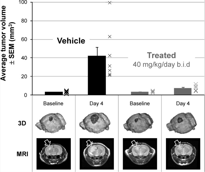 Antitumor activity in an orthotopic Ptch+/−p53−/−medulloblastoma allograft model in nude mice upon treatment with5mdiphosphate salt dosed at 40 mg/kg/day po bid or vehicle at equal dose volume.ACS Med Chem Lett. 2010 Jun 10; 1(3): 130–134. |
|---|
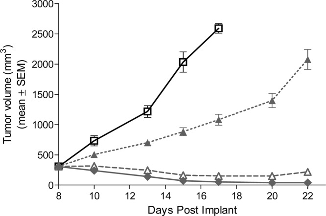 Antitumor activity upon treatment with5mdiphosphate salt or vehicle in a Ptch+/−p53−/− medulloblastoma subcutaneous allograft model in nude mice.ACS Med Chem Lett. 2010 Jun 10; 1(3): 130–134. |
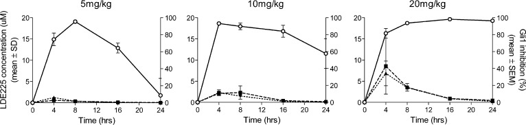
Gli1 mRNA inhibition (open circle), tumor PK (filled squares), and plasma PK (filled triangles) in Ptch+/−p53−/−medulloblastoma model after treatment with5m (Sonidegib, or erismodegib, LDE225, NVP-LDE225).ACS Med Chem Lett. 2010 Jun 10; 1(3): 130–134. |
Products are for research use only; We do not sell to patients
Copyright 2020 InvivoChem LLC | All Rights Reserved


 COA
COA