| Size | Price | Stock | Qty |
|---|---|---|---|
| 1mg |
|
||
| 5mg |
|
||
| 10mg |
|
||
| 25mg |
|
||
| 50mg |
|
||
| 100mg |
|
||
| 250mg |
|
||
| 500mg | |||
| Other Sizes |


Purity: ≥98%
| Targets |
HDAC1 ( IC50 = 9 nM ); HDAC ( IC50 = 32 nM )
Dacinostat (LAQ-824; NVP-LAQ-824) is a potent inhibitor of class I histone deacetylases (HDACs) with moderate activity against class IIb HDAC6, and no significant activity against class IIa or III HDACs. In recombinant HDAC enzyme assays: - HDAC1: IC50 = 3 nM [1,2] - HDAC2: IC50 = 4 nM [1,2] - HDAC3: IC50 = 5 nM [1,2] - HDAC6: IC50 = 25 nM [1,2] - HDAC4/5/7 (class IIa) and sirtuins (class III): IC50 > 1000 nM [1,2] |
|---|---|
| ln Vitro |
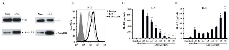
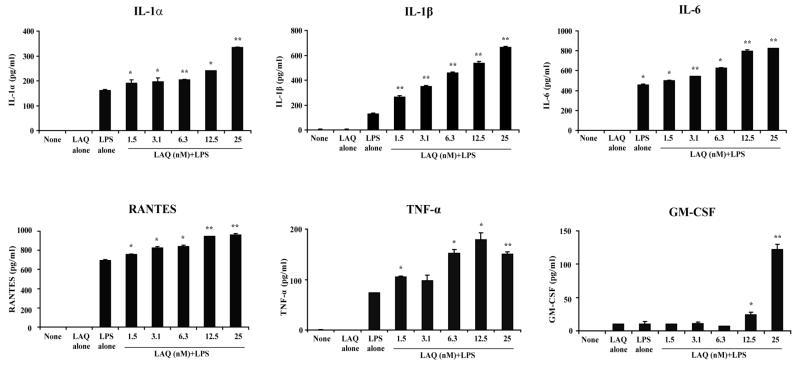
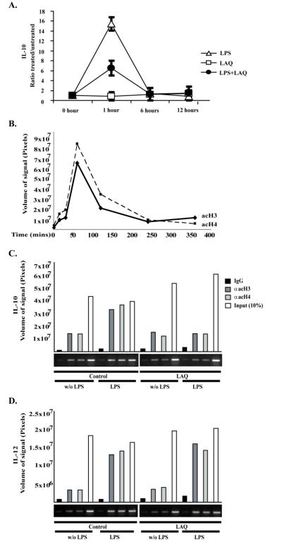
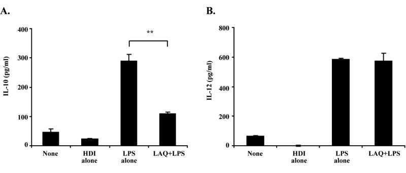
In vitro activity: LAQ824 stimulates the p21 promoter at 50% of the maximal promoter activation (AC50) of 0.30 μM, thereby initiating the expression of the gene encoding the p21 cell cycle inhibitor. With IC50 values of 0.15 μM and 0.01 μM, respectively, LAQ824 suppresses the growth of H1299, a non-small cell lung carcinoma line, and HCT116, a colon cancer cell line. Its antiproliferative action is specific to the tumor cell lines, causing only growth arrest in normal fibroblasts. Moreover, LAQ824 causes the A549 cells' p21 protein to rise in a dose-dependent manner and the Rb tumor suppressor's hypophosphorylated state to rise.[1] The IL-10 gene promoter undergoes chromatin modifications caused by LAQ824 that inhibit IL-10 production in BALB/c murine macrophages and increase the recruitment of the transcriptional repressors PU.1 and HDAC11.[2]
1. Antiproliferative activity in diverse cancer cell lines: - Human hematological cancer cell lines: Dacinostat (LAQ-824; NVP-LAQ-824) inhibited proliferation of HL-60 (acute myeloid leukemia, AML: IC50 = 0.08 μM), U937 (monocytic leukemia: IC50 = 0.1 μM), and Raji (Burkitt lymphoma: IC50 = 0.12 μM) after 72-hour MTT assay [1] - Human solid tumor cell lines: IC50 values for A549 (non-small cell lung cancer: 0.15 μM), MCF-7 (breast cancer: 0.18 μM), and HCT116 (colorectal cancer: 0.2 μM) were observed after 72-hour treatment. At 0.5 μM, cell viability was reduced by >85% in all tested lines [1] 2. Induction of histone acetylation and tumor suppressor gene expression: - In HL-60 cells, 0.1 μM Dacinostat (LAQ-824; NVP-LAQ-824) treatment for 24 hours increased acetyl-histone H3 (Lys9/14) by 4.5-fold and acetyl-histone H4 (Lys5/8/12/16) by 5.0-fold (western blot). qPCR showed a 3.2-fold upregulation of p21WAF1/CIP1 (tumor suppressor gene) and a 2.8-fold upregulation of Bax (pro-apoptotic gene) [1] 3. Inhibition of leukemic stem/progenitor cell (LSPC) function: - Primary LSPCs isolated from AML patient bone marrow were treated with Dacinostat (LAQ-824; NVP-LAQ-824) (0.05 μM, 0.1 μM, 0.2 μM) for 48 hours. Colony-forming unit (CFU) assay showed a 50% reduction in CFU-L (leukemic colonies) at 0.1 μM, and 80% reduction at 0.2 μM vs. controls [3] - Flow cytometry analysis of CD34+CD38- LSPCs (a marker of stemness) showed a 45% reduction in cell number after 0.1 μM treatment for 72 hours. Western blot detected a 3.0-fold increase in acetyl-p53 and a 2.5-fold increase in cleaved caspase-3 in these cells [3] 4. Apoptosis induction: - In U937 cells, 0.2 μM Dacinostat (LAQ-824; NVP-LAQ-824) for 48 hours induced apoptosis in 50% of cells (Annexin V-FITC/PI staining), vs. 6% in controls. Transmission electron microscopy showed typical apoptotic features (chromatin condensation, apoptotic bodies) [1] |
| ln Vivo |
LAQ824 treatment at 100 mg/kg inhibits tumor growth in HCT116 and human colon tumor xenografts in nude mice in a dose-dependent manner without causing general cytotoxicity.[1]
1. Antitumor efficacy in AML xenograft models: - Female nude mice (6-7 weeks old) were intravenously injected with 1×10⁷ HL-60 cells. When peripheral blood blast cells reached 5% (day 7), mice were randomized into 3 groups (n=6/group): vehicle (10% DMSO + 40% PEG300 + 50% PBS), Dacinostat (LAQ-824; NVP-LAQ-824) 5 mg/kg, 10 mg/kg. Drug was administered via intraperitoneal injection once daily for 14 days. Peripheral blood blast cells were reduced by 40% (5 mg/kg) and 75% (10 mg/kg) vs. vehicle. Median survival was extended from 21 days (vehicle) to 35 days (10 mg/kg group) [1] - Bone marrow analysis at study end showed a 65% reduction in leukemic infiltration in the 10 mg/kg group vs. vehicle [1] 2. Antitumor efficacy in solid tumor xenografts: - Male nude mice (6-7 weeks old) bearing HCT116 xenografts were treated with Dacinostat (LAQ-824; NVP-LAQ-824) (7.5 mg/kg, 15 mg/kg, oral gavage, once daily for 21 days) or vehicle. Tumor growth inhibition rates were 35% (7.5 mg/kg) and 65% (15 mg/kg) vs. vehicle. Tumor weights at day 21: 1.4 g (vehicle), 0.91 g (7.5 mg/kg), 0.49 g (15 mg/kg) [1] - Immunohistochemistry of HCT116 tumors showed a 3.8-fold increase in acetyl-histone H3 and a 2.2-fold increase in TUNEL-positive apoptotic cells in the 15 mg/kg group [1] 3. Modulation of LSPCs in murine leukemia model: - C57BL/6 mice were intravenously injected with 5×10⁵ MLL-AF9 leukemia cells (to induce AML). On day 10, mice were treated with Dacinostat (LAQ-824; NVP-LAQ-824) (5 mg/kg, intraperitoneal, daily for 10 days). Bone marrow CD34+CD38- LSPCs were reduced by 55% vs. vehicle controls. Secondary transplantation experiments showed that 80% of vehicle-treated recipient mice developed leukemia, vs. 20% in the dacinostat-treated group (indicating reduced LSPC self-renewal) [3] |
| Enzyme Assay |
Using the Q Sepharose Fast Flow column in ion exchange chromatography, HDAC enzymes are partially isolated from H1299 cell lysate. By using cdk2 polyclonal antibody or cdk1/cdc2 monoclonal antibody for immunoprecipitation, 500 mg of total cell lysate is used to extract enzyme complexes. Re-suspended immunoprecipitates are mixed with 1 μg of pRb recombinant protein substrate (cdk2) or 10 mL of H1 histone mixture containing 20 μg of substrate (cdc2) in kinase buffer (50 mM Hepes, pH 8, 10 mM MgCl2, 2.5 mM EDTA, 1 mM dithiothreitol, 20 mM ATP, 10 mM β-glycerophosphate, 0.1 mM NaVO4, 1 mM sodium fluoride, 50 mM ATP, 10 μCi of [γ-32P]ATP). Electrophoresis is used to separate phosphorylated Rb and H1 histones, and a PhosphorImager is used to quantify the results.
1. Recombinant class I HDAC (HDAC1/2/3) activity assay: - Recombinant human HDAC1, HDAC2, or HDAC3 enzyme was mixed with fluorogenic substrate Boc-Lys(Ac)-AMC in reaction buffer (50 mM Tris-HCl pH 8.0, 137 mM NaCl, 2.7 mM KCl, 1 mM MgCl2, 1 mM DTT). Dacinostat (LAQ-824; NVP-LAQ-824) was added at concentrations ranging from 0.1 nM to 100 nM, and the mixture was incubated at 37°C for 60 minutes. Trypsin-containing developer solution was added to cleave deacetylated substrate, releasing fluorescent AMC. Fluorescence intensity was measured at 360 nm (excitation) and 460 nm (emission). IC50 values were calculated by nonlinear regression of percentage activity (vs. vehicle) against log drug concentration using GraphPad Prism [1,2] 2. HDAC6 activity assay: - Recombinant human HDAC6 enzyme was mixed with fluorogenic substrate Tubulin-Ac-AMC (specific for HDAC6) in reaction buffer (50 mM Tris-HCl pH 7.5, 150 mM NaCl, 1 mM DTT). Dacinostat (LAQ-824; NVP-LAQ-824) (1 nM-100 nM) was added, and the mixture was incubated at 37°C for 90 minutes. Fluorescence was measured at 360 nm/460 nm, and IC50 was determined as described above [1,2] 3. Class IIa/III HDAC selectivity assay: - Recombinant HDAC4 (class IIa) and sirtuin 1 (class III) were tested with their respective fluorogenic substrates (Boc-Lys(Ac)-AMC for HDAC4, Sirt1 substrate for sirtuin 1) and Dacinostat (LAQ-824; NVP-LAQ-824) concentrations up to 10 μM. No significant inhibition (<10% activity reduction) was observed [1,2] |
| Cell Assay |
The 3-(4,5-dimethylthiazol-2-yl)-5-(3-carboxymethoxy-phenyl)-2-(4-sulfonyl)-2H-tetrazolium assay is a published procedure that is adapted to measure cell proliferation. The cells are cultivated in RPMI 1640 with 10% FBS after being seeded in 12-well dishes. TSA at different concentrations—up to 1,000 ng/mL—is present during cell culture. Trypan blue dye exclusion is used to measure the viable cell counts in a Nesbauer-type hemocytometer at 0 hours, 24 hours, and 48 hours in order to investigate the growth inhibition caused by TSA. To the RPMI 1640 medium, the same volume of ethanol is added as in the control experiment. Every experiment is carried out in triplicate and is repeated three times. Each experimental well has its average background value (treatment with medium alone) deducted; values are averaged for each compound dilution in triplicate. The percentage of growth is computed using the following formulas: If X>T0, then %Growth=(X-T0)/(GC-T0)*100; if X0, then %Growth=(X-T0)/T0*100. where X is the average value of compound-treated cells (in triplicate)-background, GC is the average value of untreated cells (in triplicate) − background, and T0 is the average value of T0 − background. In order to predict the concentration of compounds at 50% inhibition, the "% Growth" is plotted against compound concentration and used to calculate the IC50 using linear regression techniques between data points.
1. Cancer cell proliferation assay (MTT method): - Hematological (HL-60, U937, Raji) and solid tumor (A549, MCF-7, HCT116) cells were seeded in 96-well plates (3×10³-5×10³ cells/well) and incubated overnight. Dacinostat (LAQ-824; NVP-LAQ-824) (0.01 μM-1 μM) was added, and cells were cultured for 72 hours at 37°C (5% CO2). 10 μL MTT reagent (5 mg/mL) was added, incubated for 4 hours, then formazan crystals were dissolved in 100 μL DMSO. Absorbance was read at 570 nm, and cell viability (%) = (Treated absorbance / Control absorbance) × 100. IC50 values were calculated via GraphPad Prism [1] 2. Histone acetylation and gene expression assays: - HL-60 cells were seeded in 6-well plates (2×10⁵ cells/well) and treated with Dacinostat (LAQ-824; NVP-LAQ-824) (0.05 μM-0.2 μM) for 24 hours. For western blot: Cells were lysed in RIPA buffer (with protease inhibitors), 20 μg protein separated by 12% SDS-PAGE, transferred to PVDF membranes, and probed with antibodies against acetyl-histone H3, acetyl-histone H4, and β-actin. For qPCR: Total RNA was extracted, reverse-transcribed to cDNA, and amplified with primers for p21WAF1/CIP1, Bax, and GAPDH (internal control). Relative expression was calculated via 2^(-ΔΔCt) [1] 3. LSPC colony-forming and flow cytometry assays: - Primary AML patient bone marrow mononuclear cells were isolated and enriched for LSPCs via CD34+ magnetic sorting. LSPCs were seeded in methylcellulose medium with Dacinostat (LAQ-824; NVP-LAQ-824) (0.05 μM-0.2 μM) and cultured for 14 days; CFU-L colonies were counted under a microscope. For flow cytometry: LSPCs were treated with the drug for 72 hours, stained with anti-CD34-PE and anti-CD38-FITC antibodies, and analyzed via BD FACSCanto. Apoptosis was detected via Annexin V-FITC/PI staining [3] |
| Animal Protocol |
Using outbred athymic (nu/nu) female mice, the studies are carried out on-site. Following metofane anesthesia, mice receive a subcutaneous injection of a 100 μL cell suspension containing 1×10⁶ HCT116 cells into their right axillary (lateral) region. An estimated volume of 100–400 mm3 is permissible for tumors. Now, mice exhibiting tumors of a similar size range and acceptable morphology (non-necrotic) are chosen and divided into six-groups for the investigations. D5W is added to a stock solution made by dissolving NVP-LAQ824 in DMSO. This solution is then further diluted to a final DMSO concentration of 10% right before injection. The substance is injected intraperitoneally (IV) into the tail vein of tumor-bearing mice. For a total of 15 doses, NVP-LAQ824 is dosed once daily, five days a week. 5-Fluorouracil is given once a week for a total of three doses at a dose of 100 mg/kg in 0.9% saline. The vehicle is used to treat the control groups. Animals with tumors are taken out at the designated intervals.
1. HL-60 AML xenograft model: - Female nude mice (6-7 weeks old) were housed under SPF conditions. 1×10⁷ HL-60 cells (suspended in 0.2 mL PBS) were injected intravenously via tail vein. On day 7, peripheral blood smears were examined to confirm leukemic engraftment (≥5% blast cells). Mice were randomized into 3 groups (n=6/group): vehicle (10% DMSO + 40% PEG300 + 50% PBS), Dacinostat (LAQ-824; NVP-LAQ-824) 5 mg/kg, 10 mg/kg. Drug was administered via intraperitoneal injection once daily for 14 days. Peripheral blood blast cells were counted weekly via smear analysis. Mice were monitored for survival until endpoint (moribundity), and median survival was calculated via Kaplan-Meier analysis. At study end, bone marrow was harvested for leukemic infiltration analysis [1] 2. HCT116 colorectal cancer xenograft model: - Male nude mice (6-7 weeks old) were subcutaneously injected with 5×10⁶ HCT116 cells (0.1 mL PBS + 50% Matrigel) into the right flank. When tumors reached ~100 mm³, mice were randomized into 3 groups (n=6/group): vehicle, Dacinostat (LAQ-824; NVP-LAQ-824) 7.5 mg/kg, 15 mg/kg. Drug was dissolved in 0.5% carboxymethyl cellulose (CMC) and administered via oral gavage once daily for 21 days. Tumor volume (length × width² / 2) and body weight were measured twice weekly. At study end, tumors were harvested for immunohistochemistry (acetyl-histone H3, TUNEL) [1] 3. MLL-AF9 murine leukemia model: - C57BL/6 mice (female, 8 weeks old) were intravenously injected with 5×10⁵ MLL-AF9-transduced bone marrow cells (to induce AML). On day 10, mice were randomized into 2 groups (n=6/group): vehicle (10% DMSO + 40% PEG300 + 50% PBS), Dacinostat (LAQ-824; NVP-LAQ-824) 5 mg/kg. Drug was administered via intraperitoneal injection once daily for 10 days. On day 20, bone marrow was harvested, and CD34+CD38- LSPCs were quantified via flow cytometry. For secondary transplantation: 1×10⁶ bone marrow cells from primary treated mice were injected into naive C57BL/6 mice (n=5/group), and leukemia incidence was monitored for 60 days [3] |
| ADME/Pharmacokinetics |
1. Oral bioavailability in mice: Female CD1 mice (20-25 g) were administered dacinostat (LAQ-824; NVP-LAQ-824) orally (15 mg/kg) orally and intravenously (5 mg/kg). Blood samples were collected at 0.25, 0.5, 1, 2, 4, 6, 8, and 24 hours post-administration. Plasma was separated by centrifugation (3000×g, 10 min, 4℃), and drug concentration was determined by liquid chromatography-tandem mass spectrometry (LC-MS/MS). The oral bioavailability was 35%, calculated based on AUC₀₋∞ (oral: 28.5 μM·h; intravenous: 24.0 μM·h) [1]
2. Plasma pharmacokinetic parameters (mice, oral administration 15 mg/kg): - Maximum plasma concentration (Cmax) = 7.2 μM (Tmax = 1 hour) - Terminal half-life (t₁/₂) = 4.5 hours - Area under the concentration-time curve (AUC₀₋∞) = 28.5 μM·h - Apparent clearance (CL/F) = 1.8 L/kg/h [1] 3. Tissue distribution (mice, oral administration 15 mg/kg, 1 hour after administration): - Highest concentration: liver (18.5 μM), kidney (15.2 μM), tumor (HCT116) xenograft: 12.8 μM) - Moderate concentration: lung (8.6 μM), spleen (7.3 μM) - Low concentration: brain (0.9 μM), plasma (7.2 μM) - Tumor/plasma concentration ratio = 1.8 [1] 4. Metabolism: - In human liver microsomes, dacinostat (LAQ-824; NVP-LAQ-824) is mainly metabolized by CYP3A4 (65% of total metabolism) and CYP2C19 (20%). Metabolism of CYP1A2, CYP2C9 or CYP2D6 is not significant. The major metabolites were identified by LC-MS/MS as hydroxylated and N-dealkylated products [1] |
| Toxicity/Toxicokinetics |
1. Acute toxicity in mice: - Female CD1 mice (20-25 g) were administered a single oral dose of dacinostat (LAQ-824; NVP-LAQ-824) (25 mg/kg, 50 mg/kg, 100 mg/kg, 200 mg/kg; n=6 per group). No deaths occurred in the ≤50 mg/kg group; 1 out of 6 mice died in the 100 mg/kg group; 3 out of 6 mice died in the 200 mg/kg group. At the 50 mg/kg dose, a transient decrease in body weight (7% of initial body weight) was observed on day 2, which was fully recovered by day 5. No clinical symptoms (drowsiness, diarrhea) were observed at doses ≤25 mg/kg [1]
2. Chronic toxicity in rats: - Male Sprague-Dawley rats (250-300 g) were treated with Dacinostat (LAQ-824; NVP-LAQ-824) (5 mg/kg, 10 mg/kg, 20 mg/kg, by gavage, once daily for 28 days; n=8 per group). No deaths or significant changes in body weight were observed. Serum biochemistry: No changes were observed in ALT, AST, creatinine or BUN. Hematology: No changes were observed in WBC, RBC, platelets or hemoglobin. Histopathology: No abnormal lesions were observed in the liver, kidneys, spleen, heart, or lungs [1] 3. Plasma protein binding rate: - Dacinostat (LAQ-824; NVP-LAQ-824) was added to human plasma (0.1 μM, 1 μM, 10 μM) and incubated at 37°C for 30 minutes. Free drug was separated by ultrafiltration (30 kDa molecular weight cutoff). LC-MS/MS showed that plasma protein binding rate was >97% at all concentrations [1,2] 4. Toxicity in mouse leukemia model: - In the MLL-AF9 AML model (5 mg/kg intraperitoneal injection, 10 days), dacinostat (LAQ-824; NVP-LAQ-824) did not cause significant weight loss (<3% compared to the solvent control group) or clinical toxicity. Bone marrow analysis showed that the number of normal hematopoietic progenitor cells (CFU-GM, CFU-E) was not reduced compared with the vector group [3] |
| References |
|
| Additional Infomation |
N-Hydroxy-3-[4-[[2-hydroxyethyl-[2-(1H-indol-3-yl)ethyl]amino]methyl]phenyl]-2-acrylamide is a tryptamine compound. Dacinolacta is a novel histone deacetylase inhibitor. 1. Mechanism of action: Dacinolacta (LAQ-824; NVP-LAQ-824) exerts its anticancer effect by selectively inhibiting class I HDAC and HDAC6, leading to the accumulation of acetylated histones and non-histone proteins (e.g., p53). This epigenetic modification can relax chromatin, upregulate tumor suppressor genes (p21WAF1/CIP1) and pro-apoptotic genes (Bax), inhibit cancer cell proliferation, induce apoptosis, and impair the self-renewal capacity of leukemia stem cells [1,3]
2. Preclinical advantages: Compared with trachostatin A (TSA), dacinnostatin (LAQ-824; NVP-LAQ-824) has higher oral bioavailability (35% vs. <10% in mice), a longer half-life (4.5 hours vs. 2.0 hours), and higher selectivity for class I HDACs, thereby reducing toxicities associated with class IIa HDAC inhibition (e.g., cardiotoxicity) [1] 3. Potential clinical indications: Based on preclinical data, ... NVP-LAQ-824 is a candidate drug for the treatment of hematologic malignancies (acute myeloid leukemia, lymphoma) and solid tumors (colorectal cancer, non-small cell lung cancer). Its ability to target leukemia stem cells supports its application in the prevention of acute myeloid leukemia relapse [1,3]. 4. Structure-activity relationship (SAR): Dacinostat (LAQ-824; NVP-LAQ-824) is a histone deacetylase (HDAC) inhibitor based on hydroxyxamic acid; SAR studies have shown that the piperidine-3-ylindole moiety enhances the selectivity and oral absorption of class I HDACs compared to analogs without this structure [2]. |
| Molecular Formula |
C22H25N3O3
|
|
|---|---|---|
| Molecular Weight |
379.46
|
|
| Exact Mass |
379.189
|
|
| Elemental Analysis |
C, 69.64; H, 6.64; N, 11.07; O, 12.65
|
|
| CAS # |
404951-53-7
|
|
| Related CAS # |
|
|
| PubChem CID |
6445533
|
|
| Appearance |
Light yellow to yellow solid powder
|
|
| Density |
1.3±0.1 g/cm3
|
|
| Index of Refraction |
1.693
|
|
| LogP |
3.41
|
|
| Hydrogen Bond Donor Count |
4
|
|
| Hydrogen Bond Acceptor Count |
4
|
|
| Rotatable Bond Count |
9
|
|
| Heavy Atom Count |
28
|
|
| Complexity |
505
|
|
| Defined Atom Stereocenter Count |
0
|
|
| SMILES |
O([H])C([H])([H])C([H])([H])N(C([H])([H])C1C([H])=C([H])C(/C(/[H])=C(\[H])/C(N([H])O[H])=O)=C([H])C=1[H])C([H])([H])C([H])([H])C1=C([H])N([H])C2=C([H])C([H])=C([H])C([H])=C12
|
|
| InChi Key |
BWDQBBCUWLSASG-MDZDMXLPSA-N
|
|
| InChi Code |
InChI=1S/C22H25N3O3/c26-14-13-25(12-11-19-15-23-21-4-2-1-3-20(19)21)16-18-7-5-17(6-8-18)9-10-22(27)24-28/h1-10,15,23,26,28H,11-14,16H2,(H,24,27)/b10-9+
|
|
| Chemical Name |
(E)-N-hydroxy-3-[4-[[2-hydroxyethyl-[2-(1H-indol-3-yl)ethyl]amino]methyl]phenyl]prop-2-enamide
|
|
| Synonyms |
NVP-LAQ824; NVP-LAQ-824; NVP LAQ824; LAQ 824; LAQ824; LAQ-824; Dacinostat
|
|
| HS Tariff Code |
2934.99.9001
|
|
| Storage |
Powder -20°C 3 years 4°C 2 years In solvent -80°C 6 months -20°C 1 month |
|
| Shipping Condition |
Room temperature (This product is stable at ambient temperature for a few days during ordinary shipping and time spent in Customs)
|
| Solubility (In Vitro) |
DMSO: ~76 mg/mL (~200.3 mM)
Water: 1 mg/mL (Insoluble) Ethanol: 1 mg/mL |
|---|---|
| Solubility (In Vivo) |
Solubility in Formulation 1: ≥ 2.5 mg/mL (6.59 mM) (saturation unknown) in 10% DMSO + 40% PEG300 + 5% Tween80 + 45% Saline (add these co-solvents sequentially from left to right, and one by one), clear solution.
For example, if 1 mL of working solution is to be prepared, you can add 100 μL of 25.0 mg/mL clear DMSO stock solution to 400 μL PEG300 and mix evenly; then add 50 μL Tween-80 to the above solution and mix evenly; then add 450 μL normal saline to adjust the volume to 1 mL. Preparation of saline: Dissolve 0.9 g of sodium chloride in 100 mL ddH₂ O to obtain a clear solution. Solubility in Formulation 2: ≥ 2.5 mg/mL (6.59 mM) (saturation unknown) in 10% DMSO + 90% (20% SBE-β-CD in Saline) (add these co-solvents sequentially from left to right, and one by one), clear solution. For example, if 1 mL of working solution is to be prepared, you can add 100 μL of 25.0 mg/mL clear DMSO stock solution to 900 μL of 20% SBE-β-CD physiological saline solution and mix evenly. Preparation of 20% SBE-β-CD in Saline (4°C,1 week): Dissolve 2 g SBE-β-CD in 10 mL saline to obtain a clear solution. View More
Solubility in Formulation 3: ≥ 2.5 mg/mL (6.59 mM) (saturation unknown) in 10% DMSO + 90% Corn Oil (add these co-solvents sequentially from left to right, and one by one), clear solution. Solubility in Formulation 4: 1% DMSO+30% polyethylene glycol+1% Tween 80: 30mg/mL |
| Preparing Stock Solutions | 1 mg | 5 mg | 10 mg | |
| 1 mM | 2.6353 mL | 13.1766 mL | 26.3532 mL | |
| 5 mM | 0.5271 mL | 2.6353 mL | 5.2706 mL | |
| 10 mM | 0.2635 mL | 1.3177 mL | 2.6353 mL |
*Note: Please select an appropriate solvent for the preparation of stock solution based on your experiment needs. For most products, DMSO can be used for preparing stock solutions (e.g. 5 mM, 10 mM, or 20 mM concentration); some products with high aqueous solubility may be dissolved in water directly. Solubility information is available at the above Solubility Data section. Once the stock solution is prepared, aliquot it to routine usage volumes and store at -20°C or -80°C. Avoid repeated freeze and thaw cycles.
Calculation results
Working concentration: mg/mL;
Method for preparing DMSO stock solution: mg drug pre-dissolved in μL DMSO (stock solution concentration mg/mL). Please contact us first if the concentration exceeds the DMSO solubility of the batch of drug.
Method for preparing in vivo formulation::Take μL DMSO stock solution, next add μL PEG300, mix and clarify, next addμL Tween 80, mix and clarify, next add μL ddH2O,mix and clarify.
(1) Please be sure that the solution is clear before the addition of next solvent. Dissolution methods like vortex, ultrasound or warming and heat may be used to aid dissolving.
(2) Be sure to add the solvent(s) in order.
 |
|---|
|
|
Products are for research use only; We do not sell to patients
Copyright 2020 InvivoChem LLC | All Rights Reserved


 COA
COA