| Size | Price | Stock | Qty |
|---|---|---|---|
| 2mg |
|
||
| 5mg |
|
||
| 10mg |
|
||
| 25mg |
|
||
| 50mg |
|
||
| 100mg |
|
||
| 250mg |
|
||
| 500mg | |||
| Other Sizes |


Purity: ≥98%
| Targets |
Akt1 (IC50 = 5 nM); Akt3 (IC50 = 8 nM); Akt2 (IC50 = 18 nM); PKA (IC50 = 3100 nM)
Selective ATP-competitive inhibitor of Akt isoforms, including Ipatasertib (GDC0068; RG7440). It exhibits potent inhibitory activity against Akt1 with an IC50 of approximately 5 nM, Akt2 with an IC50 of approximately 18 nM, and Akt3 with an IC50 of approximately 12 nM. It shows high selectivity over other kinases (e.g., >100-fold selectivity against PKA, PKC, and other AGC kinases) [1] - Ipatasertib (GDC0068; RG7440) maintains selective inhibition of Akt1 (IC50: 5 nM), Akt2 (IC50: 18 nM), and Akt3 (IC50: 12 nM) in kinase panels, with minimal activity against non-Akt kinases, confirming its specificity for the Akt family [2] - In cellular assays, Ipatasertib (GDC0068; RG7440) inhibits phosphorylated Akt (p-Akt) at Ser473 and Thr308, downstream of Akt isoforms, without affecting the total Akt protein level; no new IC50 values for Akt isoforms are provided beyond those reported in [1] [3] |
|---|---|
| ln Vitro |
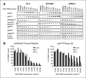
GDC-0068 only inhibits 3 kinases by >70% at 1 μM concentration when tested against a large panel of 230 kinases (PRKG1α, PRKG1β, and p70S6K, with IC50 values of 98 nM, 69 nM, and 860 nM, respectively). GDC-0068 has an IC50 of 3.1 μM and exhibits >100-fold selectivity for Akt over PKA. With IC50 values of 157 nM, 197 nM, and 208 nM, respectively, GDC-0068 treatment inhibits the phosphorylation of the Akt substrate, PRAS40, in LNCaP, PC3, and BT474M1 cells. Additionally, the Akt-signaling-driven cancer cell lines that have defects in the tumor suppressor PTEN, oncogenic mutations in PIK3CA, and HER2 amplification are all selectively inhibited by GDC-0068, with the strongest effects seen in the HER2+ and Luminal subtypes. [1-3]
Ipatasertib (GDC0068; RG7440) inhibits the proliferation of multiple human tumor cell lines with activated Akt signaling. For example, in PC-3 (prostate cancer) cells, the antiproliferative IC50 is approximately 0.8 μM; in LNCaP (prostate cancer) cells, the IC50 is approximately 1.2 μM; and in MDA-MB-468 (breast cancer) cells, the IC50 is approximately 0.6 μM. Western blot analysis shows that treatment with 1-5 μM Ipatasertib (GDC0068; RG7440) for 4 hours reduces p-Akt (Ser473 and Thr308) levels by >70%, and also decreases the phosphorylation of downstream Akt substrates, such as p-FOXO1 (Ser256) and p-GSK3β (Ser9), by 60%-80% [1] - In a panel of 24 human tumor cell lines, Ipatasertib (GDC0068; RG7440) demonstrates antiproliferative activity with IC50 values ranging from 0.3 μM to 3.5 μM, with the highest potency in cell lines with PTEN loss or PI3K mutation (e.g., BT-474 breast cancer cells, IC50: 0.3 μM). Combination treatment with Ipatasertib (GDC0068; RG7440) (0.5 μM) and paclitaxel (10 nM) enhances antiproliferative effects in MDA-MB-231 breast cancer cells, resulting in a 40% greater reduction in cell viability compared to single-agent treatment. Clone formation assays show that 0.5 μM Ipatasertib (GDC0068; RG7440) reduces the clone formation rate of PC-3 cells by >60% after 14 days of culture [2] - Ipatasertib (GDC0068; RG7440) induces apoptosis in HCT116 (colorectal cancer) cells and MCF-7 (breast cancer) cells. At a concentration of 2 μM, treatment for 24 hours increases the apoptotic rate (Annexin V-positive cells) by 30%-40% compared to the control. Western blot analysis reveals that Ipatasertib (GDC0068; RG7440) (1-3 μM) upregulates the expression of PUMA (a pro-apoptotic protein) by 2-3 fold and activates Caspase-3/7 (cleaved Caspase-3 levels increase by >2 fold). Real-time PCR shows that 2 μM Ipatasertib (GDC0068; RG7440) increases PUMA mRNA expression by 3.5 fold in HCT116 cells. Additionally, knockdown of FoxO3a or NF-κB p65 abolishes the Ipatasertib (GDC0068; RG7440)-induced PUMA upregulation, indicating that FoxO3a and NF-κB mediate this effect [3] |
| ln Vivo |
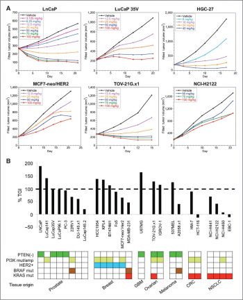
GDC-0068 oral administration causes the down-regulation of p-PRAS40 in PC3 prostate tumor xenograft models. GDC-0068 treatment in BT474-Tr xenografts lowers pS6 and peIF4G levels, relocalizes FOXO3a to the nucleus, and causes feedback upregulation of HER3 and pERK. In numerous xenograft tumor models, including the PTEN-deficient prostate cancer models LNCaP and PC3, the PIK3CA H1047R mutant breast cancer model KPL-4, and the MCF7-neo/HER2 tumor model, administration of GDC-0068 demonstrates potent antitumor efficacy.[1-3]
In nude mouse xenograft models of PC-3 prostate cancer, oral administration of Ipatasertib (GDC0068; RG7440) at doses of 30 mg/kg/day and 100 mg/kg/day for 21 days results in tumor growth inhibition (TGI) of 45% and 72%, respectively, compared to the vehicle control. No significant weight loss (≤5%) is observed in treated mice. Western blot analysis of tumor tissues shows that 100 mg/kg/day Ipatasertib (GDC0068; RG7440) reduces p-Akt (Ser473) levels by >80% and p-GSK3β (Ser9) levels by 70% [1] - In MDA-MB-468 breast cancer xenograft models (nude mice), Ipatasertib (GDC0068; RG7440) (100 mg/kg/day, oral) alone induces a TGI of 65% after 28 days. Combination treatment with Ipatasertib (GDC0068; RG7440) (100 mg/kg/day) and docetaxel (10 mg/kg/week, intraperitoneal injection) results in a TGI of 90% and significantly prolongs the median survival of mice (from 42 days in the vehicle group to 68 days in the combination group). Immunohistochemistry (IHC) of tumor tissues shows that Ipatasertib (GDC0068; RG7440) treatment reduces p-Akt (Ser473) staining intensity by >70% [2] - In HCT116 colorectal cancer xenograft models (nude mice), oral administration of Ipatasertib (GDC0068; RG7440) (100 mg/kg/day) for 14 days increases PUMA protein expression (detected by IHC) in tumor tissues by 2.5 fold and increases the number of apoptotic cells (TUNEL-positive) by 3 fold compared to the vehicle group. No significant toxicity (e.g., weight loss, organ damage) is observed in treated mice [3] |
| Enzyme Assay |
Enzymatic Assays[1]
The assay for the determination of Akt1/2/3 and PKA kinase activity employs the IMAP fluorescence polarization (FP) phosphorylation detection reagent to detect fluorescently labeled peptide substrates that have been phosphorylated by the respective kinases. The Akt enzymes employed in these studies consisted of recombinant baculovirus expressed, amino-terminal, polyhistidine-tagged, full-length, wild-type human forms and were obtained commercially. The PKA enzyme employed in these studies consisted of the recombinant untagged human isolated catalytic subunit of PKA expressed in Escherichia coli obtained commercially. Inhibitor, enzyme (9 nM Akt1 or 100 pM PKA), and substrate (100 nM Crosstide) were incubated with 5 μM ATP in assay buffer (10 mM Tris–HCl (pH 7.2), 10 mM MgCl2, 0.1% BSA (w/v), final DMSO 2% (v/v)) for 60 min at ambient temperature in a 5 μL reaction volume. Reactions were initiated by addition of enzyme + peptide substrate to ATP solutions. IMAP binding reagent (15 μL) was added to terminate the reaction, and the stopped reactions were incubated for a minimum of 30 min at room temperature (rt). For Akt kinase activity assay (HTRF-based): Recombinant human Akt isoforms (Akt1, Akt2, Akt3) are diluted in assay buffer (containing Tris-HCl, MgCl2, DTT, and BSA) to a final concentration of 1 nM. The substrate (biotinylated GSK3β peptide, final concentration 2 μM) and ATP (final concentration 10 μM, near the Km value of Akt) are added to the reaction mixture, followed by different concentrations of Ipatasertib (GDC0068; RG7440) (ranging from 0.1 nM to 10 μM). The reaction is incubated at 30°C for 60 minutes, then stopped by adding a detection mixture (streptavidin-conjugated Eu3+ and anti-phospho-GSK3β antibody-conjugated XL665). The fluorescence resonance energy transfer (FRET) signal is measured at 665 nm and 620 nm, and the ratio of 665/620 is used to calculate the percentage of kinase activity inhibition. IC50 values are determined by fitting the inhibition curves with a four-parameter logistic model [1] - For kinase selectivity assay: A panel of 130 recombinant human kinases (including AGC family kinases, tyrosine kinases, and serine/threonine kinases) is used. Each kinase is incubated with its specific substrate, ATP (at Km concentration), and Ipatasertib (GDC0068; RG7440) at a final concentration of 1 μM. Kinase activity is detected using a radioactive method (33P-ATP incorporation into the substrate). The percentage of kinase activity remaining is calculated relative to the vehicle control, and kinases with <10% remaining activity are considered potential off-targets. Results show that only Akt1, Akt2, and Akt3 have <10% remaining activity, confirming the selectivity of Ipatasertib (GDC0068; RG7440) [1] - For p-Akt inhibition assay in cell lysates: Tumor cells (e.g., PC-3) are treated with Ipatasertib (GDC0068; RG7440) for 4 hours, then lysed in RIPA buffer (containing protease and phosphatase inhibitors). The lysates are centrifuged, and the supernatant is collected. Equal amounts of protein (20 μg) are separated by SDS-PAGE and transferred to PVDF membranes. The membranes are blocked with 5% non-fat milk, then incubated with primary antibodies against p-Akt (Ser473), p-Akt (Thr308), or total Akt overnight at 4°C. After washing, the membranes are incubated with HRP-conjugated secondary antibodies for 1 hour at room temperature. The signal is detected using ECL substrate, and the band intensity is quantified using ImageJ software. The percentage of p-Akt inhibition is calculated relative to the vehicle control [2] |
| Cell Assay |
Inhibition of cellular viability was measured in LNCaP cells plated in black, clear-bottomed 96-well plates at a density of 5000 cells/well and subsequently treated with 0–10 μM 28 (ipatasertib) for 72 h at 37 °C and 5% CO2. The extent of cell proliferation was determined by measuring the reduction of resazurin to resorufin as described in the manufacturer’s protocol using an excitation wavelength of 560 nm and an emission wavelength of 590 nm. Dose–response curves were generated using the four-parameter logistic model, and 50% inhibitory concentration (IC50) values were determined from these curve fits.[1]
Briefly, after ipatasertib treatment, HCT116 WT or p53−/− were fixed with 1% formaldehyde and lysed in warm SDS lysis buffer. The genomic DNA was obtained and sheared to 200–1000 bp by sonication on ice. Samples were precleared with Protein A-Agarose/Salmon Sperm DNA (50% Slurry) for 1 h at 4 °C with agitation. Then anti-FoxO3a antibody or anti-p65 antibody was added and incubated overnight on a shaker at 4 °C. Normal rabbit IgG was used as a negative control. The protein agarose/salmon sperm DNA (50% slurry) bead was then added to precipitate the antibody/protein/DNA complexes. After washed with serial wash buffers, DNA–protein immunocomplexes were eluted from the beads by elution buffer (1% SDS, 0.1 M NaHCO3) for 30 min. Finally, the protein–DNA cross-links were reversed to release DNA by incubation with 0.2 M NaCl at 65 °C for 4 h[3]. Antiproliferative assay (MTT method): Human tumor cells are seeded in 96-well plates at a density of 2×10^3 cells/well and cultured overnight. Different concentrations of Ipatasertib (GDC0068; RG7440) (ranging from 0.01 μM to 10 μM) are added to each well, with 3 replicates per concentration. The plates are incubated at 37°C in a 5% CO2 incubator for 72 hours. After incubation, 20 μL of MTT solution (5 mg/mL) is added to each well, and the plates are incubated for another 4 hours. The supernatant is removed, and 150 μL of DMSO is added to dissolve the formazan crystals. The absorbance is measured at 570 nm using a microplate reader. The cell viability is calculated relative to the vehicle control, and the IC50 is determined using GraphPad Prism software [1] - Clone formation assay: PC-3 cells are seeded in 6-well plates at a density of 5×10^2 cells/well and cultured overnight. Ipatasertib (GDC0068; RG7440) is added at final concentrations of 0.1 μM, 0.5 μM, and 1 μM, with a vehicle control group. The plates are incubated at 37°C in a 5% CO2 incubator for 14 days, with the medium and drug refreshed every 3 days. After incubation, the cells are fixed with 4% paraformaldehyde for 15 minutes, stained with 0.1% crystal violet for 30 minutes, and then washed with water. Colonies containing >50 cells are counted under a microscope. The clone formation rate is calculated as (number of colonies in drug group / number of colonies in control group) × 100% [2] - Apoptosis assay (Annexin V-FITC/PI double staining): HCT116 cells are seeded in 6-well plates at a density of 2×10^5 cells/well and cultured overnight. Ipatasertib (GDC0068; RG7440) is added at a final concentration of 2 μM, and the cells are incubated for 24 hours. The cells are harvested by trypsinization, washed twice with cold PBS, and resuspended in 1× binding buffer. Annexin V-FITC (5 μL) and PI (10 μL) are added to the cell suspension, which is then incubated in the dark at room temperature for 15 minutes. The apoptotic rate is detected using a flow cytometer, and the data are analyzed using FlowJo software [3] - Real-time PCR for PUMA mRNA detection: HCT116 cells are treated with 2 μM Ipatasertib (GDC0068; RG7440) for 12 hours. Total RNA is extracted using an RNA extraction kit, and the RNA concentration and purity are determined using a NanoDrop spectrophotometer. Reverse transcription is performed to synthesize cDNA using a reverse transcriptase kit. Real-time PCR is carried out using a SYBR Green PCR master mix, with PUMA-specific primers and GAPDH-specific primers (internal control). The reaction conditions are: 95°C for 10 minutes, followed by 40 cycles of 95°C for 15 seconds and 60°C for 1 minute. The relative expression level of PUMA mRNA is calculated using the 2^(-ΔΔCt) method [3] |
| Animal Protocol |
Female nude mice bearing LNCaP, PC3, KPL-4, or MCF7 tumor xenografts
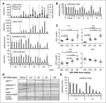
~100 mg/kg/day Orally For in vivo tumor xenograft studies, female nu/nu (nude) mice were inoculated subcutaneously in the right hind flank with PC3 cells suspended in Hank’s balanced salt solution (HBSS). When tumors reached a mean volume of 150 mm3, the animals were size matched and distributed into treatment groups consisting of 10 animals/group. Tumor volume was calculated as follows: tumor size (mm3) = (longer measurement × (shorter measurement)2) × 0.5. Following data analysis, p values were determined using Dunnett’s t test with JMP statistical software, version 7.0 (SAS Institute). Mouse body weights were recorded twice weekly using an Adventura Pro AV812 scale (Ohaus Corp.). Mice were promptly euthanized when the tumor volume exceeded 2000 mm3 or if body weight loss was ≥20% of the starting weight per IACUC protocol guidelines.[1] For PK/PD studies, blood and tumor samples were collected at 1, 3, 8, and 24 h after a single dose of ipatasertib from PC3 tumor bearing mice. Blood samples (approximately 800 μL) were collected from each animal at the scheduled sample collection time by terminal cardiac puncture into tubes containing K2EDTA as an anticoagulant and centrifuged at 1500–2000g to isolate plasma. The concentration of ipatasertib in each plasma sample was determined by a nonvalidated LC/MS/MS assay in the DMPK Bioanalytical Department at Genentech. The assay lower limit of quantitation (LLOQ) was 0.005 μM. Tumor samples were dissociated in Tris lysis buffer containing 150 mM NaCl, 20 mM Tris (pH 7.5), 1 mM EDTA, 1 mM EGTA, and 1% Triton X-100. Protein concentrations were determined using the BCA Protein Assay Kit. The human enzyme-linked immunosorbent assay (ELISA) kits were used to determine the levels of total PRAS40 and PRAS40 phosphorylated at Thr246 (p-PRAS40). The assay quantifies protein levels on the basis of measurements of absorbance. The colored product is directly proportional to the concentration of p-PRAS40 and tPRAS40 present in the specimen. The Meso Scale Discovery Multi-Spot Biomarker Detection System was used to determine the levels of total S6RP and S6RP phosphorylated at Ser235/236 (pS6RP). These assays quantify protein levels on the basis of measurements of electrochemiluminescence intensity. Levels of phosphorylated protein were normalized to total protein levels in ipatasertib-treated tumors and compared to the vehicle control.[1] In vivo efficacy was evaluated in multiple tumor cell line- and patient-derived xenograft models. Cells or tumor fragments were implanted subcutaneously into the flank of immunocompromised mice. Female or male nude (nu/nu) or severe combined immunodeficient mice (SCID)/beige mice were used. For the MCF7-neo/HER2 model, 17β-estradiol pellets (0.36 mg/pellet, 60-day release, no. SE-121) were implanted into the dorsal shoulder before cell inoculation. Male mice were castrated before implantation of tumor fragments. After implantation of tumor cells or fragments into mice, tumors were monitored until they reached mean tumor volumes of 180 to 350 mm3 and distributed into groups of 8 to 10 animals/group. ipatasertib/GDC-0068 was formulated in 0.5% methylcellulose/0.2% Tween-80 (MCT) and administered daily (QD), via oral (per os; PO) gavage.[2] HCT116 WT and PUMA−/− were harvested, and 1 × 10~6 cells in 0.2 ml of medium were implanted subcutaneously into the back of athymic nude female mice. Female 5-week-old nude mice were housed in a sterile environment with microisolator cages and allowed access to water and chow ad libitum. Mice were treated daily with ipatasertib/GDC-0068 at 40 mg/kg by oral gavage for 21 days treatment after 7 days. Calipers were used to monitor the tumor growth, volume was calculated by the formula: 0.5 × length × width2. Mice were euthanized when tumors reached ~1.0 cm3 in size. Tumors were dissected and fixed in 10% formalin and embedded in paraffin.[3] PC-3 prostate cancer xenograft model in nude mice: Male nude mice (6-8 weeks old) are used. Logarithmically growing PC-3 cells (1×10^6 cells in 0.1 mL of PBS/matrigel mixture, 1:1) are subcutaneously injected into the right dorsal flank of each mouse. When the tumor volume reaches 100-200 mm³, the mice are randomly divided into 3 groups (n=6 per group): vehicle control group, Ipatasertib (GDC0068; RG7440) 30 mg/kg group, and Ipatasertib (GDC0068; RG7440) 100 mg/kg group. Ipatasertib (GDC0068; RG7440) is dissolved in a vehicle consisting of 0.5% methylcellulose and 0.1% Tween 80, and administered orally once daily. The vehicle control group receives the same volume of vehicle. Tumor volume (calculated as length × width² × 0.5) and mouse body weight are measured twice a week. After 21 days of treatment, the mice are euthanized, and tumor tissues are collected for western blot analysis [1] - MDA-MB-468 breast cancer xenograft combination model: Female nude mice (6-8 weeks old) are subcutaneously injected with MDA-MB-468 cells (2×10^6 cells in 0.1 mL of PBS/matrigel, 1:1) into the right dorsal flank. When tumors reach 150-200 mm³, mice are divided into 4 groups (n=8 per group): vehicle control, Ipatasertib (GDC0068; RG7440) alone (100 mg/kg, oral, once daily), docetaxel alone (10 mg/kg, intraperitoneal injection, once weekly), and combination of Ipatasertib (GDC0068; RG7440) and docetaxel. The treatment lasts for 28 days. Tumor volume and body weight are measured twice a week. Mouse survival is recorded daily, and the median survival time is calculated. After treatment, tumor tissues are collected for immunohistochemistry (IHC) analysis of p-Akt [2] - HCT116 colorectal cancer xenograft mechanism model: Nude mice (6-8 weeks old) are subcutaneously injected with HCT116 cells (1.5×10^6 cells in 0.1 mL of PBS/matrigel, 1:1). When tumors reach 100-150 mm³, mice are divided into 2 groups (n=6 per group): vehicle control and Ipatasertib (GDC0068; RG7440) 100 mg/kg (oral, once daily). Treatment continues for 14 days. Tumor volume and body weight are measured every 3 days. After euthanasia, tumor tissues are fixed in 4% paraformaldehyde, embedded in paraffin, and sectioned. IHC is performed to detect PUMA protein expression (using anti-PUMA antibody), and TUNEL staining is used to detect apoptotic cells. The number of PUMA-positive cells and TUNEL-positive cells is counted under a microscope [3] |
| ADME/Pharmacokinetics |
In vitro metabolism: Human liver microsomes (0.5 mg/mL) were incubated with ipatatatinib (GDC0068; RG7440) (1 μM) and NADPH (1 mM) at 37°C for 0-60 minutes. The reaction was terminated by adding acetonitrile containing an internal standard. After centrifugation, the supernatant was analyzed by LC-MS/MS to detect metabolites. The results showed that ipatatatinib (GDC0068; RG7440) was mainly metabolized by CYP3A4, producing three major oxidative metabolites (M1, M2 and M3). The half-life of ipatatatinib (GDC0068; RG7440) in human liver microsomes was about 45 minutes [1] - Plasma protein binding: ipatatatinib (GDC0068; RG7440) (1 μM) was incubated with human, rat and canine plasma (0.5 mL) at 37°C for 1 hour. Then the sample was ultrafiltered using a centrifugal ultrafiltration tube. The concentration of ipatatatinib (GDC0068; RG7440) in the filtrate (free drug) and plasma (total drug) was determined by liquid chromatography-tandem mass spectrometry (LC-MS/MS). The plasma protein binding rate was calculated as: [(total drug concentration - free drug concentration) / total drug concentration] × 100%. The results showed that the plasma protein binding rate was >95% in human, rat and canine plasma[1]
- Pharmacokinetics in rats: Male Sprague-Dawley rats (n=3 at each time point) were given a single oral dose of ipatatatinib (GDC0068; RG7440) (10 mg/kg, dissolved in 0.5% methylcellulose/0.1% Tween 80) or intravenous injection (2 mg/kg, dissolved in DMSO/physiological saline, 1:9). Blood samples were collected at 0.25, 0.5, 1, 2, 4, 6, 8, 12 and 24 hours after administration. Plasma was separated by centrifugation and the concentration of ipatatatinib (GDC0068; RG7440) was determined by liquid chromatography-tandem mass spectrometry (LC-MS/MS). Pharmacokinetic parameters were calculated using a non-compartmental model: oral bioavailability (F) was 35%, Tmax (oral) was 1.5 hours, Cmax (oral) was 1.2 μg/mL, and terminal half-life (t1/2) was 5.2 hours [1] - Pharmacokinetics in mice: Female nude mice (n=3 at each time point) were given a single oral dose of ipatatatinib (GDC0068; RG7440) (100 mg/kg). Blood samples were collected at 0.25, 0.5, 1, 2, 4, 6, 8 and 12 hours. Plasma concentration analysis showed that Tmax was 1 hour, Cmax was 5.1 μg/mL, and t1/2 was 3.1 hours [2] |
| Toxicity/Toxicokinetics |
Acute toxicity test in mice: Female ICR mice (n=5 per group) were administered a single oral dose of 500, 1000, or 2000 mg/kg of Ipatasertib (GDC0068; RG7440). Mice were observed for 14 days, and mortality, clinical symptoms, and weight changes were recorded. No deaths were observed in any group. In the 1000 and 2000 mg/kg dose groups, mild and transient weight loss (≤5%) was observed in the first 3 days, but weight returned to normal by day 7. No other toxic clinical symptoms (e.g., somnolence, diarrhea) were observed [1].
- Repeated-dose toxicity in rats: Male Sprague-Dawley rats (n=6 per group) were administered Ipatasertib (GDC0068; RG7440) orally at doses of 10, 30, or 100 mg/kg/day for 28 days. The excipient control group was given 0.5% methylcellulose/0.1% Tween 80. Weight was measured weekly, and blood samples were collected at the end of treatment for clinical chemical analysis. At a dose of 100 mg/kg/day, serum ALT (1.5-fold higher than the control group) and AST (1.3-fold higher than the control group) were slightly elevated, but no histopathological changes in liver tissue were detected. No significant changes in body weight, hematological parameters or other clinical chemistry indicators were observed in the 10 mg/kg/day and 30 mg/kg/day dose groups. No toxicity-related deaths occurred [1] - Toxicokinetics in dogs: Beagles (n=4 per group) were orally administered 5, 20 or 50 mg/kg/day of ipatatatinib (GDC0068; RG7440) for 90 days. Plasma samples were collected on day 1 and day 90 to determine the concentration of ipatatatinib (GDC0068; RG7440). Cmax and AUC0-24h increased proportionally with increasing dose, and no accumulation was observed (accumulation ratio <1.2). No significant changes in body weight, clinical symptoms, or laboratory parameters were observed in any of the groups, indicating good tolerability at doses up to 50 mg/kg/day [2]. |
| References |
|
| Additional Infomation |
(2S)-2-(4-chlorophenyl)-1-[4-[(5R,7R)-7-hydroxy-5-methyl-6,7-dihydro-5H-cyclopenta[d]pyrimidin-4-yl]-1-piperazinyl]-3-(propyl-2-ylamino)-1-propanone is an N-arylpiperazine compound. Ipatatatinib has been used in trials for the treatment of various cancers, tumors, solid tumors, breast cancer, and gastric cancer. Ipatatatinib is an orally bioavailable serine/threonine protein kinase Akt (protein kinase B) inhibitor with potential antitumor activity. Ipatatatinib binds to and inhibits Akt activity in a non-ATP-competitive manner, which may lead to inhibition of the PI3K/Akt signaling pathway and tumor cell proliferation, and induce tumor cell apoptosis. Activation of the PI3K/Akt signaling pathway is generally associated with tumorigenesis, while dysregulation of the PI3K/Akt signaling pathway may lead to tumor resistance to multiple antitumor drugs.
Drug Indications> Treatment of breast cancer, treatment of prostate cancer Ipatasertib (GDC0068; RG7440) was developed as a targeted therapy for the treatment of tumors with abnormal Akt activation, which is usually caused by gene alterations such as PTEN deletion, PI3K mutation or Akt amplification. These changes lead to uncontrolled cell proliferation and survival, making Akt a key therapeutic target in oncology [1] - In preclinical studies, Ipatasertib (GDC0068; RG7440) has shown efficacy against a variety of Akt-activated tumor types, including prostate cancer, breast cancer, colorectal cancer and lung cancer, supporting its potential for widespread use in cancer treatment [2] - The mechanism by which Ipatasertib (GDC0068; RG7440) induces apoptosis involves dual regulation of PUMA: inhibition of Akt releases FoxO3a from cytoplasmic isolation, allowing it to translocate to the nucleus and transactivate PUMA; at the same time, Ipatasertib (GDC0068; RG7440) activates NF-κB, which directly binds to the PUMA promoter and enhances PUMA transcription. This dual mechanism contributes to the potent apoptotic effect of ipatatinib (GDC0068; RG7440) on tumor cells [3] |
| Molecular Formula |
C24H32CLN5O2
|
|---|---|
| Molecular Weight |
457.9962
|
| Exact Mass |
457.224
|
| Elemental Analysis |
C, 62.94; H, 7.04; Cl, 7.74; N, 15.29; O, 6.99
|
| CAS # |
1001264-89-6
|
| Related CAS # |
Ipatasertib dihydrochloride;1396257-94-5; Ipatasertib;1001264-89-6; 1489263-16-2 (HCl); 1491138-24-9; 1491138-23-8 (besylate)
|
| PubChem CID |
24788740
|
| Appearance |
White to light yellow solid powder
|
| Density |
1.3±0.1 g/cm3
|
| Boiling Point |
669.4±55.0 °C at 760 mmHg
|
| Flash Point |
358.7±31.5 °C
|
| Vapour Pressure |
0.0±2.1 mmHg at 25°C
|
| Index of Refraction |
1.603
|
| LogP |
1.71
|
| Hydrogen Bond Donor Count |
2
|
| Hydrogen Bond Acceptor Count |
6
|
| Rotatable Bond Count |
6
|
| Heavy Atom Count |
32
|
| Complexity |
622
|
| Defined Atom Stereocenter Count |
3
|
| SMILES |
ClC1C([H])=C([H])C(=C([H])C=1[H])[C@@]([H])(C([H])([H])N([H])C([H])(C([H])([H])[H])C([H])([H])[H])C(N1C([H])([H])C([H])([H])N(C([H])([H])C1([H])[H])C1C2=C([C@@]([H])(C([H])([H])[C@@]2([H])C([H])([H])[H])O[H])N=C([H])N=1)=O
|
| InChi Key |
GRZXWCHAXNAUHY-NSISKUIASA-N
|
| InChi Code |
InChI=1S/C24H32ClN5O2/c1-15(2)26-13-19(17-4-6-18(25)7-5-17)24(32)30-10-8-29(9-11-30)23-21-16(3)12-20(31)22(21)27-14-28-23/h4-7,14-16,19-20,26,31H,8-13H2,1-3H3/t16-,19-,20-/m1/s1
|
| Chemical Name |
(2S)-2-(4-chlorophenyl)-1-[4-[(5R,7R)-7-hydroxy-5-methyl-6,7-dihydro-5H-cyclopenta[d]pyrimidin-4-yl]piperazin-1-yl]-3-(propan-2-ylamino)propan-1-one
|
| Synonyms |
GDC0068; GDC 0068; GDC-0068; RG-7440; 1001264-89-6; Ipatasertib (GDC-0068); RG7440; (S)-2-(4-chlorophenyl)-1-(4-((5R,7R)-7-hydroxy-5-methyl-6,7-dihydro-5H-cyclopenta[d]pyrimidin-4-yl)piperazin-1-yl)-3-(isopropylamino)propan-1-one; RG-7440; GDC0068; RG 7440; RG7440; Ipatasertib
|
| HS Tariff Code |
2934.99.9001
|
| Storage |
Powder -20°C 3 years 4°C 2 years In solvent -80°C 6 months -20°C 1 month |
| Shipping Condition |
Room temperature (This product is stable at ambient temperature for a few days during ordinary shipping and time spent in Customs)
|
| Solubility (In Vitro) |
DMSO: ~92 mg/mL (~200.9 mM)
Water: <1 mg/mL (Insoluble) Ethanol: ~92 mg/mL (~200.9 mM) |
|---|---|
| Solubility (In Vivo) |
Solubility in Formulation 1: ≥ 2.08 mg/mL (4.54 mM) (saturation unknown) in 10% DMSO + 40% PEG300 + 5% Tween80 + 45% Saline (add these co-solvents sequentially from left to right, and one by one), clear solution.
For example, if 1 mL of working solution is to be prepared, you can add 100 μL of 20.8 mg/mL clear DMSO stock solution to 400 μL PEG300 and mix evenly; then add 50 μL Tween-80 to the above solution and mix evenly; then add 450 μL normal saline to adjust the volume to 1 mL. Preparation of saline: Dissolve 0.9 g of sodium chloride in 100 mL ddH₂ O to obtain a clear solution. Solubility in Formulation 2: ≥ 2.08 mg/mL (4.54 mM) (saturation unknown) in 10% DMSO + 90% (20% SBE-β-CD in Saline) (add these co-solvents sequentially from left to right, and one by one), clear solution. For example, if 1 mL of working solution is to be prepared, you can add 100 μL of 20.8 mg/mL clear DMSO stock solution to 900 μL of 20% SBE-β-CD physiological saline solution and mix evenly. Preparation of 20% SBE-β-CD in Saline (4°C,1 week): Dissolve 2 g SBE-β-CD in 10 mL saline to obtain a clear solution. View More
Solubility in Formulation 3: ≥ 2.08 mg/mL (4.54 mM) (saturation unknown) in 10% DMSO + 90% Corn Oil (add these co-solvents sequentially from left to right, and one by one), clear solution. Solubility in Formulation 4: 5% DMSO+40% PEG 300+5%Tween80+ 50%ddH2O: 92mg/ml Solubility in Formulation 5: 10 mg/mL (21.83 mM) in 0.5% MC 0.5% Tween-80 (add these co-solvents sequentially from left to right, and one by one), suspension solution; with ultrasonication. |
| Preparing Stock Solutions | 1 mg | 5 mg | 10 mg | |
| 1 mM | 2.1834 mL | 10.9170 mL | 21.8341 mL | |
| 5 mM | 0.4367 mL | 2.1834 mL | 4.3668 mL | |
| 10 mM | 0.2183 mL | 1.0917 mL | 2.1834 mL |
*Note: Please select an appropriate solvent for the preparation of stock solution based on your experiment needs. For most products, DMSO can be used for preparing stock solutions (e.g. 5 mM, 10 mM, or 20 mM concentration); some products with high aqueous solubility may be dissolved in water directly. Solubility information is available at the above Solubility Data section. Once the stock solution is prepared, aliquot it to routine usage volumes and store at -20°C or -80°C. Avoid repeated freeze and thaw cycles.
Calculation results
Working concentration: mg/mL;
Method for preparing DMSO stock solution: mg drug pre-dissolved in μL DMSO (stock solution concentration mg/mL). Please contact us first if the concentration exceeds the DMSO solubility of the batch of drug.
Method for preparing in vivo formulation::Take μL DMSO stock solution, next add μL PEG300, mix and clarify, next addμL Tween 80, mix and clarify, next add μL ddH2O,mix and clarify.
(1) Please be sure that the solution is clear before the addition of next solvent. Dissolution methods like vortex, ultrasound or warming and heat may be used to aid dissolving.
(2) Be sure to add the solvent(s) in order.
Targeted Therapy Directed by Genetic Testing in Treating Patients With Advanced Refractory Solid Tumors, Lymphomas, or Multiple Myeloma (The MATCH Screening Trial)
CTID: NCT02465060
Phase: Phase 2 Status: Active, not recruiting
Date: 2024-11-18
Dose-dependent effect of GDC-0068 on Akt pathway biomarkers. Clin Canc Res 2013, 19:1760–1772. |
Single agent efficacy of GDC-0068 in human tumor xenograft models. |
Pharmacokinetic (PK) and pharmacodynamic (PD) relationship of GDC-0068 in xenograft models. |
Products are for research use only; We do not sell to patients
Copyright 2020 InvivoChem LLC | All Rights Reserved


 COA
COA