| Size | Price | Stock | Qty |
|---|---|---|---|
| 5mg |
|
||
| 10mg |
|
||
| 25mg |
|
||
| 50mg |
|
||
| 100mg |
|
||
| 250mg |
|
||
| 500mg | |||
| Other Sizes |
Purity: ≥98%
ICG-001 (ICG001; ICG 001) is a novel and potent inhibitor of β-catenin/TCF mediated transcription. It potently and selectively antagonizes Wnt/β-catenin/TCF-mediated transcription and specifically binds to element-binding protein (CBP) with IC50 of 3 μM, but is not the related transcriptional coactivator p300. ICG-001 works by specifically binding to cyclic AMP response element-binding protein with an IC50 of 3 μM. ICG-001 modulates Wnt signaling and increased the expression of genes beneficial for cardiac regeneration in epicardial cells. ICG-001 binds cAMP-responsive element binding (CREB)-binding protein (CBP) to disrupt its interaction with β-catenin and inhibit CBP function as a coactivator of Wnt/β-catenin-mediated transcription.
| Targets |
β-catenin/TCF mediated transcription; β-catenin/CBP interaction; CBP (IC50 = 3 μM); CREB - binding protein (CBP) [3]
|
|---|---|
| ln Vitro |
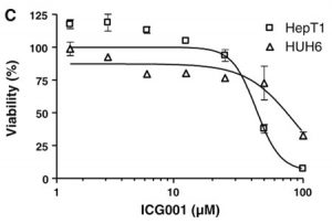
In MCF7 cells, leptin-induced EMT, invasion, and tumor sphere formation are inhibited by ICG-001 (5 μM) [1]. In presenilin-1 mutant cells, ICG-001 can phenotypically rescue normal nerve growth factor (NGF)-induced neuronal differentiation and neuronal outgrowth, highlighting the significance of neuronal differentiation and the function of the TCF/β-catenin signaling pathway in neurite outgrowth [2]. In SW480 cells, the steady-state levels of survivin and cyclin D1 RNA and protein were decreased by ICG-001 (25 μM) treatment; these two proteins are increased by β-catenin. ICG-001 inhibits the development of colon cancer cells in vitro and specifically triggers apoptosis in transformed cells but not in normal colon cells [3].
- Selectively induces apoptosis in transformed colorectal cells but not in normal colon cells, and reduces the in - vitro growth of colon carcinoma cells. It specifically binds to CBP, down - regulates β - catenin/T cell factor (TCF) signaling, and selectively inhibits the β - catenin/TCF reporter construct TOPFlash, without affecting the FOPFlash construct. The mechanism is that it disrupts the interaction between β - catenin and CBP, rather than the interaction between β - catenin and p300 [3] - Inhibits leptin - induced epithelial - mesenchymal transition (EMT), invasion, and tumorsphere formation in breast cancer cells. Treating MCF7 cells with ICG - 001 can block the leptin - induced increase in β - catenin accumulation and nuclear translocation, thus reducing the recruitment of β - catenin to the promoter. The mechanism is related to inhibiting the phosphorylation of glycogen synthase kinase 3β (GSK3β) via Akt activation induced by leptin, and reducing the expression of Wnt1 and MTA1 [1] |
| ln Vivo |
In mice, ICG-001 (5 mg/kg daily) strongly suppresses β-catenin signaling while preserving epithelial cells [2]. The development of polyps in the colon and small intestine was reduced by 42% after 9 weeks of administration of a water-soluble version of ICG-001. This impact was comparable to that of the nonsteroidal anti-inflammatory medication MK-231, which has repeatedly demonstrated efficacy in this model. In the SW620 nude mouse tumor regression xenograft model, ICG-001 (150 mg/kg, i.v.) caused a notable reduction in tumor volume over the course of 19 days of therapy without causing death or weight loss [3].
- Efficacious in the Min mouse and nude mouse xenograft models of colon cancer, which can reduce the tumor growth [3] - Inhibits Wnt/β - catenin/CBP signaling in a mouse model of pulmonary fibrosis, which is helpful to reverse pulmonary fibrosis [2] - Enzyme Assay: - Incubate nuclear extracts of SW480 cells with streptavidin - agarose beads coated with ICG - 002, and then add ICG - 001. After eluting the bound proteins, use anti - CBP antibody for immunoblotting or silver staining to confirm that CBP is the target of ICG - 001 [3] - Transfect full - length CBP, p300 or β - catenin plasmids together with TOPFlash in SW480 cells, treat them with ICG - 001, and then perform luciferase assay after 24 hours to verify that ICG - 001 specifically acts on CBP rather than p300 [3] |
| Enzyme Assay |
Coimmunoprecipitations [4]
For β-catenin-CBP/P300/E-cadherin/N-cadherin coimmunoprecipitations, MGC-803 cells were incubated 48 h with or without ICG-001 and 100 μg protein extract was diluted to 1 mL in coimmunoprecipitation (Co-IP) buffer. 2 μg of β-catenin antibody was added to the protein samples and the mix were incubated overnight at 4 °C with rotation. 20 μL of 50% protein A/G-agarose bead slurry (equilibrated in Co-IP buffer) were added, and after 2 h–incubation at 4 °C, the beads were washed 4 times with Co-IP buffer (1 ml per wash) and diluted with 1× loading buffer. Western blotting was performed, and CBP, P300, E-cadherin, N-cadherin were detected. Affinity Purification.[3] Cells were lysed in protein-binding buffer [PBB, 20 mM Hepes, pH 7.9/100 mM NaCl/0.5 mM EDTA/0.5% Nonidet P-40/6 mM MgCl2/5 mM 2-mercaptoethanol/one tablet of Complete protease inhibitor mixture]. Biotinylated ICG-002 was bound overnight at room temperature to a 50% slurry of streptavidin-agarose beads in buffer containing 50% DMSO and 50% PBB. Beads were washed to remove unbound ICG-002 and then incubated with whole-cell lysates. Proteins eluted, either specifically with 100 μM ICG-001 or by boiling in SDS, were immunoblotted and silver stained. |
| Cell Assay |
RLE-6TN Cell qPCR Studies. [3]
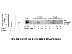
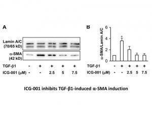
To evaluate effects of ICG-001 on α-SMA and collagen type 1 expression, RLE-6TN cells were treated with TGF-β1 (0.25 ng/mL) in the presence or absence of ICG-001 (5.0 μM). After 24 h, cells were harvested and mRNA isolated for analysis by qPCR. RNA was reverse-transcribed using SuperScript reverse transcriptase. Quantitative PCR was performed with SYBR-Green PCR using Real-Time PCR System HT7900. The amplification protocol was set as follows: 95 °C denaturation for 10 min followed by 40 cycles of 15-s denaturation at 95 °C, 1 min of annealing/extension, and data collection at 60 °C. Primer pairs used are as following: α-SMA forward 5′-ATGGCTCCGGGCTCTGTAA-3′ and reverse 5′-ACAGCCCTGGGAGCATCA-3′; collagen 1α forward 5′-TTGACCCTAACCAAGGATGC-3′ and reverse 5′-CACCCCTTCTGCGTTGTATT-3′. IPF Lung Fibroblasts. [3] Primary fibroblast cultures were derived from lung tissue from a patient with IPF undergoing transplant surgery, following informed consent and ethics approval from relevant institutions, as previously described. One cell line (CCL-134) established from a patient with IPF was obtained from ATCC. The IPF fibroblasts were treated with ICG-001(5 μM) or DMSO control for 48 h, after which mRNA was isolated for analysis by qPCR. - Colon cancer cell experiment: Culture colon cancer cells, add ICG - 001, and observe the cell apoptosis and growth status, and detect the activity of β - catenin/TCF signaling pathway through reporter gene assay [3] - Breast cancer cell experiment: Serum - starve MCF7 cells for 16 h, then treat them with 100 ng/ml leptin or not, and at the same time, add ICG - 001 or not. Observe the morphological changes of cells, detect the accumulation and nuclear translocation of β - catenin by immunofluorescence or Western blot, and evaluate the EMT, invasion and tumorsphere formation ability of cells [1] |
| Animal Protocol |
Dissolved in water; 300 mg/kg; oral administration
Seven-week-old male C57BL/6J-Apc Min/+ Bleomycin-Induced Pulmonary Fibrosis in Mice.[2] All animal use procedures were approved by the University of Washington Animal Care Committee. In prevention of pulmonary fibrosis studies, C57BL/6J mice and BAT-gal transgenic mice that express the Escherichia coli lacZ gene encoding β-galactosidase under the control of β-catenin/TCF responsive elements produced from B6D2F1 mice had s.c. placement of miniosmotic pumps (200 μL Alzet Model 2001 osmotic pumps, 1.0 ± 0.15 μL/h delivery rate; Durect Corporation) containing ICG-001 (5 mg/kg per day in saline), dexamethasone-water soluble (1 mg/kg per day in saline), or saline control on day 0. Twenty-four hours later (day 1), 0.08 units bleomycin in 50 μL of saline was administered intranasally. Control groups received 50 μL of saline intranasally on day 1. Mice were killed on day 10. In intervention studies during the inflammatory and early fibrogenic response period, C57BL/6J mice received bleomycin (0.08 units) or saline intranasally on day 0 followed on day 5 by twice daily oral treatment with pirfenidone [5-methyl-1-phenyl-2-(IH)-pyridone, 400 mg/kg/d in 0.5% carboxymethylcellulose], or miniosmotic pump infusion of ICG-001 (5 mg/kg per day) or saline control for a 10-d treatment period. In long-term reversal of established fibrosis studies, groups of mice administered bleomycin (0.08 units intranasally) on day 0, had implantation on day 21 of miniosmotic pumps containing either ICG-001 (5 mg/kg per day) or saline, and killed on day 42. Lungs were obtained for histopathology, qPCR, and collagen assay. Min Mouse Model. [3] Seven-week-old male C57BL/6J-ApcMin/+ and WT C57BL/6J mice were treated orally for 9 weeks with ICG-001 (300 mg/kg per day) or vehicle (1% carboxymethylcellulose), once daily, six times per week. Sulindac was administered in drinking water (160 ppm, dissolved in 8 mM Na2PO4 buffer, pH 7.6). At 16 weeks, the polyp number was counted manually by using a dissecting microscope. In vivo xenograft and treatment experiments [4] For in vivo studies, 4–6 week old male nude mice were used. MGC-803 cells (2 × 10⁶ cells in 200 μl PBS) were subcutaneously injected into right flank of the nude mice to establish tumors. After 7 days, 50 mg/kg/day ICG-001 was delivered into the tumors. PBS was used as the control. Treatment continued for a total of 4 weeks. The tumor size was measured using digital caliper and tumor volume was calculated with the following formula: volume = 0.5 × width2 × length. - Colon cancer mouse model: Use Min mice and nude mice, inoculate colon cancer cells to establish xenograft models, and then administer ICG - 001 to observe the tumor growth situation, but the specific administration route, frequency and dosage are not described in the literature [3] - Pulmonary fibrosis mouse model: The literature does not describe the specific animal experiment operation process of using ICG - 001 to treat pulmonary fibrosis mouse model [2] |
| References |
|
| Additional Infomation |
(6S,9aS)-6-[(4-hydroxyphenyl)methyl]-8-(1-naphthylmethyl)-4,7-dioxo-N-(phenylmethyl)-3,6,9,9a-tetrahydro-2H-pyrazino[1,2-a]pyrimidine-1-carboxamide is a peptide. Disruptions in the adipocyte cytokine spectrum, particularly elevated leptin levels, are a major cause of breast cancer progression and metastasis; however, the underlying mechanisms are not fully understood. In particular, whether leptin is involved in epithelial-mesenchymal transition (EMT) remains unknown. This article provides molecular evidence that leptin induces a morphological shift in breast cancer cells from epithelial cells to spindle-shaped mesenchymal cells. To investigate potential downstream mediators regulating leptin-induced EMT, we found functional interactions between leptin, metastasis-associated protein 1 (MTA1), and components of the Wnt1 signaling pathway. Leptin increases β-catenin accumulation and nuclear translocation, thereby promoting promoter recruitment. Silencing β-catenin or using the small molecule inhibitor ICG-001 inhibited leptin-induced EMT, invasion, and tumor spheroid formation. Mechanistically, leptin significantly reduces the formation of the GSK3β-LKB1-Axin complex by activating Akt-stimulated phosphorylation of glycogen synthase kinase 3β (GSK3β), thereby increasing β-catenin accumulation. Leptin treatment also increases Wnt1 expression, further promoting GSK3β phosphorylation. Inhibition of Wnt1 blocks leptin-stimulated GSK3β phosphorylation. We also found that leptin increases the expression of MTA1, an important regulator of the Wnt1 signaling pathway. MTA1 is a key component of leptin-mediated Wnt/β-catenin pathway regulation, as silencing MTA1 inhibits leptin-induced Wnt1 expression, GSK3β phosphorylation, and β-catenin activation. In addition, analysis of leptin-treated breast tumors showed increased expression of Wnt1, pGSK3β and vimentin, increased accumulation of β-catenin in the nucleus, and decreased expression of E-cadherin, providing in vivo evidence for previously undiscovered interactions between leptin and the MTA1/Wnt signaling pathway during epithelial-mesenchymal transition (EMT) in breast cancer cells. [2]
Idiopathic pulmonary fibrosis (IPF)/common interstitial pneumonia is a serious disease characterized by progressive lung scarring and destruction. Anti-inflammatory therapies, including corticosteroids, have limited efficacy against this ultimately fatal disease. There is an urgent need to find novel drugs that can interact with key molecular pathways in the pathogenesis of pulmonary fibrosis to prevent or reverse the progression of pulmonary fibrosis in these patients. Due to the aberrant activation of the Wnt/β-catenin signaling pathway in the lungs of patients with idiopathic pulmonary fibrosis (IPF), we selected ICG-001 as a target to intervene in pulmonary fibrosis using this pathway. ICG-001 is a small molecule that specifically inhibits the transcription of T-cytokines/β-catenin in a cyclic adenosine monophosphate response element-binding protein (CBP)-dependent manner. ICG-001 selectively blocks the interaction of β-catenin/CBP without interfering with the interaction of β-catenin/p300. We report here that ICG-001 (5 mg/kg/day) significantly inhibits the β-catenin signaling pathway, alleviates bleomycin-induced pulmonary fibrosis in mice, and protects lung epithelial cells. ICG-001 in combination with bleomycin prevents fibrosis, while late administration reverses existing fibrosis and significantly improves survival. Since there is currently no effective treatment for idiopathic pulmonary fibrosis (IPF), selective inhibition of Wnt/β-catenin-dependent transcription suggests a potential unique treatment for pulmonary fibrosis. [2] Most colorectal cancers have hereditary and somatic mutations in adenomatous polyposis (APC) that lead to the activation of β-catenin response genes. To identify small molecule antagonists of this pathway, we screened transformed colorectal cancer cells using a secondary structure template compound library to look for compounds that could inhibit β-catenin response reporter genes. We identified ICG-001, a small molecule that downregulates the β-catenin/T cytokine signaling pathway by specifically binding to cyclic adenosine monophosphate (cAMP) response element-binding protein. ICG-001 selectively induces apoptosis in transformed cells but not in normal colon cells, inhibits the in vitro growth of colon cancer cells, and is effective in Min mice and nude mouse xenograft models of colon cancer. [3] Background: ICG-001 is a small molecule that binds to CREB-binding protein (CBP), thereby disrupting its interaction with β-catenin and inhibiting the function of CBP as a Wnt/β-catenin-mediated transcriptional coactivator. Given its ability to inhibit the Wnt/β-catenin signaling pathway, ICG-001 has been used to exert its anticancer effects in certain tumor types. This study investigated ICG-001 and its potential role in the treatment of gastric cancer (GC). Methods: Gastric cancer cell lines SGC-7901, MGC-803, BGC-823, and MKN-45 were used in both in vitro and in vivo experiments. In vitro, cell proliferation, tumor spheroid formation, metastasis, tumorigenesis, and resistance to chemotherapeutic drugs were assessed using the MTT assay, colony formation assay, flow cytometry, migration and invasion assays, and tumor spheroid culture. In vivo experiments were conducted using a thymic nude mouse subcutaneous xenograft model. Changes in RNA and protein levels were detected by qRT-PCR, Western blot, immunoprecipitation, and immunofluorescence analysis. Results: This study demonstrates that ICG-001 significantly inhibits the growth and metastasis of various gastric cancer cell lines, induces apoptosis, and enhances the inhibitory effect on tumor spheroids in vitro. The mechanism may be that ICG-001 can effectively block the binding of β-catenin to CBP and N-cadherin, while promoting the binding of β-catenin to P300 and E-cadherin, rather than changing the distribution and expression of β-catenin. Conclusion: Our results show that ICG-001 can inhibit the growth and metastasis of gastric cancer cell lines, reduce their stem cell-like characteristics and chemotherapy resistance, suggesting that ICG-001 is a potential small molecule therapeutic for gastric cancer. Keywords: gastric cancer; growth; ICG-001; stem cell-like characteristics; Wnt/β-catenin signaling pathway. [4] ICG-001 is a small molecule inhibitor designed to target the Wnt/β-catenin signaling pathway, which is activated by mutations in most colorectal cancers. ICG-001 can inhibit β-catenin/TCF-mediated gene transcription by binding to CBP and disrupting the interaction between β-catenin and CBP. [3] |
| Molecular Formula |
C33H32N4O4
|
|---|---|
| Molecular Weight |
548.63
|
| Exact Mass |
548.242
|
| Elemental Analysis |
C, 72.24; H, 5.88; N, 10.21; O, 11.66
|
| CAS # |
780757-88-2
|
| Related CAS # |
1422253-38-0 (PRI-724);847591-62-2 (deleted);780757-88-2 (ICG001);
|
| PubChem CID |
11238147
|
| Appearance |
White to off-white solid powder
|
| Density |
1.4±0.1 g/cm3
|
| Boiling Point |
895.6±65.0 °C at 760 mmHg
|
| Melting Point |
133-134ºC
|
| Flash Point |
495.4±34.3 °C
|
| Vapour Pressure |
0.0±0.3 mmHg at 25°C
|
| Index of Refraction |
1.722
|
| LogP |
4.01
|
| Hydrogen Bond Donor Count |
2
|
| Hydrogen Bond Acceptor Count |
4
|
| Rotatable Bond Count |
6
|
| Heavy Atom Count |
41
|
| Complexity |
930
|
| Defined Atom Stereocenter Count |
2
|
| SMILES |
C1CN([C@H]2CN(C(=O)[C@@H](N2C1=O)CC3=CC=C(C=C3)O)CC4=CC=CC5=CC=CC=C54)C(=O)NCC6=CC=CC=C6
|
| InChi Key |
HQWTUOLCGKIECB-IHLOFXLRSA-N
|
| InChi Code |
InChI=1S/C33H32N4O4/c38-27-15-13-23(14-16-27)19-29-32(40)35(21-26-11-6-10-25-9-4-5-12-28(25)26)22-30-36(18-17-31(39)37(29)30)33(41)34-20-24-7-2-1-3-8-24/h1-16,29-30,38H,17-22H2,(H,34,41)/t29-,30+/m1/s1
|
| Chemical Name |
rel-(6R,9aR)-Hexahydro-6-[(4-hydroxyphenyl)methyl]-8-(1-naphthalenylmethyl)-4,7-dioxo-N-(phenylmethyl)-2H-pyrazino[1,2-a]pyrimidine-1(6H)-carboxamide
|
| Synonyms |
ICG-001; ICG 001; ICG-001; 847591-62-2; 780757-88-2; (S,S)-ICG 001; (6S,9aS)-N-benzyl-6-(4-hydroxybenzyl)-8-(naphthalen-1-ylmethyl)-4,7-dioxooctahydro-1H-pyrazino[1,2-a]pyrimidine-1-carboxamide; (6S,9aS)-6-(4-hydroxybenzyl)-N-benzyl-8-(naphthalen-1-ylmethyl)-4,7-dioxo-hexahydro-2H-pyrazino[1,2-a]pyrimidine-1(6H)-carboxamide; ICG001
|
| HS Tariff Code |
2934.99.9001
|
| Storage |
Powder -20°C 3 years 4°C 2 years In solvent -80°C 6 months -20°C 1 month |
| Shipping Condition |
Room temperature (This product is stable at ambient temperature for a few days during ordinary shipping and time spent in Customs)
|
| Solubility (In Vitro) |
DMSO: 100 mg/mL (182.3 mM)
Water: 1 mg/mL (Insoluble) Ethanol: 10 mg/mL (18.2 mM) |
|---|---|
| Solubility (In Vivo) |
Solubility in Formulation 1: ≥ 2.5 mg/mL (4.56 mM) (saturation unknown) in 10% DMSO + 40% PEG300 + 5% Tween80 + 45% Saline (add these co-solvents sequentially from left to right, and one by one), clear solution.
For example, if 1 mL of working solution is to be prepared, you can add 100 μL of 25.0 mg/mL clear DMSO stock solution to 400 μL PEG300 and mix evenly; then add 50 μL Tween-80 to the above solution and mix evenly; then add 450 μL normal saline to adjust the volume to 1 mL. Preparation of saline: Dissolve 0.9 g of sodium chloride in 100 mL ddH₂ O to obtain a clear solution. Solubility in Formulation 2: 2.5 mg/mL (4.56 mM) in 10% DMSO + 90% (20% SBE-β-CD in Saline) (add these co-solvents sequentially from left to right, and one by one), suspension solution; with heating and sonication. For example, if 1 mL of working solution is to be prepared, you can add 100 μL of 25.0 mg/mL clear DMSO stock solution to 900 μL of 20% SBE-β-CD physiological saline solution and mix evenly. Preparation of 20% SBE-β-CD in Saline (4°C,1 week): Dissolve 2 g SBE-β-CD in 10 mL saline to obtain a clear solution. View More
Solubility in Formulation 3: ≥ 2.5 mg/mL (4.56 mM) (saturation unknown) in 10% DMSO + 90% Corn Oil (add these co-solvents sequentially from left to right, and one by one), clear solution. Solubility in Formulation 4: 1.67 mg/mL (3.04 mM) in 15% Cremophor EL + 85% Saline (add these co-solvents sequentially from left to right, and one by one), suspension solution; with ultrasonication. Preparation of saline: Dissolve 0.9 g of sodium chloride in 100 mL ddH₂ O to obtain a clear solution. Solubility in Formulation 5: 1.67 mg/mL (3.04 mM) in 17% Polyethylene glycol 12-hydroxystearate in Saline (add these co-solvents sequentially from left to right, and one by one), suspension solution; with ultrasonication. Preparation of saline: Dissolve 0.9 g of sodium chloride in 100 mL ddH₂ O to obtain a clear solution. |
| Preparing Stock Solutions | 1 mg | 5 mg | 10 mg | |
| 1 mM | 1.8227 mL | 9.1136 mL | 18.2272 mL | |
| 5 mM | 0.3645 mL | 1.8227 mL | 3.6454 mL | |
| 10 mM | 0.1823 mL | 0.9114 mL | 1.8227 mL |
*Note: Please select an appropriate solvent for the preparation of stock solution based on your experiment needs. For most products, DMSO can be used for preparing stock solutions (e.g. 5 mM, 10 mM, or 20 mM concentration); some products with high aqueous solubility may be dissolved in water directly. Solubility information is available at the above Solubility Data section. Once the stock solution is prepared, aliquot it to routine usage volumes and store at -20°C or -80°C. Avoid repeated freeze and thaw cycles.
Calculation results
Working concentration: mg/mL;
Method for preparing DMSO stock solution: mg drug pre-dissolved in μL DMSO (stock solution concentration mg/mL). Please contact us first if the concentration exceeds the DMSO solubility of the batch of drug.
Method for preparing in vivo formulation::Take μL DMSO stock solution, next add μL PEG300, mix and clarify, next addμL Tween 80, mix and clarify, next add μL ddH2O,mix and clarify.
(1) Please be sure that the solution is clear before the addition of next solvent. Dissolution methods like vortex, ultrasound or warming and heat may be used to aid dissolving.
(2) Be sure to add the solvent(s) in order.
|
|---|
|
|
Products are for research use only; We do not sell to patients
Copyright 2020 InvivoChem LLC | All Rights Reserved


 COA
COA