| Size | Price | Stock | Qty |
|---|---|---|---|
| 5mg |
|
||
| 10mg |
|
||
| 25mg |
|
||
| 50mg |
|
||
| 100mg |
|
||
| 250mg |
|
||
| 500mg | |||
| Other Sizes |


Purity: ≥98%
| Targets |
MLXBIR3SG (Ki = 14 nM); cIAP1-BIR3 (Ki = 17 nM); XIAP-BIR3 (Ki = 28 nM); cIAP2-BIR3 (Ki = 43 nM); XIAP-BIR2 (Ki = 112 nM)
The target of GDC-0152 (RG7419) is Inhibitor of Apoptosis Proteins (IAPs), a family of anti-apoptotic proteins including cIAP1, cIAP2, and XIAP; it acts as a Smac mimetic to competitively bind to the BIR3 domain of IAPs. - For human cIAP1 BIR3 domain (fluorescence polarization binding assay): Ki = 0.8 nM [1] - For human cIAP2 BIR3 domain (same assay as cIAP1): Ki = 1.5 nM [1] - For human XIAP BIR3 domain (HTRF binding assay): IC₅₀ = 35 nM [1] |
|---|---|
| ln Vitro |
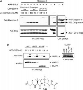
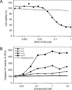
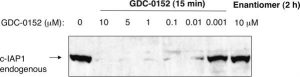
GDC-0152 can block protein−protein interactions that involve IAP proteins and pro-apoptotic molecules. GDC-0152 is demonstrated to disrupt the association of ML-IAP, cIAP1, and cIAP2 with Smac as well as XIAP binding to partially processed caspase-9 using transiently transfected HEK293T cells. The endogenous association of ML-IAP and Smac is effectively also eliminated by GDC-0152 in melanoma SK-MEL28 cells. When compared to healthy human mammary epithelial cells (HMEC), GDC-0152 had no effect on HMEC but reduced cell viability in the MDA-MB-231 breast cancer cell line. It has been discovered that GDC-0152 activates caspases 3 and 7 in a dose- and time-dependent manner. In A2058 melanoma cells, GDC-0152 has been shown to cause cIAP1 to degrade rapidly. Its affinity for cIAP1 is supported by the fact that it successfully induces cIAP1 degradation at concentrations as low as 10 nM.
1. Antiproliferative activity against cancer cell lines: GDC-0152 (RG7419) (0.01–1000 nM) exhibited potent antiproliferative effects on a panel of human solid tumor cell lines with high IAP expression. GI₅₀ values were: 12 nM (A549, non-small cell lung cancer), 8 nM (MDA-MB-231, triple-negative breast cancer), 15 nM (HCT116, colorectal cancer), 9 nM (SK-OV-3, ovarian cancer). In contrast, it had minimal activity on normal human fibroblasts (GI₅₀ > 1000 nM) [1] 2. Induction of cIAP1/2 degradation: Treatment of MDA-MB-231 cells with GDC-0152 (RG7419) (1–50 nM) for 4 hours induced dose-dependent degradation of cIAP1 and cIAP2 (detected by western blot). At 10 nM, cIAP1 protein levels were reduced by >90% vs. control; cIAP2 was reduced by 75%. No significant change in XIAP protein levels was observed (consistent with its lower binding affinity for XIAP) [1] 3. Activation of apoptotic signaling: GDC-0152 (RG7419) (5–50 nM) induced apoptosis in A549 cells. After 24-hour treatment with 20 nM, flow cytometry (Annexin V-FITC/PI staining) showed apoptotic cells increased from 4% (control) to 42%. Western blot revealed cleavage of caspase-3 (p17 active fragment) and PARP (89 kDa cleaved fragment), with maximal cleavage at 20 nM [1] 4. Enhancement of TNF-α-induced apoptosis: In HCT116 cells, GDC-0152 (RG7419) (1–10 nM) synergized with TNF-α (10 ng/mL) to induce apoptosis. At 5 nM + TNF-α, apoptotic cells increased to 65% vs. 8% (TNF-α alone) or 12% (drug alone), indicating potentiation of TNF-α-mediated cell death [1] |
| ln Vivo |
GDC-0152 has moderate predicted hepatic clearance based on metabolic stability assays carried out using human liver microsomes.Over the range of concentrations investigated (0.1–100 μM), plasma protein binding of GDC-0152 is moderate and comparable in mice (88–91%), rats (89–91%), dogs (81–90%), monkeys (76–85%), and humans (75–83%); higher plasma protein binding is observed in rabbits (95–96%). In all tested species, GDC-0152 does not preferentially distribute to red blood cells with blood-plasma partition ratios ranging from 0.6 to 1.1. GDC-0152's pharmacokinetics are achieved with a Cmax of 53.7 μM and an AUC of 203.5 h•μM. [1]
1. Antitumor efficacy in A549 lung cancer xenografts: Female athymic nude mice (6–8 weeks old) were subcutaneously injected with 5×10⁶ A549 cells. When tumors reached 100–150 mm³, mice were randomized into 4 groups (n=6/group): vehicle, 10 mg/kg GDC-0152 (RG7419), 25 mg/kg GDC-0152 (RG7419), 50 mg/kg GDC-0152 (RG7419). The drug was administered via intravenous injection every 3 days for 21 days (7 total doses). The 50 mg/kg group achieved 85% tumor growth inhibition (TGI); tumor weight was reduced by 78% vs. vehicle. No complete tumor regression was observed [1] 2. Pharmacodynamic effects in tumor tissues: Tumor tissues from the A549 xenograft model (50 mg/kg group) were collected 24 hours after the last dose. Western blot showed cIAP1 protein levels were reduced by 80% vs. vehicle; immunohistochemistry (IHC) revealed increased cleaved caspase-3 staining (4-fold higher positive cells) in tumor sections, confirming in vivo activation of apoptotic signaling [1] 3. Efficacy in MDA-MB-231 breast cancer xenografts: Female nude mice bearing MDA-MB-231 xenografts (120–160 mm³) were treated with GDC-0152 (RG7419) (50 mg/kg, iv, q3d for 18 days). TGI was 72%, and tumor weight was 35% of the vehicle group. No significant metastasis to lung or liver was observed in the treatment group [1] |
| Enzyme Assay |
The IAP protein constructs are added to wells containing serial dilutions of the antagonists or the peptide AVPW, as appropriate, and the Hid-FAM probe or AVP-diPhe-FAM probe in the polarization buffer to determine the inhibitory constants (Ki) for the antagonists. After 30 minutes, samples are read. The IC50 values are calculated by fitting the data to a 4-parameter equation using software, and fluorescence polarization values are plotted as a function of antagonist concentration. Based on the IC50 values, the antagonists' Ki values are calculated.
1. Fluorescence Polarization (FP) Assay for cIAP1/cIAP2 BIR3 Binding: Recombinant human cIAP1 BIR3 or cIAP2 BIR3 domain (20 nM) was incubated with a fluorescently labeled Smac peptide (5 nM, labeled with FITC at the N-terminus) and serial concentrations of GDC-0152 (RG7419) (0.001–100 nM) in assay buffer (50 mM Tris-HCl pH 7.5, 150 mM NaCl, 0.01% Tween-20, 1 mM DTT) at 25°C for 60 minutes. FP signals were measured using a microplate reader (excitation 485 nm, emission 535 nm). Ki values were calculated using a competitive binding model based on the reduction in FP signal (due to drug displacement of the Smac peptide) [1] 2. HTRF Assay for XIAP BIR3 Binding: The assay was performed in 384-well plates using recombinant human XIAP BIR3 domain (50 nM) and a biotinylated Smac peptide (10 nM). GDC-0152 (RG7419) concentrations ranged from 0.1–1000 nM. After 1-hour incubation at 37°C, streptavidin-conjugated Eu³⁺ cryptate and anti-XIAP antibody conjugated with XL665 were added. FRET signals were measured at 620 nm and 665 nm; IC₅₀ was calculated as the concentration inhibiting 50% of the Smac-XIAP BIR3 interaction [1] 3. Caspase-3 Activation Assay (XIAP Inhibition Reversal): Recombinant XIAP (10 nM) was pre-incubated with GDC-0152 (RG7419) (0.1–1000 nM) for 30 minutes, then mixed with recombinant caspase-3 (5 nM) and a fluorogenic caspase-3 substrate (Ac-DEVD-AMC, 50 μM) in assay buffer (20 mM HEPES pH 7.4, 100 mM NaCl, 10 mM DTT). Fluorescence (excitation 380 nm, emission 460 nm) was measured every 10 minutes for 2 hours. The EC₅₀ for reversing XIAP-mediated caspase-3 inhibition was 42 nM [1] |
| Cell Assay |
GDC-0152 is used to treat HMECs and MDA-MB-231 breast cancer cells in the recommended concentrations. The CellTiter-Glo luminescent cell viability assay is used to determine cell death 72 hours after the start of treatment.
1. Antiproliferative Assay (GI₅₀ Determination): Cancer cells (A549, MDA-MB-231, HCT116) were seeded in 96-well plates at a density of 1000–2000 cells/well and incubated overnight (37°C, 5% CO₂). GDC-0152 (RG7419) was added at serial concentrations (0.01–1000 nM), and cells were cultured for 72 hours. Cell viability was measured using the CellTiter-Glo Luminescent Cell Viability Assay (luminescence intensity proportional to ATP content). GI₅₀ was defined as the concentration inhibiting cell growth by 50% vs. the vehicle control [1] 2. Western Blot for IAP Degradation and Apoptosis Markers: MDA-MB-231 or A549 cells were seeded in 6-well plates (5×10⁵ cells/well) and grown to 70% confluence. GDC-0152 (RG7419) (1–50 nM) was added, and cells were incubated for 4–24 hours. Cells were lysed in RIPA buffer containing protease inhibitors; lysates were separated by 12% SDS-PAGE and transferred to PVDF membranes. Membranes were blocked with 5% non-fat milk, incubated overnight at 4°C with primary antibodies (cIAP1, cIAP2, XIAP, cleaved caspase-3, cleaved PARP, β-actin), then with HRP-conjugated secondary antibodies. Bands were visualized using ECL chemiluminescence [1] 3. Flow Cytometry for Apoptosis Detection: A549 cells were seeded in 12-well plates (2×10⁵ cells/well) and treated with GDC-0152 (RG7419) (5–50 nM) for 24 hours. Cells were harvested, washed with cold PBS, and stained with Annexin V-FITC and propidium iodide (PI) for 15 minutes at room temperature (dark). Stained cells were analyzed using a flow cytometer; apoptotic cells were defined as Annexin V-positive (PI-negative: early apoptosis; PI-positive: late apoptosis) [1] 4. TNF-α Synergy Assay: HCT116 cells were treated with GDC-0152 (RG7419) (1–10 nM) + TNF-α (10 ng/mL) for 24 hours. Apoptosis was measured by flow cytometry (Annexin V-FITC/PI) and cleaved caspase-3 western blot. Synergy was calculated using the combination index (CI) < 0.8 (indicating synergism) [1] |
| Animal Protocol |
phosphate-buffered saline; 10, 50, 100 mg/kg; Oral Human-tumor xenograft mouse models of MDA-MB-231 breast cancer
1. A549 Lung Cancer Xenograft Model: Female athymic nude mice (6–8 weeks old, 18–22 g) were acclimated to the laboratory (12 h light/dark cycle, 22±2°C) for 7 days. A549 cells (5×10⁶ cells in 0.2 mL PBS/matrigel 1:1) were subcutaneously injected into the right flank. When tumors reached 100–150 mm³ (≈10 days post-injection), mice were randomized into 4 groups (n=6/group). GDC-0152 (RG7419) was formulated in a vehicle consisting of 10% DMSO, 30% cremophor EL, and 60% normal saline. Doses were 10, 25, 50 mg/kg, administered via intravenous injection (tail vein) every 3 days for 21 days (7 doses total). The vehicle group received the same volume of vehicle. Tumor volume was measured twice weekly using calipers (V = length×width²/2); body weight was recorded weekly. At study end, mice were euthanized, tumors were excised, weighed, and stored at -80°C for western blot or fixed in 4% paraformaldehyde for IHC [1] 2. MDA-MB-231 Breast Cancer Xenograft Model: Female nude mice were injected subcutaneously with 4×10⁶ MDA-MB-231 cells (PBS/matrigel 1:1). When tumors reached 120–160 mm³, mice were treated with GDC-0152 (RG7419) (50 mg/kg, iv, q3d for 18 days; n=6/group) or vehicle (n=6). Tumor volume and body weight were monitored as described above. At study end, lungs and livers were harvested to assess metastasis (H&E staining) [1] 3. Pharmacodynamic Tissue Collection: For the A549 model, 3 mice per group were euthanized 24 hours after the last dose. Tumors were divided into two parts: one frozen in liquid nitrogen for western blot, the other fixed in 4% paraformaldehyde, embedded in paraffin, and sectioned (5 μm) for IHC (cleaved caspase-3 antibody) [1] |
| ADME/Pharmacokinetics |
1. Pharmacokinetics in mice (intravenous administration): Male CD1 mice (n=3 at each time point) were administered a single intravenous injection of GDC-0152 (RG7419) (25 mg/kg, dissolved in 10% DMSO/30% Cremophor EL/60% saline). Blood samples (0.15 mL) were collected from the tail vein at 0.083, 0.25, 0.5, 1, 2, 4, 6, 8, and 12 hours post-administration. Plasma was separated by centrifugation (3000×g, 10 min, 4℃) and stored at -80℃. Drug concentrations were determined by liquid chromatography-tandem mass spectrometry (LC-MS/MS). Pharmacokinetic parameters (non-compartmental model analysis): terminal half-life (t₁/₂) = 2.8 h, clearance (CL) = 15.2 mL/min/kg, steady-state volume of distribution (Vdss) = 8.1 L/kg [1]
2. Plasma protein binding: Human plasma and mouse plasma (500 μL) were mixed with GDC-0152 (RG7419) (0.1–10 μM) and dialyzed at 37°C for 4 h using a dialysis membrane with a molecular weight cutoff of 12–14 kDa. The concentration of free drug in the dialysate was determined by LC-MS/MS. Plasma protein binding rate: 97.2% (human), 96.8% (mouse) [1] 3. In vitro metabolism (liver microsomes): GDC-0152 (RG7419) (1 μM) was incubated with human liver microsomes (HLM) or mouse liver microsomes (MLM) in the presence of NADPH (1 mM) at 37°C. Samples were collected at 0, 5, 10, 20, 30 and 60 minutes. Drug concentration was determined by LC-MS/MS. Half-life (t₁/₂): 45 min (HLM), 38 min (MLM); Intrinsic clearance (CLint): 32 μL/min/mg protein (HLM), 38 μL/min/mg protein (MLM). CYP inhibition screening results showed that CYP1A2, 2C9, 2C19, 2D6 and 3A4 were not significantly inhibited (IC₅₀ > 50 μM) [1] 4. Oral bioavailability: Male CD-1 mice (n=3 at each time point) were given a single oral dose of GDC-0152 (RG7419) (100 mg/kg, dissolved in 0.5% methylcellulose/0.2% Tween-80). Plasma concentrations at all time points were below the lower limit of quantitation (LLOQ = 1 ng/mL), indicating poor oral bioavailability (<1%) [1] |
| Toxicity/Toxicokinetics |
1. Acute toxicity in mice: Male and female CD-1 mice (n=4 per sex per dose group) were administered a single intravenous injection of GDC-0152 (RG7419) (75, 100, 150 mg/kg). Mice were observed for 14 days. The maximum tolerated dose (MTD) was 100 mg/kg; 150 mg/kg resulted in a 50% mortality rate (2 out of 4 mice per sex died), accompanied by lethargy and ataxia (appearing 1 hour after administration). At a dose of 100 mg/kg, transient weight loss was observed (maximum 4.5%, recovered on day 3); no other toxic symptoms were observed [1]
2. Subacute toxicity in xenograft models: In the A549 and MDA-MB-231 xenograft studies (50 mg/kg, intravenous, every 3 days for 21/18 days), GDC-0152 (RG7419) did not cause significant weight loss (<5%) or abnormal clinical symptoms (e.g., diarrhea, piloerection). Serum samples collected at the end of the study showed no significant changes in ALT, AST (liver function), BUN, or creatinine (kidney function) compared to the vector group [1]. 3. Hematologic toxicity: In mice treated with 50 mg/kg GDC-0152 (RG7419) (intravenous injection every 3 days for 21 days), complete blood counts (CBCs) showed no significant changes in white blood cells (WBCs), red blood cells (RBCs), or platelets compared to the vector group, indicating no bone marrow suppression [1]. |
| References | |
| Additional Infomation |
GDC-0152 has been used in clinical trials for the treatment of solid tumors. The Smac mimic GDC-0152 is the second mitochondrial caspase activator (Smac) mimic and an IAP (inhibitor of apoptosis protein) inhibitor with potential antitumor activity. GDC-0152 binds to the Smac binding groove on IAPs, including the direct caspase inhibitor X-linked IAP (XIAP) and cellular IAPs 1 and 2, thereby inhibiting their activity and promoting the induction of apoptosis through apoptosis signaling pathways. IAPs are overexpressed in various cancer cell types and inhibit apoptosis by binding to and inhibiting the activity of active caspases -3, -7, and -9 through their baculoviral lAP repeat (BIR) domains. The endogenous IAP antagonist Smac binds to IAPs via its four-amino acid motif at the N-terminus.
1. Background: GDC-0152 (RG7419) is a potent small molecule Smac mimic and a selective inhibitor of IAP proteins, which has been developed as a clinical candidate for cancer treatment. IAP proteins are overexpressed in a variety of human cancers, and they inhibit apoptosis by binding to and inhibiting caspases; Smac mimics counteract this inhibition by displacing caspases from IAPs [1] 2. Mechanism of action: GDC-0152 (RG7419) binds to the BIR3 domains of cIAP1 and cIAP2 with high affinity, inducing their autoubiquitination and proteasome degradation. The degradation of cIAP releases TNF receptor-associated factor 2 (TRAF2) and activates the non-canonical NF-κB pathway, while simultaneously relieving caspase inhibition—these effects together lead to apoptosis of cancer cells. Its low affinity for XIAP limits off-target effects [1] 3. Clinical candidate status: GDC-0152 (RG7419) has been advanced to the preclinical development stage due to its strong in vitro and in vivo antitumor activity, good pharmacokinetic characteristics (low clearance, moderate half-life) and manageable toxicity. Due to poor oral bioavailability, the drug was designed for intravenous administration and its potential use in solid tumors (e.g., lung cancer, breast cancer, colorectal cancer) was evaluated [1] 4. Therapeutic synergy: Preclinical data showed that GDC-0152 (RG7419) can synergize with TNF-α and other cancer therapies (e.g., chemotherapy, immunotherapy) by enhancing apoptosis signaling, suggesting that combination therapy has the potential to improve clinical efficacy [1] |
| Molecular Formula |
C25H34N6O3S
|
|---|---|
| Molecular Weight |
498.64
|
| Exact Mass |
498.241
|
| Elemental Analysis |
C, 60.22; H, 6.87; N, 16.85; O, 9.63; S, 6.43
|
| CAS # |
873652-48-3
|
| Related CAS # |
873652-48-3;873581-21-6 (HCl);
|
| PubChem CID |
46940575
|
| Appearance |
White to off-white solid powder
|
| Density |
1.3±0.1 g/cm3
|
| Index of Refraction |
1.606
|
| LogP |
2.09
|
| Hydrogen Bond Donor Count |
3
|
| Hydrogen Bond Acceptor Count |
7
|
| Rotatable Bond Count |
8
|
| Heavy Atom Count |
35
|
| Complexity |
743
|
| Defined Atom Stereocenter Count |
3
|
| SMILES |
C(N1CCC[C@H]1C(=O)NC1SN=NC=1C1C=CC=CC=1)(=O)[C@H](C1CCCCC1)NC(=O)[C@H](C)NC
|
| InChi Key |
WZRFLSDVFPIXOV-LRQRDZAKSA-N
|
| InChi Code |
InChI=1S/C25H34N6O3S/c1-16(26-2)22(32)27-21(18-12-7-4-8-13-18)25(34)31-15-9-14-19(31)23(33)28-24-20(29-30-35-24)17-10-5-3-6-11-17/h3,5-6,10-11,16,18-19,21,26H,4,7-9,12-15H2,1-2H3,(H,27,32)(H,28,33)/t16-,19-,21-/m0/s1
|
| Chemical Name |
(2S)-1-[(2S)-2-cyclohexyl-2-[[(2S)-2-(methylamino)propanoyl]amino]acetyl]-N-(4-phenylthiadiazol-5-yl)pyrrolidine-2-carboxamide
|
| Synonyms |
GDC0152; GDC 0152; GDC0152; RG-7419; RG7419; RG 7419
|
| HS Tariff Code |
2934.99.9001
|
| Storage |
Powder -20°C 3 years 4°C 2 years In solvent -80°C 6 months -20°C 1 month |
| Shipping Condition |
Room temperature (This product is stable at ambient temperature for a few days during ordinary shipping and time spent in Customs)
|
| Solubility (In Vitro) |
|
|||
|---|---|---|---|---|
| Solubility (In Vivo) |
Solubility in Formulation 1: ≥ 2.5 mg/mL (5.01 mM) (saturation unknown) in 10% DMSO + 40% PEG300 + 5% Tween80 + 45% Saline (add these co-solvents sequentially from left to right, and one by one), clear solution.
For example, if 1 mL of working solution is to be prepared, you can add 100 μL of 25.0 mg/mL clear DMSO stock solution to 400 μL PEG300 and mix evenly; then add 50 μL Tween-80 to the above solution and mix evenly; then add 450 μL normal saline to adjust the volume to 1 mL. Preparation of saline: Dissolve 0.9 g of sodium chloride in 100 mL ddH₂ O to obtain a clear solution. Solubility in Formulation 2: ≥ 2.5 mg/mL (5.01 mM) (saturation unknown) in 10% DMSO + 90% (20% SBE-β-CD in Saline) (add these co-solvents sequentially from left to right, and one by one), clear solution. For example, if 1 mL of working solution is to be prepared, you can add 100 μL of 25.0 mg/mL clear DMSO stock solution to 900 μL of 20% SBE-β-CD physiological saline solution and mix evenly. Preparation of 20% SBE-β-CD in Saline (4°C,1 week): Dissolve 2 g SBE-β-CD in 10 mL saline to obtain a clear solution. View More
Solubility in Formulation 3: ≥ 2.5 mg/mL (5.01 mM) (saturation unknown) in 10% DMSO + 90% Corn Oil (add these co-solvents sequentially from left to right, and one by one), clear solution. Solubility in Formulation 4: 30% Propylene glycol , 5% Tween 80 , 65% D5W: 5mg/mL |
| Preparing Stock Solutions | 1 mg | 5 mg | 10 mg | |
| 1 mM | 2.0055 mL | 10.0273 mL | 20.0545 mL | |
| 5 mM | 0.4011 mL | 2.0055 mL | 4.0109 mL | |
| 10 mM | 0.2005 mL | 1.0027 mL | 2.0055 mL |
*Note: Please select an appropriate solvent for the preparation of stock solution based on your experiment needs. For most products, DMSO can be used for preparing stock solutions (e.g. 5 mM, 10 mM, or 20 mM concentration); some products with high aqueous solubility may be dissolved in water directly. Solubility information is available at the above Solubility Data section. Once the stock solution is prepared, aliquot it to routine usage volumes and store at -20°C or -80°C. Avoid repeated freeze and thaw cycles.
Calculation results
Working concentration: mg/mL;
Method for preparing DMSO stock solution: mg drug pre-dissolved in μL DMSO (stock solution concentration mg/mL). Please contact us first if the concentration exceeds the DMSO solubility of the batch of drug.
Method for preparing in vivo formulation::Take μL DMSO stock solution, next add μL PEG300, mix and clarify, next addμL Tween 80, mix and clarify, next add μL ddH2O,mix and clarify.
(1) Please be sure that the solution is clear before the addition of next solvent. Dissolution methods like vortex, ultrasound or warming and heat may be used to aid dissolving.
(2) Be sure to add the solvent(s) in order.
| NCT Number | Recruitment | interventions | Conditions | Sponsor/Collaborators | Start Date | Phases |
| NCT00977067 | Terminated | Drug: GDC-0152 | Solid Cancers | Genentech, Inc. | June 2007 | Phase 1 |
|
|---|
|
|
Products are for research use only; We do not sell to patients
Copyright 2020 InvivoChem LLC | All Rights Reserved


 COA
COA