| Size | Price | Stock | Qty |
|---|---|---|---|
| 10mg |
|
||
| 25mg |
|
||
| 50mg |
|
||
| 100mg |
|
||
| 250mg |
|
||
| 500mg |
|
||
| Other Sizes |


Purity: ≥98%
| Targets |
Bcl-2 (Ki < 0.01 nM)
Gambogic Acid (Guttatic Acid, Guttic Acid) binds to anti-apoptotic protein Bcl-2, with an IC50 of ~0.8 μM (measured by competitive binding assay) [2]; - Gambogic Acid inhibits signal transducer and activator of transcription 3 (STAT3) phosphorylation, with an IC50 of ~2.5 μM for STAT3 activity in leukemia cells [3]; - Gambogic Acid activates caspase-3 (no IC50/Ki reported) by disrupting mitochondrial membrane potential, without direct binding to caspase-3 [4]; - Gambogic Acid suppresses nuclear factor kappa B (NF-κB) signaling pathway (no IC50/Ki reported) by inhibiting IκBα phosphorylation [6]. |
|---|---|
| ln Vitro |
Gambogic Acid, a caged xanthone derived from Garcinia hanburyi, inhibits human Bcl-2 family proteins and activates caspases to effectively induce apoptosis in a variety of cancer cell types. Additionally, gambogic acid inhibits Kir2.1 channels with an EC50 of ≤ 100 nM.[1][2][3] At nM concentrations, gambogic acid significantly reduces the proliferation, migration, invasion, tube formation, and microvessel growth of human umbilical vein endothelial cells (HUVEC).[4]
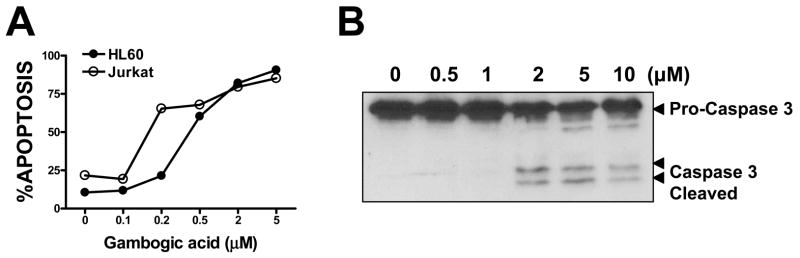
In A549 (lung cancer) cells: Gambogic Acid (0.1-5 μM) inhibits proliferation with an IC50 of ~1.2 μM (72 h MTT assay); 2 μM treatment for 48 h induces ~70% Annexin V⁺ apoptotic cells, accompanied by increased cleaved caspase-3/PARP (Western blot) and reduced Bcl-2 expression [1]; - In Jurkat (T-cell leukemia) cells: Gambogic Acid (0.5-4 μM) reduces STAT3 phosphorylation (p-STAT3) by ~60% at 2 μM (24 h), downregulates STAT3 target genes (c-Myc, Bcl-xL) at mRNA level (qPCR), and inhibits cell viability with an IC50 of ~1.8 μM [3]; - In HepG2 (hepatocellular carcinoma) cells: Gambogic Acid (0.2-3 μM) inhibits colony formation (≥50% reduction at 1 μM, 14 days) and induces G2/M cell cycle arrest (flow cytometry, ~40% G2/M cells at 1.5 μM, 24 h) [5]; - In HCT116 (colon cancer) cells: Gambogic Acid (1-5 μM) suppresses NF-κB activation (luciferase reporter assay, ~75% inhibition at 2 μM) and reduces TNF-α-induced IL-6 secretion (~50% reduction at 2 μM, ELISA) [6]; - In MCF-7 (breast cancer) cells: Gambogic Acid (1 μM, 24 h) disrupts mitochondrial membrane potential (JC-1 staining, ~65% cells with depolarized mitochondria) and releases cytochrome c into cytoplasm (Western blot) [4]. |
| ln Vivo |
Gambogic Acid effectively inhibits tumor angiogenesis and suppressed tumor growth with low side effects using metronomic chemotherapy with Gambogic Acid.[4] Gambogic acid has a variety of useful properties, such as the induction of apoptosis, inhibition of proliferation, and prevention of tumor angiogenesis and cancer metastasis.[5] Gambogic acid effectively inhibits tumor growth in both animal tumor models and human clinical trials with few adverse effects and little toxicity to the immune and hematopoietic systems. It is possible for gambogic acid to cause tumor-specific toxicity and tissue-specific proteasome inhibition.[6] 45 mg/kg (i.p.) is the mice LD50.[7]
In nude mouse xenograft model of A549 lung cancer: Female nude mice (6-8 weeks old) were subcutaneously inoculated with 5×10⁶ A549 cells. When tumors reached ~100 mm³, Gambogic Acid (10 mg/kg, intraperitoneal injection, i.p.) was administered once daily for 14 days. Tumor volume was reduced by ~60% vs. vehicle, and IHC showed increased cleaved caspase-3 in tumor tissues [1]; - In BALB/c mouse model of leukemia (Jurkat cell xenograft): Mice were intravenously injected with 1×10⁷ Jurkat cells. Gambogic Acid (5 mg/kg, i.p.) was given every other day for 10 days, prolonging median survival from 18 days (vehicle) to 28 days and reducing peripheral blood leukemia cell count by ~55% [3]; - In C57BL/6 mouse model of colon cancer (MC38 cells): Gambogic Acid (15 mg/kg, oral gavage) once daily for 12 days reduced tumor weight by ~45% vs. vehicle, with no significant effect on body weight [6]; - In SD rat model of hepatocellular carcinoma (HepG2 xenograft): Gambogic Acid (8 mg/kg, i.p.) once daily for 16 days inhibited tumor growth by ~50% and downregulated p-STAT3 in tumor tissues (Western blot) [5]. |
| Enzyme Assay |
Time-Resolved-Fluorescence Resonance Energy Transfer (TR-FRET) Assays[1]
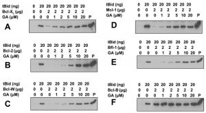
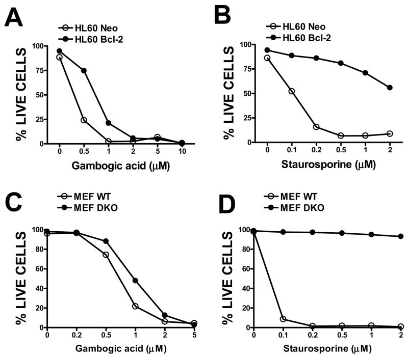
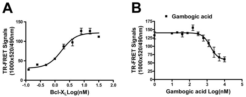
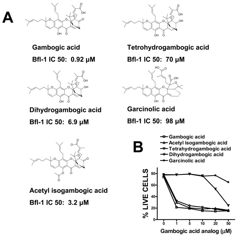
\For TR-FRET assays, GST-Bcl-XL and anti-GST-terbium were mixed together with the FITC-Bad BH3 peptide in PBS containing 0.005% tween 20 in 96 well plates in a total volume of 20 μl per well. After incubation at room temperature for 30 min, 2 μl of gambogic acid-containing solutions were added to the reaction mixtures containing 10 nM of Bcl-XL, 10 nM of FITC-Bad BH3 peptide and 2 nM of anti-GST-terbium for 30 min at room temperature. TR-FRET signals were measured with a SpectraMax M5 plate reader using the following settings: excitation at 330nm, emission for FITC signal at 490 nm, and emission for terbium signal at 520 nm.\ \Mitochondria Purification and Protein Release Assays[1] \HeLa cells were pelleted by centrifugation, and then washed once in HM buffer (10 mM HEPES, pH 7.4, 250 mM mannitol, 10 mM KCl, 5 mM MgCl2, 1 mM EGTA), containing 1 mM PMSF and a mixture of protease inhibitors. The cell pellet was then homogenized in HM buffer by 50 strokes of a dounce homogenizer, using a B-type pestle. The homogenate was centrifuged twice at 600g for 5 min to remove nuclei and debris. The resulting supernatant was centrifuged at 10,000g for 10 min, and the resulting mitochondria-containing pellet was washed twice with the HM buffer.[1] \\For mitochondrial protein release assays, 10 μl of mitochondria (50 μg) were added into a final volume of 50 μl HM buffer containing gambogic acid, tBid or tBid pre-incubated with gambogic acid or Bcl-2 family proteins at 30°C for 15 min. The reactions were further incubated at 30°C for 40–60 min, then mitochondria were pelleted by centrifugation and the supernatants were collected, boiled in Laemmli sample buffer, and analyzed by SDS-PAGE/immunoblotting using anti-SMAC antibody. The natural product gambogic acid (GA) has been reported to have cytotoxic activity against tumor cells in culture and was identified as an active compound in a cell-based high-throughput screening assay for activators of caspases, proteases involved in apoptosis. Using the antiapoptotic Bcl-2 family protein, Bfl-1, as a target for screening of a library of natural products, we identified GA as a competitive inhibitor that displaced BH3 peptides from Bfl-1 in a fluorescence polarization assay. Analysis of competition for BH3 peptide binding revealed that GA inhibits all six human Bcl-2 family proteins to various extents, with Mcl-1 and Bcl-B the most potently inhibited [concentrations required for 50% inhibition (IC(50)), < 1 micromol/L]. Competition for BH3 peptide binding was also confirmed using a time-resolved fluorescence resonance energy transfer assay. GA functionally inhibited the antiapoptotic Bcl-2 family proteins as shown by experiments using isolated mitochondria in which recombinant purified Bcl-2 family proteins suppress SMAC release in vitro, showing that GA neutralizes their suppressive effects on mitochondria in a concentration-dependent manner. GA killed tumor cell lines via an apoptotic mechanism, whereas analogues of GA with greatly reduced potency at BH3 peptide displacement showed little or no cytotoxic activity. However, GA retained cytotoxic activity against bax-/-bak-/- cells in which antiapoptotic Bcl-2 family proteins lack a cytoprotective phenotype, implying that GA also has additional targets that contribute to its cytotoxic mechanism. Altogether, the findings suggest that suppression of antiapoptotic Bcl-2 family proteins may be among the cytotoxic mechanisms by which GA kills tumor cells.[1] gambogic acid (GA), a xanthone derived from the resin of the Garcinia hanburyi, has been recently demonstrated to bind transferrin receptor and exhibit potential anticancer effects through a signaling mechanism that is not fully understood. Because of the critical role of NF-kappaB signaling pathway, we investigated the effects of GA on NF-kappaB-mediated cellular responses and NF-kappaB-regulated gene products in human leukemia cancer cells. Treatment of cells with GA enhanced apoptosis induced by tumor necrosis factor (TNF) and chemotherapeutic agents, inhibited the expression of gene products involved in antiapoptosis (IAP1 and IAP2, Bcl-2, Bcl-x(L), and TRAF1), proliferation (cyclin D1 and c-Myc), invasion (COX-2 and MMP-9), and angiogenesis (VEGF), all of which are known to be regulated by NF-kappaB. GA suppressed NF-kappaB activation induced by various inflammatory agents and carcinogens and this, accompanied by the inhibition of TAK1/TAB1-mediated IKK activation, inhibited IkappaBalpha phosphorylation and degradation, suppressed p65 phosphorylation and nuclear translocation, and finally abrogated NF-kappaB-dependent reporter gene expression. The NF-kappaB activation induced by TNFR1, TRADD, TRAF2, NIK, TAK1/TAB1, and IKKbeta was also inhibited. The effect of GA mediated through transferrin receptor as down-regulation of the receptor by RNA interference reversed its effects on NF-kappaB and apoptosis. Overall our results demonstrate that GA inhibits NF-kappaB signaling pathway and potentiates apoptosis through its interaction with the transferrin receptor.[3] Bcl-2 Binding Assay: Reaction mixture (25 mM Tris-HCl pH 7.5, 100 mM NaCl, 0.1% BSA) was mixed with recombinant Bcl-2 protein (0.5 μg/well), fluorescently labeled BH3 peptide (50 nM), and serial concentrations of Gambogic Acid (0.1-10 μM) in 96-well plates. After incubation at 37°C for 90 minutes, fluorescence polarization (FP) was measured. IC50 was calculated by fitting dose-response curves to inhibit BH3-Bcl-2 binding [2]; - STAT3 Kinase Assay: Recombinant STAT3 (0.2 μg/well) and ATP (100 μM) were mixed with reaction buffer (50 mM Tris-HCl pH 7.4, 10 mM MgCl₂, 1 mM DTT) and Gambogic Acid (0.5-20 μM). Incubation at 30°C for 60 minutes was followed by adding phospho-STAT3 antibody (ELISA-based detection). Absorbance at 450 nm was measured, and IC50 for STAT3 inhibition was determined [3]; - NF-κB Luciferase Assay: HCT116 cells transfected with NF-κB luciferase reporter plasmid were treated with Gambogic Acid (0.5-5 μM) for 24 h, then lysed. Luciferase substrate was added, and luminescence was measured. Inhibition rate of NF-κB activity was calculated relative to vehicle [6]. |
| Cell Assay |
MTT assay is used to assess how treatments such as gambogic acid, CDDP alone, or both together, affect in vitro cell viability. In 96-well culture plates, the cells (2×10⁴ cells per mL) are seeded. Following an overnight incubation, gambogic acid is applied to NCI-H460, A549, and NCI-H1299 cells in the following concentrations: 0.125, 0.25, 0.25, 0.5, 1, 2, and 4 μM, 0.44, 0.88, 1.75, 3.5, 7, 10.5 and 14 μM, and 0.44, 0.88, 1.75, 4, 8, 12, and 16 μM respectively. In NSCLC cells, three sequences are tested for the combined treatment: (a) Gambogic Acid followed by CDDP cells are exposed to Gambogic Acid for 48 h, and then after washout of Gambogic Acid, cells are treated with CDDP for an additional 48 h; (b) CDDP followed by Gambogic Acid cells are exposed to CDDP for 48 h, and then after washout of CDDP, cells are treated with Gambogic Acid for an additional 48 h; and (c) concurrent treatment cells are exposed to both Gambogic Acid and ADM for 48 h. The nature of the drug interaction is analysed by using the combination index (CI)[2].
Cell Viability Assay (MTT): A549 cells were seeded in 96-well plates (5×10³ cells/well) and cultured overnight. Gambogic Acid (0.1-5 μM) was added, and cells were incubated for 72 h. MTT reagent (0.5 mg/mL) was added for 4 h; formazan was dissolved in DMSO, and absorbance at 570 nm was measured. IC50 was calculated via GraphPad Prism [1]; - Apoptosis Assay (Annexin V/PI): Jurkat cells were treated with Gambogic Acid (2 μM) for 24 h. Cells were harvested, washed with PBS, stained with Annexin V-FITC and PI for 15 minutes (dark), and analyzed by flow cytometry to quantify apoptotic cells [3]; - Mitochondrial Membrane Potential Assay (JC-1): MCF-7 cells were seeded on coverslips, treated with Gambogic Acid (1 μM) for 24 h, and incubated with JC-1 dye (5 μM) for 30 minutes. Fluorescent images were captured (red for intact mitochondria, green for depolarized mitochondria), and the red/green ratio was calculated [4]; - Colony Formation Assay: HepG2 cells were seeded in 6-well plates (2×10³ cells/well) and treated with Gambogic Acid (0.5-2 μM) for 24 h. Fresh medium was added, and cells were cultured for 14 days. Colonies were stained with crystal violet, counted, and the inhibition rate was calculated [5]. |
| Animal Protocol |
Mice: A549 viable cells (5×10⁶/100 μL PBS per mouse) are subcutaneously injected into the right flank of male SCID mice that are 7 to 8 weeks old in order to assess the in vivo antitumor activity of gambogic acid combined with CDDP. The mice are randomly assigned to one of four treatment groups when the tumor volume reaches 100 mm3, including control (saline only, n=5), gambogic acid (3.0 mg/kg every two days, intravenously; n=6), CDDP (4 mg/kg every week, intravenously; n=6), and sequential combination (CDDP treatment one day before gambogic acid treatment, n=6). To help detect any additive effects of combination therapy with platinum-based agents and gambogic acid, CDDP (4 mg/kg, weekly) is typically administered at doses lower than the maximum tolerated dose. Once every two days, a caliper is used to measure the tumor's size. Once every two days, body weight is measured. The tumors are removed after 14 days, and the mice are then put to death. They are then kept at -80°C for future research.
A549 Lung Cancer Xenograft Protocol: Female nude mice (6-8 weeks old, n=6/group) were subcutaneously injected with 5×10⁶ A549 cells (PBS:Matrigel=1:1) into the right flank. When tumors reached ~100 mm³: Gambogic Acid was dissolved in 10% DMSO + 40% PEG300 + 50% normal saline, administered at 10 mg/kg (i.p., once daily, 14 days); vehicle group received the same solvent. Tumor volume (length×width²/2) and body weight were recorded every 2 days [1]; - Leukemia Mouse Protocol: BALB/c mice (7-9 weeks old, n=5/group) were intravenously injected with 1×10⁷ Jurkat cells. Three days later, Gambogic Acid (5 mg/kg, i.p., dissolved in 5% DMSO + 95% sesame oil) was given every other day for 10 days. Peripheral blood was collected to count leukemia cells, and survival was monitored [3]; - Colon Cancer Oral Administration Protocol: C57BL/6 mice (6-8 weeks old, n=6/group) were subcutaneously inoculated with 2×10⁶ MC38 cells. When tumors reached ~120 mm³, Gambogic Acid (15 mg/kg, oral gavage, dissolved in 0.5% CMC-Na + 0.1% Tween 80) was administered once daily for 12 days. Tumors were weighed at sacrifice [6]. |
| ADME/Pharmacokinetics |
In SD rats: the half-life (t1/2) of gambogey acid (10 mg/kg, intravenous injection) was approximately 2.3 hours; the bioavailability of oral (20 mg/kg) was approximately 15% (as determined by high performance liquid chromatography) [7]; - In mice: gambogey acid was mainly distributed in the liver and kidneys (1 hour after intraperitoneal injection of 5 mg/kg, the tissue concentration was approximately 2-3 times higher than the plasma concentration) [7]; - In vitro metabolism (rat liver microsomes): gambogey acid was metabolized by cytochrome P450 3A4 (CYP3A4), and the metabolic clearance rate was approximately 0.8 mL/min/mg protein [7]
|
| Toxicity/Toxicokinetics |
Intraperitoneal LD50 in rats: 88 mg/kg liver: other changes. Indian Journal of Experimental Biology, 5(96), 1967 [PMID:6062434]
Intravenous LD50 in rats: 107 mg/kg liver: other changes. Indian Journal of Experimental Biology, 5(96), 1967 [PMID:6062434] Subcutaneous LD50 in mice: 354 mg/kg. CRC Handbook of Antibiotic Compounds, Vol. 1-, Berdy, J., Boca Raton, FL, CRC Press, 1980, 8(1)(331), 1982 LD50 in mammals (unspecified species): 55 mg/kg. Zhongliu Cancer Review, edited by Yu, R. et al., Shanghai Science and Technology Press, People's Republic of China, 1994, -(220), 1994 In ICR mice: the median lethal dose (LD50) of gamboge is approximately 45 mg/kg. mg/kg (intraperitoneal injection) and 120 mg/kg (oral administration); lethal doses of mice showed liver congestion (H&E staining) [7]; - In nude mice treated with gamboge (10 mg/kg, intraperitoneal injection, 14 days): serum ALT (~42 U/L vs. carrier ~40 U/L), AST (~58 U/L vs. carrier ~55 U/L) or BUN (~18 mg/dL vs. carrier ~17 mg/dL) showed no significant changes [1]; - In vitro cytotoxicity to normal cells: gamboge (2 μM) reduced the survival rate of normal human lung fibroblasts (MRC-5) by <15% (72-hour MTT assay) [1]; - The plasma protein binding rate of gamboge was approximately 92% (measured by rat plasma ultrafiltration) [7]. |
| References | |
| Additional Infomation |
beta-Guttiferin hanburyi has been reported to exist in plants of the genus Garcinia hanburyi, and related data have been reported. Garcinia hanburyi acid (2) is a natural product isolated from the resin of plants of the genus Garcinia hanburyi. Using our high-throughput screening method based on cells and caspase, we found that it is an effective apoptosis inducer. In the caspase activation experiment of T47D breast cancer cells, the EC50 value of garcinia hanburyi acid was 0.78 μM. We further characterized the apoptosis-inducing activity of garcinia hanburyi acid in human breast tumor cells T47D by nuclear fragmentation experiments and flow cytometry analysis. The study found that the apoptosis induced by garcinia hanburyi acid was independent of the cell cycle, which is different from paclitaxel, which arrests cells in the G2/M phase. To understand the structure-activity relationship (SAR) of garcinia hanburyi acid, we synthesized derivatives of compound 2 with various functional groups modified by different functional groups. The structure-activity relationship of gamboge hanburyi determined by caspase activation assay showed that the carbon-carbon double bond at positions 9 and 10 of the α,β-unsaturated ketone is crucial to its biological activity, while the 6-hydroxy and 30-carboxyl groups can tolerate various modifications. Conventional growth inhibition assay confirmed the importance of the carbon-carbon double bond at positions 9 and 10. The high efficiency, novel mechanism of action, easy isolation and abundant sources of compound 2 as an apoptosis inducer, as well as its easy chemical modification, make gamboge hanburyi an ideal molecule for the development of anticancer drugs. [2] Gamboge hanburyi is the main active ingredient of gamboge hanburyi. It has been previously reported that it can activate apoptosis in various cancer cell lines by targeting transferrin receptor and regulating nuclear factor-κB signaling pathway. Whether GA inhibits angiogenesis (which is crucial for cancer and other human diseases) is still unclear. In this study, nanomolar concentrations of GA significantly inhibited the proliferation, migration, invasion, tubular formation and microvascular growth of human umbilical vein endothelial cells (HUVEC). In xenograft prostate cancer models, we found that metronid chemotherapy combined with GA can effectively inhibit tumor angiogenesis and tumor growth with fewer side effects. GA’s effects on activating apoptosis, inhibiting cell proliferation and migration in HUVECs are better than those in human prostate cancer cells (PC3), suggesting that GA may be a potential anticancer drug that can exert a low chemical toxicity effect by inhibiting angiogenesis. In addition, we found that GA can inhibit the activation of vascular endothelial growth factor receptor 2 and its downstream protein kinases (such as c-Src, focal adhesion kinase and AKT). These data together suggest that GA inhibits angiogenesis and may become a feasible candidate drug in anti-angiogenic and anticancer therapies. [4] Garcinia cambogia (GA) is a cage-like xanthones extracted from Garcinia hanburyi and has a strong apoptosis-inducing effect in a variety of cancer cells. GA’s unique efficacy makes it a novel anticancer drug. More and more studies are dedicated to elucidating the molecular mechanism of GA’s anticancer effect, and it has been reported that GA treatment affects multiple key signaling pathways. This review summarizes the various functional roles of gambogey acid in cancer cells, including inducing apoptosis, inhibiting cell proliferation, and preventing cancer metastasis and tumor angiogenesis. [5]
Gambogey acid is a natural compound isolated from the resin of gamboge plants. Its anticancer activity is mainly achieved by targeting multiple signaling pathways (Bcl-2, STAT3, NF-κB). [2][3][6]; - Garcinia cambogia enhances the efficacy of cisplatin in A549 cells: the combined use of garcinia cambogia (0.5 μM) and cisplatin (1 μM) increased the apoptosis rate from about 30% (cisplatin alone) to about 65%[1]; - No FDA approval or clinical trial data for garcinia cambogia has been reported in the cited literature; it is mainly used as a tool for preclinical anticancer research[1][2][3][4][5][6][7]; - Garcinia cambogia induces apoptosis in a p53-independent manner: it shows similar cytotoxicity in p53 wild-type (HCT116) and p53-deficient (HCT116 p53⁻/⁻) cells[4]. |
| Molecular Formula |
C38H44O8
|
|
|---|---|---|
| Molecular Weight |
628.75
|
|
| Exact Mass |
628.303
|
|
| Elemental Analysis |
C, 72.59; H, 7.05; O, 20.36
|
|
| CAS # |
2752-65-0
|
|
| Related CAS # |
|
|
| PubChem CID |
9852185
|
|
| Appearance |
Yellow solid powder
|
|
| Density |
1.3±0.1 g/cm3
|
|
| Boiling Point |
808.9±65.0 °C at 760 mmHg
|
|
| Melting Point |
88.5°C
|
|
| Flash Point |
251.4±27.8 °C
|
|
| Vapour Pressure |
0.0±3.0 mmHg at 25°C
|
|
| Index of Refraction |
1.627
|
|
| Source |
Originated from plants: Guttiferae Garcinia hanburyi Hook. f.
|
|
| LogP |
10.3
|
|
| Hydrogen Bond Donor Count |
2
|
|
| Hydrogen Bond Acceptor Count |
8
|
|
| Rotatable Bond Count |
8
|
|
| Heavy Atom Count |
46
|
|
| Complexity |
1490
|
|
| Defined Atom Stereocenter Count |
5
|
|
| SMILES |
O1C(C([H])([H])[H])(C([H])([H])[H])C2([H])C([H])([H])C3([H])C([H])=C4C(C5C(=C6C([H])=C([H])C(C([H])([H])[H])(C([H])([H])C([H])([H])/C(/[H])=C(\C([H])([H])[H])/C([H])([H])[H])OC6=C(C([H])([H])/C(/[H])=C(\C([H])([H])[H])/C([H])([H])[H])C=5OC24C1(C([H])([H])C([H])=C(C(=O)O[H])C([H])([H])[H])C3=O)O[H])=O
|
|
| InChi Key |
GEZHEQNLKAOMCA-RRZNCOCZSA-N
|
|
| InChi Code |
InChI=1S/C38H44O8/c1-20(2)10-9-15-36(8)16-14-24-29(39)28-30(40)26-18-23-19-27-35(6,7)46-37(33(23)41,17-13-22(5)34(42)43)38(26,27)45-32(28)25(31(24)44-36)12-11-21(3)4/h10-11,13-14,16,18,23,27,39H,9,12,15,17,19H2,1-8H3,(H,42,43)/b22-13-/t23-,27+,36-,37+,38-/m1/s1
|
|
| Chemical Name |
(Z)-4-[(1S,2S,8R,17S,19R)-12-hydroxy-8,21,21-trimethyl-5-(3-methylbut-2-enyl)-8-(4-methylpent-3-enyl)-14,18-dioxo-3,7,20-trioxahexacyclo[15.4.1.02,15.02,19.04,13.06,11]docosa-4(13),5,9,11,15-pentaen-19-yl]-2-methylbut-2-enoic acid
|
|
| Synonyms |
|
|
| HS Tariff Code |
2934.99.9001
|
|
| Storage |
Powder -20°C 3 years 4°C 2 years In solvent -80°C 6 months -20°C 1 month |
|
| Shipping Condition |
Room temperature (This product is stable at ambient temperature for a few days during ordinary shipping and time spent in Customs)
|
| Solubility (In Vitro) |
DMSO: ~100 mg/mL (159 mM)
Water: 1 mg/mL (Insoluble) Ethanol: N/A |
|---|---|
| Solubility (In Vivo) |
Solubility in Formulation 1: ≥ 2.5 mg/mL (3.98 mM) (saturation unknown) in 10% DMSO + 40% PEG300 + 5% Tween80 + 45% Saline (add these co-solvents sequentially from left to right, and one by one), clear solution.
For example, if 1 mL of working solution is to be prepared, you can add 100 μL of 25.0 mg/mL clear DMSO stock solution to 400 μL PEG300 and mix evenly; then add 50 μL Tween-80 to the above solution and mix evenly; then add 450 μL normal saline to adjust the volume to 1 mL. Preparation of saline: Dissolve 0.9 g of sodium chloride in 100 mL ddH₂ O to obtain a clear solution. Solubility in Formulation 2: ≥ 2.5 mg/mL (3.98 mM) (saturation unknown) in 10% DMSO + 90% Corn Oil (add these co-solvents sequentially from left to right, and one by one), clear solution. For example, if 1 mL of working solution is to be prepared, you can add 100 μL of 25.0 mg/mL clear DMSO stock solution to 900 μL of corn oil and mix evenly. View More
Solubility in Formulation 3: 2% DMSO+40% PEG 300+2% Tween 80+ddH2O: 4mg/mL |
| Preparing Stock Solutions | 1 mg | 5 mg | 10 mg | |
| 1 mM | 1.5905 mL | 7.9523 mL | 15.9046 mL | |
| 5 mM | 0.3181 mL | 1.5905 mL | 3.1809 mL | |
| 10 mM | 0.1590 mL | 0.7952 mL | 1.5905 mL |
*Note: Please select an appropriate solvent for the preparation of stock solution based on your experiment needs. For most products, DMSO can be used for preparing stock solutions (e.g. 5 mM, 10 mM, or 20 mM concentration); some products with high aqueous solubility may be dissolved in water directly. Solubility information is available at the above Solubility Data section. Once the stock solution is prepared, aliquot it to routine usage volumes and store at -20°C or -80°C. Avoid repeated freeze and thaw cycles.
Calculation results
Working concentration: mg/mL;
Method for preparing DMSO stock solution: mg drug pre-dissolved in μL DMSO (stock solution concentration mg/mL). Please contact us first if the concentration exceeds the DMSO solubility of the batch of drug.
Method for preparing in vivo formulation::Take μL DMSO stock solution, next add μL PEG300, mix and clarify, next addμL Tween 80, mix and clarify, next add μL ddH2O,mix and clarify.
(1) Please be sure that the solution is clear before the addition of next solvent. Dissolution methods like vortex, ultrasound or warming and heat may be used to aid dissolving.
(2) Be sure to add the solvent(s) in order.
|
|---|
|
 |
 |
|---|
 |
 |
Products are for research use only; We do not sell to patients
Copyright 2020 InvivoChem LLC | All Rights Reserved


 COA
COA