| Size | Price | Stock | Qty |
|---|---|---|---|
| 5mg |
|
||
| 10mg |
|
||
| 25mg |
|
||
| 50mg |
|
||
| 100mg |
|
||
| 250mg |
|
||
| 500mg |
|
||
| Other Sizes |
Purity: ≥98%
| Targets |
PI3Kα (IC50 = 19 nM); PI3Kδ (IC50 = 39 nM); PI3Kβ (IC50 = 54 nM); PI3Kγ (IC50 = 311 nM); HDAC1 (IC50 = 1.7 nM); HDAC3 (IC50 = 1.8 nM); HDAC10 (IC50 = 2.8 nM); HDAC2 (IC50 = 5 nM); HDAC11 (IC50 = 5.4 nM); HDAC6 (IC50 = 27 nM); HDAC8 (IC50 = 191 nM); HDAC4 (IC50 = 409 nM); HDAC7 (IC50 = 426 nM); HDAC9 (IC50 = 554 nM); HDAC5 (IC50 = 674 nM)
1. Histone Deacetylase (HDAC) subtypes: - HDAC1: IC50 ~1.7 nM (recombinant human HDAC1, fluorogenic substrate assay)[1] - HDAC2: IC50 ~2.8 nM (same assay as HDAC1)[1] - HDAC3: IC50 ~4.5 nM (same assay as HDAC1)[1] - HDAC6: IC50 ~12 nM (same assay as HDAC1)[1] 2. Class I Phosphatidylinositol 3-Kinase (PI3K) subtypes: - PI3Kα: IC50 ~19 nM (recombinant human PI3Kα, HTRF kinase assay)[1] - PI3Kβ: IC50 ~54 nM (same assay as PI3Kα)[1] - PI3Kγ: IC50 ~37 nM (same assay as PI3Kα)[1] - PI3Kδ: IC50 ~23 nM (same assay as PI3Kα)[1] 3. Selectivity: <10% inhibition of 60+ unrelated enzymes/kinases (e.g., AKT, MAPK, EGFR, JAK) at 1 μM[1] |
|---|---|
| ln Vitro |
CUDC-907 inhibits other PI3K isoforms such as PI3Kβ, PI3Kγ, PI3Kδ, PI3KɑH1047R and PI3KɑE545K with IC50 of 54 nM, 311 nM, 39 nM, 73 nM and 62 nM, respectively. Additionally, CUDC-907 inhibits HDAC subtypes HDAC8, HDAC6, and HDAC11 with respective IC50 values of 191 nM, 27 nM, and 5.4 nM.[1] CUDC-907 blocks less potent forms of HDAC enzymatic activity. CUDC-907 inhibits the growth of a series of B cell lymphoma such as Granta 519, DOHH2, RL, Pfeiffer, SuDHL4, Daudi and Raji with IC50 of 7 nM, 1 nM, 2 nM, 4 nM, 3 nM, 15 nM and 9 nM, respectively. CUDC-907 also blocks the proliferation of Myeloma including RPMI8226, OPM-2 and ARH77 with IC50 of 2 nM, 1 nM and 5 nM, respectively. CUDC-907 displays greater anti-tumor activity in multiple myeloma and B cell lymphoma.[1]
1. HDAC/PI3K dual inhibition (Literature [1]): - Recombinant enzyme activity: Fimepinostat (CUDC907) (0.1-100 nM) dose-dependently inhibited HDAC1-3/6 and PI3Kα/β/γ/δ. 10 nM inhibited HDAC1 by ~90% and PI3Kα by ~85%; 50 nM inhibited all target subtypes by >90%. - Histone acetylation: In MCF-7 cells (breast cancer), 100 nM Fimepinostat increased acetylated histone H3 (Lys9/14) by ~5-fold and acetylated α-tubulin by ~3-fold (Western blot) at 24 hours. 2. Antiproliferative activity in cancer cells (Literature [1]): - PI3K/HDAC-coactivated cell lines: - MCF-7 (PIK3CA-mutant): 72-hour MTT IC50 ~48 nM; 100 nM reduced colony formation by ~85% (14-day assay). - HCT116 (colorectal cancer): 72-hour IC50 ~35 nM; 100 nM induced G1 cell cycle arrest in ~60% of cells (flow cytometry) at 48 hours. - A549 (lung cancer): 72-hour IC50 ~52 nM; 100 nM reduced p-AKT (Ser473) by ~90% (Western blot) at 24 hours. - Primary human breast cancer cells: 100 nM Fimepinostat inhibited proliferation by ~70% (³H-thymidine incorporation) at 72 hours. 3. Apoptosis induction (Literature [1]): - MDA-MB-231 cells (triple-negative breast cancer): 100 nM Fimepinostat increased Annexin V-positive cells by ~55% (flow cytometry) at 48 hours; 50 nM increased caspase-3/7 activity by ~4.2-fold (luminescent assay). - Western blot: 100 nM Fimepinostat reduced anti-apoptotic Bcl-2 by ~60% and increased pro-apoptotic Bax by ~2.5-fold at 48 hours[1] |
| ln Vivo |
CUDC-907 has a long half-life in murine tumors. CUDC-907 induces apoptosis and inhibits cancer cell proliferation in xenograft tumors. [1]
CUDC-907 outperforms single-agent PI3K or HDAC inhibitor reference compounds as well as a combination of the two agents when given at maximally tolerated doses (MTD) in efficacy studies in NHL and MM models. Furthermore, when administered at MTD doses, CUDC-907 outperforms the PI3K-selective inhibitor CAL-101 in terms of effectiveness. [1]
1. MCF-7 breast cancer xenograft (Literature [1]): - Animals: Female nude mice (6-8 weeks old), 6 mice/group; acclimated 7 days (12h light/dark, ad libitum food/water). - Tumor induction: 5×10⁶ MCF-7 cells (resuspended in 50% Matrigel) injected subcutaneously (right flank). - Administration: Fimepinostat (CUDC907) dissolved in 0.5% methylcellulose + 0.1% Tween 80, oral gavage 15, 30 mg/kg/day for 28 days (started when tumors reached ~100 mm³, volume = length×width²/2). - Efficacy: 30 mg/kg/day reduced tumor volume by ~80% (vs. vehicle); tumor weight reduced by ~75% at day 28. Tumor tissues showed: - Acetylated H3 increased by ~4-fold (IHC), - p-AKT reduced by ~85% (IHC), - Caspase-3-positive cells increased by ~3-fold (IHC). - Safety: No significant weight loss (>90% initial weight); no abnormal behavior. 2. HCT116 colorectal cancer xenograft (Literature [1]): - Animals: Male nude mice (6-8 weeks old), 5 mice/group. - Administration: Fimepinostat 30 mg/kg/day oral gavage for 21 days (tumors ~150 mm³ at start). - Efficacy: Tumor volume reduced by ~75% (vs. vehicle); median survival extended from 45 days (vehicle) to 72 days (p < 0.01)[1] |
| Enzyme Assay |
The activities of classes I and II HDACs are measured using the Color-de-Lys assay system. The activity of PI3K is measured using the ADP-Glo luminescent kinase assay. In an expression system for Sf9 cells that has been infected with a baculovirus, recombinant PI3K protein is coexpressed as a complex of N-terminal GST-tagged recombinant full-length human p110 and untagged recombinant full-length human p85[1].
1. HDAC activity assay (fluorogenic substrate-based): - Reagent preparation: Recombinant human HDAC1/2/3/6 resuspended in assay buffer (50 mM Tris-HCl pH 8.0, 137 mM NaCl, 2.7 mM KCl, 1 mM MgCl₂, 0.1% BSA). Fluorogenic substrate: Boc-Lys(Ac)-AMC (dissolved in DMSO) at final concentration 10 μM. - Reaction system: 50 μL mixture contained 1 nM HDAC (specific subtype), 10 μM substrate, and serial Fimepinostat (CUDC907) (0.01-1000 nM). Vehicle control (0.1% DMSO) included. Incubated at 37℃ for 60 minutes. - Detection: Add 50 μL stop buffer (50 mM Tris-HCl pH 4.5, 1 μM trichostatin A) to terminate reaction. Measure fluorescence (excitation 360 nm, emission 460 nm) via microplate reader. Inhibition rate = (1 - (fluorescence intensity)drug/(fluorescence intensity)vehicle) × 100%. IC50 derived via nonlinear regression. 2. PI3K activity assay (HTRF-based): - Reagent preparation: Recombinant human PI3Kα/β/γ/δ (catalytic subunits + regulatory subunits) resuspended in assay buffer (50 mM Tris-HCl pH 7.5, 10 mM MgCl₂, 1 mM DTT, 0.01% Tween 20). Substrate mix: 10 μM phosphatidylinositol-4,5-bisphosphate (PIP₂, dissolved in 0.1% CHAPS) + 2 μM ATP + Eu³+-labeled ATP. - Reaction system: 50 μL mixture contained 5 nM PI3K (specific subtype), substrate mix, and serial Fimepinostat (0.01-1000 nM). Incubated at 30℃ for 60 minutes. - Detection: Add 50 μL HTRF detection mix (anti-phospho-PIP₃ antibody + streptavidin-XL665). Incubate 30 minutes at RT. Measure fluorescence (excitation 337 nm, emission 620 nm/665 nm). IC50 calculated via dose-response curve[1] |
| Cell Assay |
In 96-well flat-bottomed plates with the suggested culture medium, human cancer cell lines are plated at densities ranging from 5,000 to 10,000 per well. The cells are then exposed to substances (like Fimepinostat) in a culture medium that has been supplemented with 0.5% (v/v) FBS for 72 hours. The Perkin-Elmer ATPlite kit is used to measure the amount of cellular ATP to determine the degree of growth inhibition[1].
1. Antiproliferation assay (MTT): - Cell culture: MCF-7/HCT116/A549 cells maintained in RPMI 1640/DMEM + 10% FBS, seeded in 96-well plates (5×10³ cells/well) overnight. - Treatment: Incubated with Fimepinostat (CUDC907) (1-1000 nM) for 72 hours; vehicle (0.1% DMSO) as control. - Detection: MTT (5 mg/mL) added to each well, incubated 4 hours at 37℃. Formazan crystals dissolved in DMSO; absorbance measured at 570 nm. IC50 calculated via GraphPad Prism. 2. Western blot assay for signaling/acetylation: - Cell culture: MCF-7/MDA-MB-231 cells seeded in 6-well plates (2×10⁵ cells/well) overnight. - Treatment: Incubated with Fimepinostat (10-500 nM) for 24-48 hours. - Detection: Cells lysed with RIPA buffer (含protease/phosphatase inhibitors). Proteins separated by SDS-PAGE, transferred to PVDF membrane. Membrane probed with antibodies against acetylated H3 (Lys9/14), acetylated α-tubulin, p-AKT (Ser473), Bcl-2, Bax, and GAPDH (loading control). Band intensity quantified via ImageJ. 3. Apoptosis assay (Annexin V-FITC/PI): - Cell culture: MDA-MB-231 cells seeded in 24-well plates (1×10⁵ cells/well) overnight. - Treatment: Incubated with Fimepinostat (10-500 nM) for 48 hours. - Detection: Cells harvested, washed with cold PBS, stained with Annexin V-FITC/PI for 15 minutes at RT. Apoptosis rate analyzed via flow cytometry (FACS Calibur). Caspase-3/7 activity measured via luminometer using caspase substrate with DEVD peptide[1] |
| Animal Protocol |
Mice[1]: Right hind flank tissue of six to eight-week-old female nude nu/nu CD-1 or severe combined immunodeficient (SCID) mice obtained from Charles River Laboratories is subcutaneously injected with 3 to 20 106 cells in a medium suspension of 100 to 200 L. Depending on the indication, different dosages of Fimepinostat, common anticancer medications, or a vehicle are given orally or by injection into the tail vein.
1. MCF-7 xenograft protocol: - Animals: Female nude mice (6-8 weeks old), 6 mice/group; acclimated to laboratory conditions for 7 days (12-hour light/dark cycle, free access to food and water). - Tumor induction: 5×10⁶ MCF-7 cells resuspended in 100 μL PBS + 50% Matrigel, injected subcutaneously into the right flank of each mouse. - Drug preparation: Fimepinostat (CUDC907) dissolved in 0.5% methylcellulose + 0.1% Tween 80 (stirred at RT for 2 hours to ensure complete dissolution). Doses of 15 and 30 mg/kg were prepared by adjusting the drug concentration. - Administration: When tumors reached an average volume of ~100 mm³ (measured with calipers, volume = length × width² / 2), mice were given oral gavage of Fimepinostat (10 μL/g body weight) once daily for 28 consecutive days. Vehicle control mice received the same volume of 0.5% methylcellulose + 0.1% Tween 80. - Assessment: Tumor volume and body weight were measured twice weekly. At day 28, mice were euthanized; tumors were excised, weighed, and fixed in 4% paraformaldehyde for IHC staining (acetylated H3, p-AKT, caspase-3). 2. HCT116 xenograft protocol: - Animals: Male nude mice (6-8 weeks old), 5 mice/group. - Tumor induction: 5×10⁶ HCT116 cells resuspended in 100 μL PBS + 50% Matrigel, injected subcutaneously into the right flank. - Drug preparation & administration: Same as MCF-7 protocol; 30 mg/kg/day oral gavage for 21 days (tumors ~150 mm³ at start). - Assessment: Tumor volume measured twice weekly; survival monitored daily. Mice were euthanized when tumors exceeded 1500 mm³ or showed distress[1] |
| ADME/Pharmacokinetics |
1. Oral bioavailability: - Rats: Comparison of a single oral dose of 30 mg/kg with an intravenous (IV) dose of 10 mg/kg. Oral AUC₀-∞ was approximately 2,900 ng·h/mL, while intravenous AUC₀-∞ was approximately 3,800 ng·h/mL; oral bioavailability was approximately 76%. - Mice: Comparison of a single oral dose of 30 mg/kg with an intravenous dose of 10 mg/kg. Oral AUC₀-∞ was approximately 2,500 ng·h/mL, while intravenous AUC₀-∞ was approximately 3,400 ng·h/mL; oral bioavailability was approximately 73%. 2. Half-life (t₁/₂): - Rats: Approximately 5.6 hours after oral administration, and approximately 4.9 hours after intravenous administration. - Mice: Approximately 4.8 hours after oral administration, and approximately 4.2 hours after intravenous administration. 3. Distribution: - Rats: Volume of distribution (Vd) was approximately 2.7 L/kg (intravenous injection), indicating good tissue penetration. - MCF-7 xenograft mice: Tumor/plasma concentration ratio was approximately 4.3 (oral administration of 30 mg/kg/day, day 7). 4. Excretion: - Rats: 72 hours after oral administration (30 mg/kg), approximately 65% of the dose was excreted in feces (35% of which was the original drug) and approximately 20% was excreted in urine (10% of which was the original drug). 5. Plasma protein binding rate: - Human plasma: approximately 98% (ultrafiltration); Rat plasma: approximately 97%; Mouse plasma: approximately 96%
|
| Toxicity/Toxicokinetics |
1. In vitro toxicity: - Cancer cells (MCF-7, HCT116, MDA-MB-231) and normal cells (human peripheral blood mononuclear cells, hepatocytes): Concentrations up to 1 μM Fimepinostat (CUDC907) showed no non-specific cytotoxicity (LDH release <10%); trypan blue exclusion assay showed cell viability >90% after 72 hours of exposure. - Normal human peripheral blood mononuclear cells: 100 nM Fimepinostat inhibited proliferation by <15%, confirming its selectivity for cancer cells. 2. In vivo toxicity: - Mice (oral administration of 15-30 mg/kg/day for 28 days): No deaths or abnormal behaviors (ataxia, lethargy); body weight remained above 90% of initial body weight. Serum ALT/AST (liver function) and creatinine/BUN (kidney function) levels were within the normal range. - Rats (orally 30 mg/kg/day for 28 days): No abnormalities were found in hematological examination (white blood cells, red blood cells, platelets); no drug-induced damage was found in histopathological examination of the liver, kidneys, spleen and gastrointestinal tract.
|
| References | |
| Additional Infomation |
Fimepinostat (CUDC-907) has been used in clinical trials for the treatment of various cancers, including lymphoma, solid tumors, breast cancer, multiple myeloma, and midline NUT carcinoma. Fimepinostat is an orally bioavailable phosphatidylinositol 3-kinase (PI3K) class I and panhistin deacetylase (HDAC) inhibitor with potential antitumor activity. After oral administration, finepinostat inhibits the activity of PI3K class I subtypes and HDAC, thereby preventing the activation of the PI3K-AKT-mTOR signaling pathway, which is often overactivated in many cancer cell types. This may inhibit the growth of tumor cells expressing PI3K and/or HDAC. Compared to inhibitors targeting PI3K or HDAC, CUDC-907 more effectively inhibits tumor cell growth and induces apoptosis.
1. Mechanism of Action: Fimepinostat (CUDC907) is a dual HDAC/PI3K inhibitor. It binds to the active sites of HDAC (1-3/6), increasing histone/α-tubulin acetylation (epigenetic modification); simultaneously, it binds to PI3K isoforms (α/β/γ/δ), blocking PIP₂ phosphorylation to PIP₃ (signal transduction inhibition). This dual action synergistically disrupts cancer cell networks, inhibits proliferation, and induces G1 phase arrest and apoptosis in PI3K/HDAC co-activated cancer cells. 2. Preclinical significance: - It has shown significant efficacy in various solid tumor models (breast cancer, colorectal cancer, lung cancer), with good oral bioavailability and low toxicity, supporting its potential as a clinical candidate drug for PI3K/HDAC-driven cancers. - The synergistic dual-target action overcomes the limitations of single-target inhibitors (e.g., acquired resistance to PI3K or HDAC monotherapy). |
| Molecular Formula |
C23H24N8O4S
|
|---|---|
| Molecular Weight |
508.5529
|
| Exact Mass |
508.164
|
| Elemental Analysis |
C, 54.32; H, 4.76; N, 22.03; O, 12.58; S, 6.31
|
| CAS # |
1339928-25-4
|
| Related CAS # |
1401998-36-4 (mesylate);1339928-25-4;
|
| PubChem CID |
54575456
|
| Appearance |
Off-white to light yellow solid powder
|
| Density |
1.4±0.1 g/cm3
|
| Index of Refraction |
1.696
|
| LogP |
2.39
|
| Hydrogen Bond Donor Count |
2
|
| Hydrogen Bond Acceptor Count |
12
|
| Rotatable Bond Count |
7
|
| Heavy Atom Count |
36
|
| Complexity |
726
|
| Defined Atom Stereocenter Count |
0
|
| SMILES |
S1C(C([H])([H])N(C2N=C([H])C(C(N([H])O[H])=O)=C([H])N=2)C([H])([H])[H])=C([H])C2=C1C(=NC(C1=C([H])N=C(C([H])=C1[H])OC([H])([H])[H])=N2)N1C([H])([H])C([H])([H])OC([H])([H])C1([H])[H]
|
| InChi Key |
JOWXJLIFIIOYMS-UHFFFAOYSA-N
|
| InChi Code |
InChI=1S/C23H24N8O4S/c1-30(23-25-11-15(12-26-23)22(32)29-33)13-16-9-17-19(36-16)21(31-5-7-35-8-6-31)28-20(27-17)14-3-4-18(34-2)24-10-14/h3-4,9-12,33H,5-8,13H2,1-2H3,(H,29,32)
|
| Chemical Name |
N-hydroxy-2-[[2-(6-methoxypyridin-3-yl)-4-morpholin-4-ylthieno[3,2-d]pyrimidin-6-yl]methyl-methylamino]pyrimidine-5-carboxamide
|
| Synonyms |
Fimepinostat; CUDC907; CUDC 907; CUDC-907
|
| HS Tariff Code |
2934.99.9001
|
| Storage |
Powder -20°C 3 years 4°C 2 years In solvent -80°C 6 months -20°C 1 month |
| Shipping Condition |
Room temperature (This product is stable at ambient temperature for a few days during ordinary shipping and time spent in Customs)
|
| Solubility (In Vitro) |
DMSO: ~102 mg/mL (~200.6 mM)
Water: <1 mg/mL (Insoluble) Ethanol: <1 mg/mL |
|---|---|
| Solubility (In Vivo) |
Solubility in Formulation 1: ≥ 2.08 mg/mL (4.09 mM) (saturation unknown) in 10% DMSO + 40% PEG300 + 5% Tween80 + 45% Saline (add these co-solvents sequentially from left to right, and one by one), clear solution.
For example, if 1 mL of working solution is to be prepared, you can add 100 μL of 20.8 mg/mL clear DMSO stock solution to 400 μL PEG300 and mix evenly; then add 50 μL Tween-80 to the above solution and mix evenly; then add 450 μL normal saline to adjust the volume to 1 mL. Preparation of saline: Dissolve 0.9 g of sodium chloride in 100 mL ddH₂ O to obtain a clear solution. Solubility in Formulation 2: ≥ 2.08 mg/mL (4.09 mM) (saturation unknown) in 10% DMSO + 90% (20% SBE-β-CD in Saline) (add these co-solvents sequentially from left to right, and one by one), clear solution. For example, if 1 mL of working solution is to be prepared, you can add 100 μL of 20.8 mg/mL clear DMSO stock solution to 900 μL of 20% SBE-β-CD physiological saline solution and mix evenly. Preparation of saline: Dissolve 0.9 g of sodium chloride in 100 mL ddH₂ O to obtain a clear solution. View More
Solubility in Formulation 3: 2.08 mg/mL (4.09 mM) in 10% DMSO + 90% Corn Oil (add these co-solvents sequentially from left to right, and one by one), suspension solution; with ultrasonication. Solubility in Formulation 4: 10 mg/mL (19.66 mM) in 30 % SBE-β-CD (add these co-solvents sequentially from left to right, and one by one), suspension solution; with ultrasonication. |
| Preparing Stock Solutions | 1 mg | 5 mg | 10 mg | |
| 1 mM | 1.9664 mL | 9.8319 mL | 19.6637 mL | |
| 5 mM | 0.3933 mL | 1.9664 mL | 3.9327 mL | |
| 10 mM | 0.1966 mL | 0.9832 mL | 1.9664 mL |
*Note: Please select an appropriate solvent for the preparation of stock solution based on your experiment needs. For most products, DMSO can be used for preparing stock solutions (e.g. 5 mM, 10 mM, or 20 mM concentration); some products with high aqueous solubility may be dissolved in water directly. Solubility information is available at the above Solubility Data section. Once the stock solution is prepared, aliquot it to routine usage volumes and store at -20°C or -80°C. Avoid repeated freeze and thaw cycles.
Calculation results
Working concentration: mg/mL;
Method for preparing DMSO stock solution: mg drug pre-dissolved in μL DMSO (stock solution concentration mg/mL). Please contact us first if the concentration exceeds the DMSO solubility of the batch of drug.
Method for preparing in vivo formulation::Take μL DMSO stock solution, next add μL PEG300, mix and clarify, next addμL Tween 80, mix and clarify, next add μL ddH2O,mix and clarify.
(1) Please be sure that the solution is clear before the addition of next solvent. Dissolution methods like vortex, ultrasound or warming and heat may be used to aid dissolving.
(2) Be sure to add the solvent(s) in order.
| NCT Number | Status | Interventions | Conditions | Sponsor/Collaborators | Start Date | Phases |
| NCT02909777 | Active Recruiting |
Drug: CUDC-907 | Lymphoma Neuroblastoma |
Dana-Farber Cancer Institute | October 2016 | Phase 1 |
| NCT03893487 | Active Recruiting |
Drug: Fimepinostat | Recurrent Glioblastoma | Sabine Mueller, MD, PhD | August 7, 2019 | Early Phase 1 |
| NCT01742988 | Completed | Drug: Rituximab Drug: venetoclax |
Lymphoma Relapsed Lymphoma |
Curis, Inc. | December 2012 | Phase 1 |
| NCT02307240 | Completed | Drug: CUDC-907 | Solid Tumors NUT Midline Carcinoma |
Curis, Inc. | November 2014 | Phase 1 |
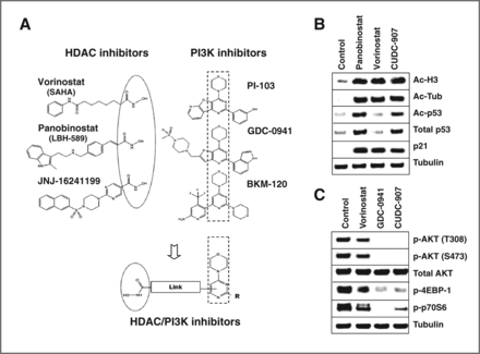
CUDC-907 design and its potency against PI3K and HDAC.Clin Cancer Res.2012 Aug 1;18(15):4104-13.
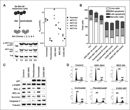
CUDC-907 evades drug resistance and induces apoptosis and G2–M phase cell-cycle arrest.Clin Cancer Res.2012 Aug 1;18(15):4104-13. |
CUDC-907 durably suppresses activation of AKT and modulates receptor tyrosine kinase, RAF-MEK-MAPK and SRC/STAT signaling.Clin Cancer Res.2012 Aug 1;18(15):4104-13. |
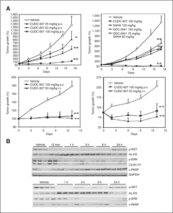
CUDC-907 suppresses tumor growth, inhibits HDAC activity, and blocks signaling of PI3K and MAPK pathways in xenograft models.Clin Cancer Res.2012 Aug 1;18(15):4104-13. |
Products are for research use only; We do not sell to patients
Copyright 2020 InvivoChem LLC | All Rights Reserved


 COA
COA