| Size | Price | Stock | Qty |
|---|---|---|---|
| 10mg |
|
||
| 25mg |
|
||
| 50mg |
|
||
| 100mg |
|
||
| 250mg |
|
||
| 500mg |
|
||
| 1g | |||
| Other Sizes |


Purity: ≥98%
| Targets |
The reported targets of FH535 and key parameters are as follows:
- Tcf/β-catenin signaling pathway: Inhibits Tcf/β-catenin-mediated transcriptional activity; half-maximal inhibitory concentration (IC50) = 1.5 μM (luciferase reporter gene assay in SW480 cells) [1] - Peroxisome proliferator-activated receptor gamma (PPARγ): Downregulates PPARγ-mediated transcriptional activity; IC50 = 2.3 μM (luciferase reporter gene assay in CV-1 cells transfected with PPARγ) [1] - Peroxisome proliferator-activated receptor delta (PPARδ): Downregulates PPARδ-mediated transcriptional activity; IC50 = 3.1 μM (luciferase reporter gene assay in CV-1 cells transfected with PPARδ) [1]; |
|---|---|
| ln Vitro |
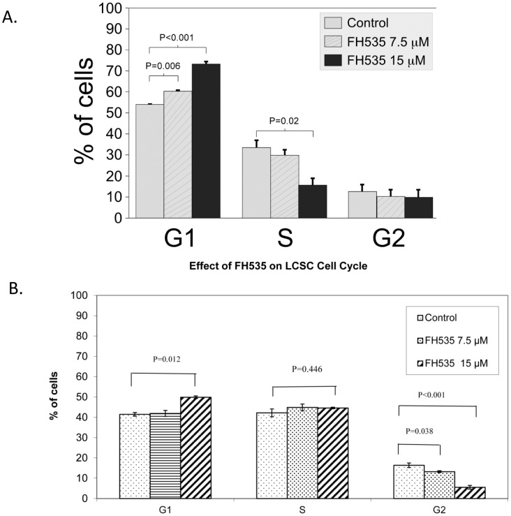
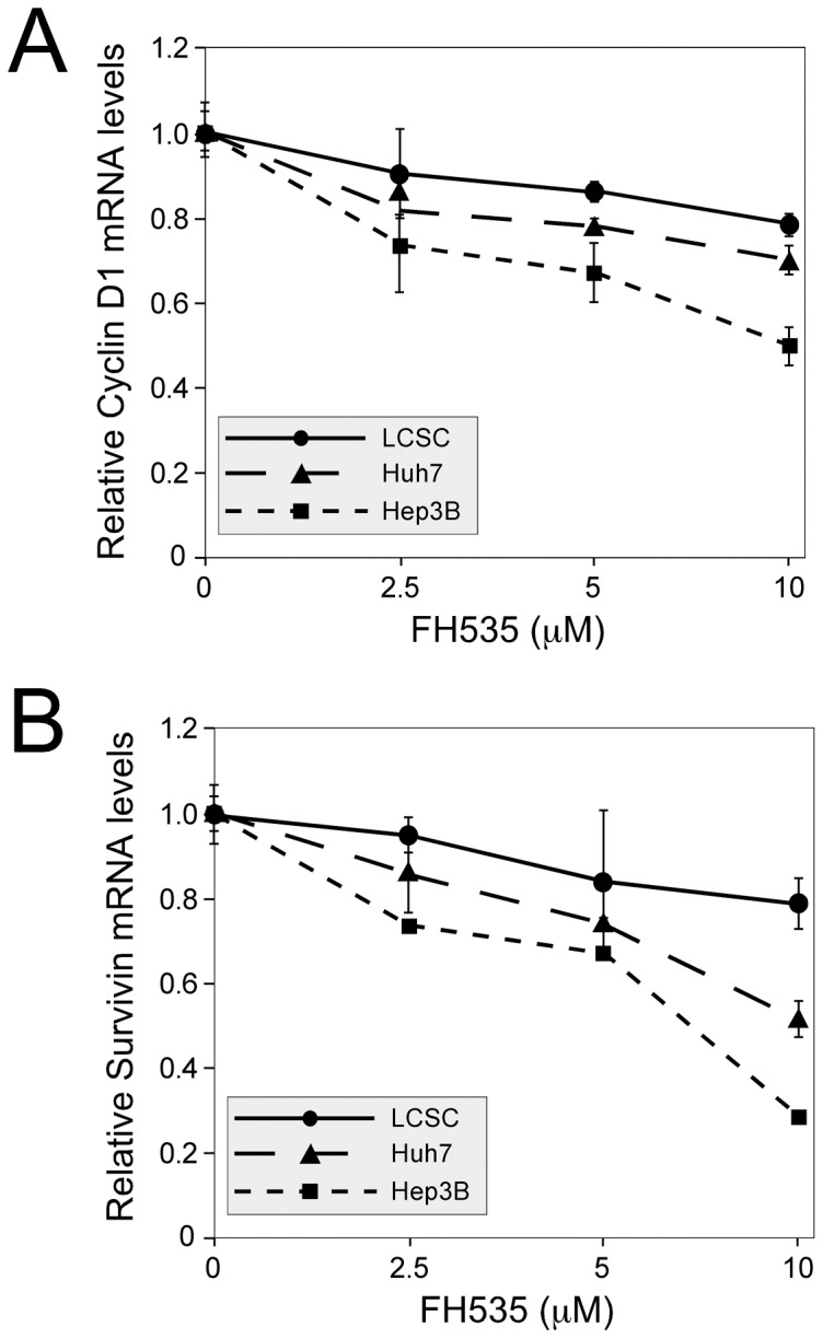
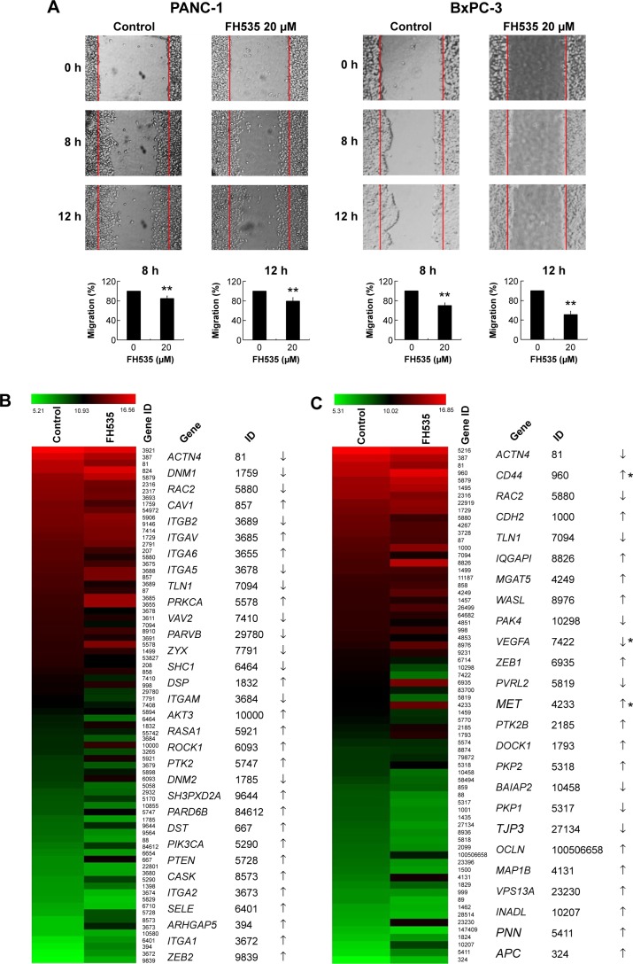
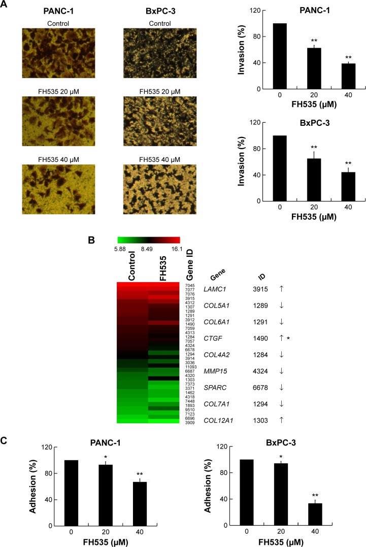
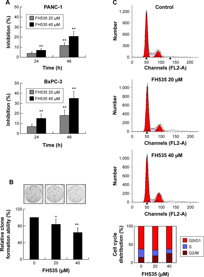
FH535 inhibits both PPAR and Wnt/β-catenin. In HCT116 cells, FH535 prevents PPARγ and PPARδ transactivation. FH535 (15 μM) action does not require cysteine residues in the PPAR ligand-binding domain, but it does depend on functional PPARδ. The coactivators GRIP1 and β-catenin are not able to bind to PPARδ and PPARγ when FH535 is present. Twelve cancer cell lines that exhibit the wnt/β-catenin pathway are toxically affected by FH535 [1]. FH535 (20 μM) suppresses the migration of pancreatic cancer cells and the β-catenin pathway in these cells. Furthermore, FH535 (20, 40 μM) suppresses the proliferation and invasion of pancreatic cancer cells [2]. In pancreatic cancer cells, FH535 suppresses genes linked to angiogenesis [3].
1. Inhibition of Tcf/β-catenin signaling and downregulation of PPAR activities: - In SW480 colon cancer cells (with constitutively active Tcf/β-catenin signaling), FH535 (0.5-5 μM) dose-dependently inhibited Tcf/β-catenin-mediated luciferase activity. At 2 μM, the inhibition rate reached 78% ± 3.2%, with an IC50 = 1.5 μM. This was accompanied by a 45% ± 4.1% reduction in c-Myc mRNA (a Tcf/β-catenin target gene) and a 38% ± 3.5% reduction in Cyclin D1 mRNA (RT-PCR) [1] - In CV-1 cells co-transfected with PPARγ/PPARδ and their respective luciferase reporter plasmids, FH535 (0.5-10 μM) inhibited rosiglitazone (PPARγ agonist)-induced PPARγ activity (IC50 = 2.3 μM) and GW501516 (PPARδ agonist)-induced PPARδ activity (IC50 = 3.1 μM). At 5 μM, PPARγ and PPARδ activities were reduced by 62% ± 3.8% and 55% ± 3.3%, respectively [1] 2. Antiproliferative and anti-metastatic effects on pancreatic cancer cells: - In PANC-1 and BxPC-3 pancreatic cancer cells, FH535 (1-10 μM) inhibited cell proliferation in a dose-dependent manner (CCK-8 assay). After 72 hours, the IC50 values were 2.8 μM (PANC-1) and 3.5 μM (BxPC-3) [2] - Transwell migration assay: FH535 (5 μM) reduced the migration rate of PANC-1 cells by 65% ± 4.2% and BxPC-3 cells by 58% ± 3.8% vs. control. Invasion assay (Matrigel-coated Transwell): 5 μM FH535 inhibited invasion by 72% ± 3.5% (PANC-1) and 68% ± 4.1% (BxPC-3) [2] - Western blot: FH535 (5 μM) increased cleaved caspase-3 levels by 2.5-fold (PANC-1) and 2.2-fold (BxPC-3), upregulated Bax by 1.8-fold, and downregulated Bcl-2 by 45% ± 3.2%, indicating induction of apoptosis [2] 3. Suppression of pancreatic cancer cell growth and angiogenesis-related genes: - In PANC-1 cells, FH535 (2.5-10 μM) reduced β-catenin nuclear translocation (immunofluorescence): nuclear β-catenin positive cells decreased from 68% ± 3.5% (control) to 22% ± 2.8% (10 μM) [3] - RT-PCR: FH535 (10 μM) downregulated angiogenesis-related genes: VEGF by 58% ± 4.1%, bFGF by 52% ± 3.8%, and MMP-9 by 65% ± 3.5% in PANC-1 cells [3]. |
| ln Vivo |
On mouse pancreatic cancer xenografts, FH535 (25 mg/kg, ip) exhibits anticancer activity. Additionally, FH535 prevents angiogenesis in xenografts of pancreatic cancer [2].
1. Inhibition of pancreatic cancer xenograft growth and angiogenesis in nude mice: - Female BALB/c nude mice (4-6 weeks old, 18-20 g) were subcutaneously injected with PANC-1 cells (5×10⁶ cells/mouse) into the right flank to establish xenograft models. When tumors reached ~100 mm³, mice were randomly divided into 2 groups (n=6): - Control group: Intraperitoneal injection of vehicle (DMSO + normal saline, DMSO ≤ 0.1%) once daily, 5 days/week for 4 weeks [3] - FH535 group: Intraperitoneal injection of 10 mg/kg/day FH535 (dissolved in vehicle) once daily, 5 days/week for 4 weeks [3] - Endpoint results: - Tumor volume: Reduced from 1250 ± 120 mm³ (control) to 480 ± 85 mm³ (FH535 group), with an inhibition rate of 61.6% [3] - Tumor weight: Decreased from 1.85 ± 0.15 g (control) to 0.72 ± 0.08 g (FH535 group) [3] - Angiogenesis: Immunohistochemistry (IHC) for CD31 (endothelial cell marker) showed that the number of CD31-positive blood vessels decreased from 32 ± 3.2 vessels/field (control) to 12 ± 2.1 vessels/field (FH535 group) [3] - Tumor tissue molecular changes: Western blot showed β-catenin protein levels reduced by 58% ± 4.2%, VEGF by 62% ± 3.8%, and PPARγ by 45% ± 3.5% in FH535-treated tumors [3] |
| Enzyme Assay |
1. Tcf/β-catenin transcriptional activity assay (luciferase reporter gene assay, Literature [1]):
- SW480 cells (which stably express a Tcf/β-catenin-responsive luciferase reporter plasmid, pTOPflash) were seeded into 24-well plates at a density of 5×10⁴ cells/well and cultured in DMEM with 10% FBS for 24 hours [1] - The medium was replaced with fresh medium containing FH535 (0.1, 0.5, 1, 2, 5 μM) or vehicle (DMSO, final concentration ≤ 0.1%). Each concentration was tested in triplicate [1] - After 24 hours of incubation at 37°C (5% CO₂), cells were lysed with passive lysis buffer. Luciferase activity was detected using a dual-luciferase reporter assay system (Renilla luciferase plasmid pRL-TK was co-transfected as internal control) [1] - Relative luciferase activity (firefly/Renilla) was calculated, and the IC50 for Tcf/β-catenin inhibition was determined to be 1.5 μM [1] 2. PPARγ/PPARδ transcriptional activity assay (luciferase reporter gene assay, Literature [1]): - CV-1 cells were seeded into 24-well plates (5×10⁴ cells/well) and cultured for 24 hours. Cells were co-transfected with: (1) PPARγ/PPARδ expression plasmid, (2) PPAR-responsive luciferase reporter plasmid (pPPRE-luc), and (3) pRL-TK [1] - After 24 hours of transfection, cells were treated with FH535 (0.5-10 μM) plus PPAR agonist (rosiglitazone for PPARγ, GW501516 for PPARδ, both 100 nM) for another 24 hours [1] - Luciferase activity was detected, and IC50 values for PPARγ (2.3 μM) and PPARδ (3.1 μM) inhibition were calculated [1]. |
| Cell Assay |
1. Pancreatic cancer cell proliferation assay (CCK-8 method, Literature [2]):
- PANC-1 and BxPC-3 cells were seeded into 96-well plates at a density of 3×10³ cells/well and cultured in RPMI 1640 with 10% FBS for 24 hours [2] - The medium was replaced with fresh medium containing FH535 (1, 2.5, 5, 7.5, 10 μM) or vehicle. Each concentration had 5 replicate wells [2] - After incubation for 24, 48, and 72 hours, 10 μL of CCK-8 reagent was added to each well and incubated for 2 hours. Absorbance at 450 nm was measured using a microplate reader, and cell viability was calculated to determine IC50 values [2] 2. Cell migration and invasion assay (Transwell, Literature [2,3]): - Migration assay: PANC-1/BxPC-3 cells (5×10⁴ cells/well) in serum-free medium were added to the upper chamber of Transwell inserts; medium containing 10% FBS was added to the lower chamber. FH535 (5 μM) was added to both chambers [2] - Invasion assay: Inserts were pre-coated with Matrigel (1:8 dilution) and dried overnight. Cells (1×10⁵ cells/well) and FH535 (5 μM) were added as in the migration assay [2] - After 24 hours (migration) or 48 hours (invasion), non-migrated/non-invaded cells on the upper surface were wiped off. Cells on the lower surface were fixed with 4% paraformaldehyde, stained with crystal violet, and counted under a microscope (5 fields/insert) [2,3] 3. Apoptosis detection (Annexin V-FITC/PI staining, Literature [2]): - PANC-1 cells were seeded into 6-well plates (2×10⁵ cells/well) and treated with FH535 (5 μM) for 48 hours [2] - Cells were trypsinized, washed with cold PBS, and resuspended in binding buffer. 5 μL of Annexin V-FITC and 5 μL of PI were added, and the mixture was incubated in the dark for 15 minutes [2] - Apoptotic rate was analyzed via flow cytometry, with early apoptosis (Annexin V⁺/PI⁻) and late apoptosis (Annexin V⁺/PI⁺) summed [2] 4. Nuclear β-catenin detection (immunofluorescence, Literature [3]): - PANC-1 cells were seeded on coverslips (1×10⁴ cells/coverslip) and treated with FH535 (10 μM) for 24 hours [3] - Cells were fixed with 4% paraformaldehyde, permeabilized with 0.1% Triton X-100, blocked with 5% BSA, and incubated with anti-β-catenin primary antibody (4°C, overnight) [3] - FITC-conjugated secondary antibody was added, and nuclei were stained with DAPI. The percentage of nuclear β-catenin-positive cells was counted under a fluorescence microscope [3]. |
| Animal Protocol |
Prepared in 100 μL DMSO/DMEM (1:1); 25 mg/kg; i.p.
Female BALB/c athymic nude mice (Four weeks old) 1. Pancreatic cancer xenograft model in nude mice: - Animals: Female BALB/c nude mice (4-6 weeks old, 18-20 g) were maintained under specific pathogen-free (SPF) conditions (22±2°C, 12-hour light/dark cycle, free access to sterile food and water) [3] - Tumor establishment: PANC-1 cells were harvested in logarithmic growth phase, resuspended in PBS (5×10⁶ cells/100 μL), and subcutaneously injected into the right flank of each mouse [3] - Grouping and administration: When tumors grew to ~100 mm³ (7 days after injection), mice were randomly divided into 2 groups (n=6): - Control group: Intraperitoneal injection of vehicle (DMSO diluted with normal saline to final DMSO concentration ≤ 0.1%), 0.2 mL/mouse, once daily, 5 days/week for 4 weeks [3] - FH535 group: Intraperitoneal injection of FH535 (10 mg/kg/day, dissolved in vehicle), 0.2 mL/mouse, once daily, 5 days/week for 4 weeks [3] - Sample collection and detection: - Tumor volume was measured every 3 days using a caliper (volume = length × width² / 2) [3] - At the end of treatment, mice were anesthetized with pentobarbital sodium (40 mg/kg, intraperitoneal injection). Tumors were excised, weighed, and divided into two parts: one fixed in 4% formalin for IHC (CD31 staining); the other stored at -80°C for Western blot analysis [3] - Serum was collected via orbital vein to detect liver function (ALT, AST) and renal function (creatinine), which were within normal ranges [3]; |
| Toxicity/Toxicokinetics |
1. In vitro cytotoxicity (references [2,3]):
- In normal human pancreatic ductal epithelial cells (HPDE6-C7), concentrations up to 10 μM of FH535 had no significant effect on cell viability (CCK-8 assay: viability >85% vs. solvent control group), indicating low toxicity to normal cells [2] - In pancreatic cancer cells (PANC-1, BxPC-3), FH535 showed selective cytotoxicity with IC50 values of 2.8-3.5 μM, which was 3-4 times higher than the viability threshold of normal cells [2,3] 2. In vivo toxicity: - Nude mice treated with 10 mg/kg FH535 daily for 4 consecutive weeks: - Body weight: No significant difference compared to the control group (weight gain: 8.5% ± 1.2% vs. 9.2% ± 1.5%) [3] - Liver and kidney function: Serum ALT (32-45 U/L), AST (80-95 U/L), and creatinine (45-60 μmol/L) were all within the normal range for nude mice [3]; Histopathology: No obvious lesions were observed in the liver, kidney, spleen, or lung tissue of FH535 treated mice [3]; |
| References |
|
| Additional Infomation |
2,5-Dichloro-N-(2-methyl-4-nitrophenyl)benzenesulfonamide is a sulfonamide.
1. Background and Mechanism of Action: - FH535 is a synthetic small molecule inhibitor initially identified as an inhibitor of the Tcf/β-catenin signaling pathway—a key oncogenic pathway activated in various cancers, such as colon and pancreatic cancer [1,3] - Its dual mechanism includes: (1) inhibiting the transcriptional activity of Tcf/β-catenin by blocking the nuclear translocation of β-catenin and its binding to Tcf protein, thereby downregulating oncogenes such as c-Myc and Cyclin D1 [1,3]; (2) downregulating the activity of PPARγ and PPARδ, two proteins overexpressed in some cancers that promote cell survival and angiogenesis [1] 2. Therapeutic Potential: - Preclinical studies have confirmed that FH535 It has anticancer effects against pancreatic cancer (the most studied indication), including inhibiting cell proliferation, migration, invasion and xenograft growth, as well as inhibiting tumor angiogenesis [2,3] - Based on in vitro data from SW480 cells, it may also be effective against other β-catenin-driven cancers (e.g., colon cancer) [1] 3. Research use: - Research use: as a tool compound to study the role of Tcf/β-catenin and PPAR signaling pathways in cancer development and to validate these pathways as therapeutic targets [1,3]; |
| Molecular Formula |
C13H10CL2N2O4S
|
|
|---|---|---|
| Molecular Weight |
361.20
|
|
| Exact Mass |
359.973
|
|
| Elemental Analysis |
C, 43.23; H, 2.79; Cl, 19.63; N, 7.76; O, 17.72; S, 8.88
|
|
| CAS # |
108409-83-2
|
|
| Related CAS # |
|
|
| PubChem CID |
3463933
|
|
| Appearance |
Light yellow to yellow solid powder
|
|
| Density |
1.6±0.1 g/cm3
|
|
| Boiling Point |
526.3±60.0 °C at 760 mmHg
|
|
| Melting Point |
141-142℃
|
|
| Flash Point |
272.1±32.9 °C
|
|
| Vapour Pressure |
0.0±1.4 mmHg at 25°C
|
|
| Index of Refraction |
1.651
|
|
| LogP |
5.12
|
|
| Hydrogen Bond Donor Count |
1
|
|
| Hydrogen Bond Acceptor Count |
5
|
|
| Rotatable Bond Count |
3
|
|
| Heavy Atom Count |
22
|
|
| Complexity |
505
|
|
| Defined Atom Stereocenter Count |
0
|
|
| SMILES |
O=S(C1=CC(Cl)=CC=C1Cl)(NC2=CC=C([N+]([O-])=O)C=C2C)=O
|
|
| InChi Key |
AXNUEXXEQGQWPA-UHFFFAOYSA-N
|
|
| InChi Code |
InChI=1S/C13H10Cl2N2O4S/c1-8-6-10(17(18)19)3-5-12(8)16-22(20,21)13-7-9(14)2-4-11(13)15/h2-7,16H,1H3
|
|
| Chemical Name |
2,5-dichloro-N-(2-methyl-4-nitrophenyl)-benzenesulfonamide
|
|
| Synonyms |
FH-535; FH535; FH 535;
|
|
| HS Tariff Code |
2934.99.9001
|
|
| Storage |
Powder -20°C 3 years 4°C 2 years In solvent -80°C 6 months -20°C 1 month |
|
| Shipping Condition |
Room temperature (This product is stable at ambient temperature for a few days during ordinary shipping and time spent in Customs)
|
| Solubility (In Vitro) |
DMSO: 72 mg/mL (199.3 mM)
Water: 1 mg/mL (Insoluble) Ethanol: 1 mg/mL |
|---|---|
| Solubility (In Vivo) |
Solubility in Formulation 1: ≥ 2.5 mg/mL (6.92 mM) (saturation unknown) in 10% DMSO + 40% PEG300 + 5% Tween80 + 45% Saline (add these co-solvents sequentially from left to right, and one by one), clear solution.
For example, if 1 mL of working solution is to be prepared, you can add 100 μL of 25.0 mg/mL clear DMSO stock solution to 400 μL PEG300 and mix evenly; then add 50 μL Tween-80 to the above solution and mix evenly; then add 450 μL normal saline to adjust the volume to 1 mL. Preparation of saline: Dissolve 0.9 g of sodium chloride in 100 mL ddH₂ O to obtain a clear solution. Solubility in Formulation 2: 2.5 mg/mL (6.92 mM) in 10% DMSO + 90% (20% SBE-β-CD in Saline) (add these co-solvents sequentially from left to right, and one by one), suspension solution; with ultrasonication. For example, if 1 mL of working solution is to be prepared, you can add 100 μL of 25.0 mg/mL clear DMSO stock solution to 900 μL of 20% SBE-β-CD physiological saline solution and mix evenly. Preparation of 20% SBE-β-CD in Saline (4°C,1 week): Dissolve 2 g SBE-β-CD in 10 mL saline to obtain a clear solution. (Please use freshly prepared in vivo formulations for optimal results.) |
| Preparing Stock Solutions | 1 mg | 5 mg | 10 mg | |
| 1 mM | 2.7685 mL | 13.8427 mL | 27.6855 mL | |
| 5 mM | 0.5537 mL | 2.7685 mL | 5.5371 mL | |
| 10 mM | 0.2769 mL | 1.3843 mL | 2.7685 mL |
*Note: Please select an appropriate solvent for the preparation of stock solution based on your experiment needs. For most products, DMSO can be used for preparing stock solutions (e.g. 5 mM, 10 mM, or 20 mM concentration); some products with high aqueous solubility may be dissolved in water directly. Solubility information is available at the above Solubility Data section. Once the stock solution is prepared, aliquot it to routine usage volumes and store at -20°C or -80°C. Avoid repeated freeze and thaw cycles.
Calculation results
Working concentration: mg/mL;
Method for preparing DMSO stock solution: mg drug pre-dissolved in μL DMSO (stock solution concentration mg/mL). Please contact us first if the concentration exceeds the DMSO solubility of the batch of drug.
Method for preparing in vivo formulation::Take μL DMSO stock solution, next add μL PEG300, mix and clarify, next addμL Tween 80, mix and clarify, next add μL ddH2O,mix and clarify.
(1) Please be sure that the solution is clear before the addition of next solvent. Dissolution methods like vortex, ultrasound or warming and heat may be used to aid dissolving.
(2) Be sure to add the solvent(s) in order.
|
|---|
|
|
FH535 inhibited pancreatic cancer cell migration.Onco Targets Ther.2015 Jul 6;8:1651-70. |
|---|
FH535 inhibited pancreatic cancer cell invasion.Onco Targets Ther.2015 Jul 6;8:1651-70. |
Inhibitory effect of FH535 on pancreatic cancer cell growth.Onco Targets Ther.2015 Jul 6;8:1651-70. |
Products are for research use only; We do not sell to patients
Copyright 2020 InvivoChem LLC | All Rights Reserved


 COA
COA