| Size | Price | Stock | Qty |
|---|---|---|---|
| 10mg |
|
||
| 25mg |
|
||
| 50mg |
|
||
| 100mg |
|
||
| 250mg |
|
||
| 500mg | |||
| Other Sizes |


Purity: ≥98%
| Targets |
dynamin (IC50 = 15 μM); HSV-1/2
Dynasore specifically targets dynamin GTPase (isoforms dynamin 1, 2, 3) with IC50 values of 15 μM (dynamin 1), 18 μM (dynamin 2), and 20 μM (dynamin 3) for inhibiting GTPase activity [1] Dynasore binds to the GTP-binding pocket of dynamin, blocking GTP hydrolysis without inhibiting other GTPases (e.g., Ras, Rho) at concentrations up to 100 μM [1] |
|---|---|
| ln Vitro |
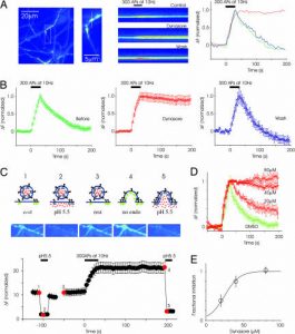
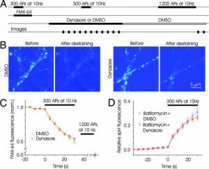
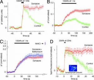
Dynasore inhibits dynamin1, dynamin2, and mitochondrial dynamin Drp1 GTPase activity, but not that of other small GTPases. When added, dynasore quickly prevents the creation of coated vesicles, thereby acting as a powerful inhibitor of the known dynamin-dependent endocytic pathway. This effect is seen within seconds. G-shaped, semi-formed pits and O-shaped, completely formed pits, which are trapped during pinch-off, are the two types of coated pit intermediates that accumulate during dynasore processing [1]. Human fetal neurons, astrocytes, primary reproductive tract cells, and epithelial and neural cells are all susceptible to HSV-1 and HSV-2 infection. Dynasore prevents these infections. When given eight hours after virus entrance, dynasore prevents newly generated viral proteins from leaving the nucleus and increases the amount of viral capsids that reach the nuclear pore [2]. Dynasore inhibits the rise in left ventricular end-diastolic pressure brought on either ischemia or reperfusion. Moreover, dynamicsore decreases infarct size and cardiac troponin I efflux upon reperfusion. Dynasore increased cardiomyocyte viability and survival in adult mouse cardiomyocytes grown under oxidative stress [3].
Dynasore (20 μM) inhibited clathrin-mediated endocytosis in HeLa and COS-7 cells, reducing transferrin internalization by 85% [1] - Dynasore (30 μM) disrupted trafficking of herpes simplex virus (HSV-1) envelope proteins (gB, gD) in Vero cells, reducing viral particle release by 70% [2] - Dynasore (10-25 μM) protected mitochondrial morphology and function in neonatal rat cardiomyocytes under oxidative stress, reducing mitochondrial ROS production by 60% and preserving ΔΨm (mitochondrial membrane potential) [3] - Dynasore (15 μM) inhibited neuronal apoptosis in rat spinal cord-derived neurons after oxygen-glucose deprivation (OGD), reducing caspase-3 activation by 55% and TUNEL-positive cells by 48% [4] - Dynasore (20 μM) suppressed astrocytic proliferation in primary rat astrocytes, reducing BrdU incorporation by 62% [4] - Dynasore (25 μM) blocked migrasome release in HT1080 fibrosarcoma cells, reducing migrasome number by 75% without affecting cell migration speed [5] - Dynasore (10-30 μM) showed minimal cytotoxicity in normal cells (HeLa, cardiomyocytes, neurons) with cell viability > 80%, but inhibited HSV-1-infected Vero cell viability (IC50 = 45 μM) [1][2][3][4] - Western blot analysis showed Dynasore (15-25 μM) increased Bcl-2/Bax ratio (2.3-fold) in OGD-treated neurons and reduced phosphorylated ERK1/2 (40%) in proliferating astrocytes [4] |
| ln Vivo |
After spinal cord injury (SCI) in rats, dynasore dramatically reduced motor dysfunction at 3, 7, and 10 days. By preventing the activation of pathways leading to mitochondrial apoptosis and astrocyte proliferation in rat neurons following spinal cord damage, Dynasore greatly improves motor function [4].
In Langendorff-perfused mouse heart models, Dynasore (10 μM, perfused for 30 minutes) improved cardiac lusitropy (diastolic function), increasing left ventricular end-diastolic volume by 25% and reducing end-diastolic pressure by 30% [3] - In rat spinal cord injury (SCI) models, intraperitoneal administration of Dynasore (5 mg/kg, q.d. for 7 days post-SCI) improved motor function recovery, increasing BBB (Basso-Beattie-Bresnahan) scores from 4.2 to 8.6 at 28 days post-injury [4] - SCI rats treated with Dynasore showed reduced neuronal apoptosis (35% fewer TUNEL-positive cells) and astrocytic scar formation (40% reduction in GFAP-positive area) at injury sites [4] - Perfused mouse hearts treated with Dynasore exhibited preserved mitochondrial morphology (65% reduction in fragmented mitochondria) and increased ATP production by 32% [3] |
| Enzyme Assay |
ATP Measurement[3]
A luminescence assa was used to quantify cardiomyocyte and Hela cell ATP content. Briefly, after Dynasore treatment and H2O2 exposure, cardiomyocytes were lysed and ATP content was measured in the cell lysates. Meanwhile, in a separate set of wells following same experimental protocol, surviving cardiomyocytes were counted using a TBE assay. Cellular ATP per single live cardiomyocyte was then calculated for each treatment condition. Similar procedures were applied to cultured non-stressed Hela cells treated with control or Dynasore. Dynamin GTPase activity assay: Recombinant dynamin (isoforms 1/2/3) was incubated with [γ-32P]GTP as substrate. Serial concentrations of Dynasore (5 μM to 100 μM) were added, and the mixture was incubated at 37°C for 60 minutes. GTP hydrolysis was quantified by measuring released 32Pi, and IC50 values were calculated from dose-response curves of inhibition [1] - Dynamin-GTP binding assay: Fluorescently labeled GTP was incubated with recombinant dynamin 2. Serial concentrations of Dynasore (10 μM to 80 μM) were added, and fluorescence polarization was measured at 25°C. Binding affinity (Kd) was derived from competition curves, showing Dynasore displaced GTP with an IC50 of 16 μM [1] |
| Cell Assay |
Isolation and Culture of Adult Mouse Cardiomyocytes[3]
Mouse ventricular myocytes were isolated from male adult C6/Black mouse (8∼12 weeks; Charles River) after dissociation with collagenase II (2 mg/ml) using a previously described method. After dissociation, cardiomyocytes were plated on laminin-precoated 35 mm2 culture dishes at a density of ∼1,500/mm2 and maintained in a humidified atmosphere of 5% CO2 at 37°C. After 1 hour of plating, cardiomyocytes were replenished with fresh medium (serum supplemented or depleted) and subjected to 2 hours of drug treatment (Dynasore or vehicle) followed by oxidative stress (30 µM H2O2 for 35 min). For ATP supplement experiments, the cells were treated with 3 mM ATP for 30 min before exposure to H2O2. Live-cell Mitochondria Imaging with Spinning Disc Confocal Microscopy[3] HeLa cells were maintained in DMEM supplemented with 10% FBS and 100 µg/ml Normocin. Cells were maintained in a humidified atmosphere of 5% CO2 at 37°C. Cells were seeded at a density of 7 × 10⁴ cells/cm2 and allowed to adhere overnight. Cells were then transduced with Organelle Lights™ Mito-RFP BacMam 1.0. Twenty-four hours after transduction, cells were pretreated with either control or 1 µM Dynasore for 1 hour before being exposed to normal conditions or 200 µM H2O2 for 15 minutes. Before and after exposure to H2O2, cells were imaged using a Nikon Ti inverted microscope, Yokogowa CSU-X1 spinning disk confocal unit with 568-nm DPSS laser source, and a high resolution Cool SNAP HQ2 camera. Images were acquired at 400 ms exposure per frame and automatically processed using a bas relief filter to highlight edges. Clathrin-mediated endocytosis assay: HeLa cells were incubated with Alexa Fluor 488-labeled transferrin and Dynasore (10-40 μM) for 30 minutes. Internalized transferrin was quantified by flow cytometry, and inhibition rates were calculated relative to vehicle controls [1] - HSV-1 trafficking assay: Vero cells infected with HSV-1 (MOI = 1) were treated with Dynasore (10-50 μM) for 12 hours. Viral envelope proteins (gB, gD) were detected by immunofluorescence, and intracellular trafficking defects were scored by confocal microscopy [2] - Mitochondrial function assay: Neonatal rat cardiomyocytes were exposed to H2O2 (200 μM) plus Dynasore (10-25 μM) for 24 hours. Mitochondrial ROS was measured by DCFH-DA staining, and ΔΨm was detected by JC-1 staining [3] - Neuronal apoptosis assay: Rat spinal cord neurons were subjected to OGD (2 hours) and treated with Dynasore (10-20 μM) for 24 hours. Apoptosis was assessed by TUNEL staining and caspase-3 activity assay [4] - Astrocyte proliferation assay: Primary rat astrocytes were treated with Dynasore (10-30 μM) for 48 hours. Proliferation was measured by BrdU incorporation and cell count [4] - Migrasome formation assay: HT1080 cells were plated on fibronectin-coated dishes and treated with Dynasore (15-30 μM) for 16 hours. Migrasomes were visualized by GFP-tagged tetraspanin 4 (TSPAN4) and counted by fluorescence microscopy [5] |
| Animal Protocol |
In the Dynasore groups, the rats are given dynasore immediately at
a dose of 1, 10, or 30 mg/kg through intraperitoneal injection after
SCI, while the rats in the sham and SCI groups receive DMSO (same volume
as dynasore groups) through intraperitoneal injection
Rats Langendorff-Perfused Heart[3] Male C6/Black mice (8∼12 weeks) were anesthetized with isoflurane (flow 3%) and 100% O2 in an anesthesia chamber and anti-coagulated with heparin (50 IU, i.p.). After cervical dislocation, hearts were rapidly excised, mounted on a Langendorff apparatus and perfused retrogradely at a constant rate of 2.6 ml/min with oxygenated Krebs-Henseleit buffer containing (mmol/L): NaCl 118, NaHCO3 24, CaCL2.2H2O 2.5, KCL 4.7, KH2PO4 1.2, MgSO4-7H2O 1.2, Glucose 11, EDTA 0.5, adjusted to a pH of 7.4. The apparatus was water-jacketed for temperature control to maintain a core temperature of the heart at 37°C. The buffer passed through a membranous “lung” made of SilasticTM Medical Grade Tubing, which was gassed continuously with 95% O2-5% CO2. Fine platinum electrodes were placed on the right atrium and apex of the left ventricle to record the electrocardiogram and heart rate throughout the experiment. A Millar MIKRO-TIP catheter transducer was inserted into the left ventricle from the left atrium to measure left ventricular pressure. Left ventricular end diastolic pressure (LVEDP), left ventricular end systolic pressure (LVESP) and heart rate were monitored and recorded continuously using PowerLab system. Left ventricular developed pressure (LVDP) was calculated by subtracting LVEDP from LVESP. Hearts were paced at 360 bpm with bipolar electrodes attached to the right atrium, using stimuli delivered from a stimulator. After the initial 15 min stabilization, hearts were excluded from further study if they exhibited one or more of the following exclusion criteria: LVEDP higher than 20 mmHg; LVDP less than 50 mmHg; intrinsic heart rate less than 280 bpm or irregular; or aortic regurgitation. The volume of the perfusate was reduced to 200 ml and allowed to recirculate. The hearts were then randomized to one of the following two treatment groups: Dynasore group (n = 8, added into the recirculating perfusate in stepwise fashion to reach a final concentration of 1 µM within 120 min of recirculation) or DMSO control group (n = 8, added in a similar manner of Dynasore). Hearts were then subjected to 30 min of global ischemia followed by 1 hour of reperfusion. Pacing was initiated after stabilization except during ischemia and was reinitiated 2 min after reperfusion. Langendorff-perfused mouse heart model: Male C57BL/6 mice (8-10 weeks old) were euthanized, and hearts were excised and perfused with Krebs-Henseleit buffer. After stabilization (20 minutes), Dynasore (10 μM) was added to the perfusate for 30 minutes. Cardiac function (left ventricular pressure, volume) was measured by a pressure-volume catheter [3] - Rat spinal cord injury (SCI) model: Female Sprague-Dawley rats (200-250 g) were subjected to contusive SCI at T9-T10. Immediately post-injury, rats were randomized into groups (n=10/group) and treated with: (1) vehicle (DMSO + saline) i.p., (2) Dynasore (5 mg/kg) i.p. once daily for 7 days. Motor function was assessed by BBB scores weekly for 28 days, and spinal cord tissues were collected for histology [4] |
| Toxicity/Toxicokinetics |
Dynasore exhibits low in vitro cytotoxicity in normal cells: HeLa cells (IC50 > 100 μM), neonatal cardiomyocytes (IC50 > 80 μM), and rat spinal cord neurons (IC50 > 75 μM) [1][3][4]
- In a repeated-dose toxicity study in rats with spinal cord injury (5 mg/kg, intraperitoneal injection, for 7 consecutive days), Dynasore did not cause significant weight loss, and histological examination showed no abnormalities in the liver, kidneys, or heart [4] - Dynasore (30 μM) does not inhibit human cytochrome P450 enzymes (CYP1A2, CYP2C9, CYP3A4) in vitro [1] |
| References | |
| Additional Infomation |
Dynasore is a carbazone formed by the condensation of the hydrazone moiety of 3,4-dihydroxybenzaldehyde hydrazone with the carboxyl group of 3-hydroxy-2-naphthoic acid. It is a cell-permeable, reversible, non-competitive GTPase inhibitor that inhibits the activity of dynein 1 and 2, as well as Drp1 (mitochondria), while having no significant effect on two other small GTPases, MxA and Cdc42. It is an EC 3.6.5.5 (dynein GTPase) inhibitor. It belongs to the catechol, naphthol, acylhydrazine, and hydrazone classes. Its function is related to 3-hydroxy-2-naphthoic acid ester. Dynein is essential for clathrin-dependent vesicle formation. Dynein is required in the late stage of membrane budding during the transition from fully formed indentation to contracting vesicle. Dynein may also play other roles in the early stages of vesicle formation. We screened about 16,000 small molecules and identified a compound called dynasore 1, which interfered with the GTPase activity of dynein 1, dynein 2 and mitochondrial dynein Drp1 in vitro, but did not interfere with the activity of other small GTPases. dynasore is a potent inhibitor of the endocytosis pathway known to be dependent on dynein, and its mechanism of action is to rapidly block the formation of coated vesicles within seconds of dynasore addition. During dynasore treatment, two types of coated indentation intermediates accumulate: U-shaped semi-formed indentations and O-shaped fully formed indentations (captured during contraction). Thus, dynein plays a role in both steps of clathrin coating formation. GTP hydrolysis may be necessary in both steps. [1]
Dynasore is a small molecule dynein GTPase activity inhibitor that inhibits the invasion of a variety of viruses, including herpes simplex virus (HSV), but its effect on other steps of the viral life cycle is not yet clear. This study aimed to verify whether dynasore is involved in viral protein transport, thereby exhibiting pleiotropic inhibitory effects against HSV infection. Dynasore inhibited HSV-1 and HSV-2 infection of human epithelial cells and neurons (including primary germ cells, human fetal neurons, and astrocytes). Similar results were obtained when cells were transfected with plasmids expressing dominant-negative dynasore. Kinetic studies showed that adding dynasore upon viral entry into cells reduced the number of viral capsids reaching the nuclear pores; adding dynasore 8 hours after viral entry blocked the transport of newly synthesized viral proteins from the nucleus to the cytoplasm. Proximity connectivity analysis indicated that dynasore treatment prevented the co-localization of VP5 and dynasore. This resulted in a reduced number of viral capsids isolated from the sucrose gradient. Compared to the control group, fewer capsids were observed in the dynasore-treated cells under electron microscopy. Furthermore, the number of infectious progeny viruses released into the culture supernatant and intercellular transmission were also reduced. In summary, these findings suggest that targeting the dynein-HSV interaction may provide a new strategy for the treatment and prevention of HSV. Importance: Herpes simplex virus (HSV) infection remains a global health problem, associated with significant morbidity, particularly in newborns and immunocompromised individuals, highlighting the need to develop new treatment and prevention approaches. Current research indicates that dynein plays a role in multiple steps of the viral life cycle and provides a new target for antiviral therapy. The dynein small molecule inhibitor Dynasore is pleiotropic against HSV-1 and HSV-2 infections and inhibits viral entry into cells, viral protein transport, and capsid formation. [2] Background: Heart failure due to diastolic dysfunction imposes a huge economic, morbid, and mortality burden in the United States. Currently, there are limited drugs available to improve diastolic dysfunction. It has recently been found that dynein-associated protein 1 (Drp1) mediates mitochondrial division during ischemia/reperfusion (I/R) injury, and that inhibition of Drp1 reduces myocardial infarction area. We hypothesized that the small-molecule, non-competitive dynein GTPase inhibitor Dynasore may have beneficial effects on cardiac physiology during ischemia/reperfusion (I/R) injury. Methods and Results: In Langendorff-perfused mouse hearts, pretreatment with 1 µM Dynasore prevented I/R-induced elevation of left ventricular end-diastolic pressure (LVEDP), indicating a significant and specific diastolic enhancement effect. Dynasore also reduced cardiac troponin I release and infarct size during reperfusion. In cultured adult mouse cardiomyocytes subjected to oxidative stress, Dynasore increased cardiomyocyte survival and viability (as determined by trypan blue exclusion) and reduced intracellular adenosine triphosphate (ATP) consumption. Furthermore, Dynasore pretreatment protected mitochondria from oxidative stress-induced breakage in cultured cells. Conclusion: Dynasore protects myocardial diastolic function and reduces cell damage through a mechanism that maintains mitochondrial morphology and intracellular ATP levels in damaged cells. The protective effects of drugs such as Dynasore on mitochondria can produce clinical benefits by positively influencing energy metabolism in diastolic dysfunction. [3] Spinal cord injury (SCI) is a common and serious neurological injury for which there is currently no effective treatment. Our previous experimental results showed that dynein-associated protein 1 (Drp1) mediates mitochondrial division during SCI, and inhibition of Drp1 has a significant protective effect in a rat model of SCI. Dynasore inhibits the activity of GTPases on the plasma membrane (dynein 1, 2) and mitochondrial membrane (Drp1). This study aimed to investigate the beneficial effects of Dynasore on a rat model of SCI and its potential mechanism. Sprague-Dawley rats were randomly divided into a sham-operated group, a spinal cord injury (SCI) group, and 1 mg, 10 mg, and 30 mg Dynasore groups. The rat SCI model was established using the Allen model. Dynasore was administered immediately by intraperitoneal injection. Motor function test results showed that Dynasore significantly improved motor dysfunction in rats at 3, 7, and 10 days after SCI (P < 0.05). Western blot results showed that Dynasore significantly reduced the expression of Drp1, dynein 1, and dynein 2, and decreased the expression of Bax, cytochrome C, and active Caspase-3 3 days after spinal cord injury (SCI), but increased the expression of Bcl-2 (P < 0.05). Notably, Dynasore inhibited the upregulation of proliferating cell nuclear antigen (PCNA) and glial fibrillary acidic protein (GAFP) expression 3 days after SCI (P < 0.05). Immunofluorescence double labeling results showed that the number of apoptotic neurons and proliferating astrocytes was significantly reduced in the Dynasore group compared with the SCI group (P < 0.05). Furthermore, Nissl staining histological assessment showed that the number of surviving neurons was significantly increased in the Dynasore group compared with the SCI group (P < 0.05). This neuroprotective effect was dose-dependent (P < 0.05). To the best of our knowledge, this is the first study to show that Dynasore significantly enhances motor function in rats with spinal cord injury (SCI), possibly by inhibiting neuronal mitochondrial apoptosis pathways and astrocyte proliferation. [4] Dynasore is a cell-permeable, reversible dynein GTPase inhibitor, a key regulator of membrane fission events such as clathrin-mediated endocytosis, mitochondrial fission, and viral transport. [1] Dynasore works by inhibiting GTP hydrolysis to block dynein-dependent membrane remodeling, thereby disrupting processes that require dynein-mediated fission. [1][5] Dynasore has multiple biological activities: antiviral (HSV-1), cardioprotective (mitochondrial protection), and neuroprotective (SCI recovery). [2][3][4] Dynasore is a valuable tool compound for studying dysin-dependent cellular processes and has potential therapeutic applications in viral infections, cardiovascular diseases, and nerve injuries.[1][2][3][4][5] |
| Molecular Formula |
C18H14N2O4
|
|
|---|---|---|
| Molecular Weight |
322.31
|
|
| Exact Mass |
322.095
|
|
| Elemental Analysis |
C, 67.08; H, 4.38; N, 8.69; O, 19.85
|
|
| CAS # |
304448-55-3
|
|
| Related CAS # |
|
|
| PubChem CID |
135533054
|
|
| Appearance |
Light brown to brown solid powder
|
|
| Density |
1.36±0.1 g/cm3
|
|
| Index of Refraction |
1.665
|
|
| LogP |
4.06
|
|
| Hydrogen Bond Donor Count |
4
|
|
| Hydrogen Bond Acceptor Count |
5
|
|
| Rotatable Bond Count |
3
|
|
| Heavy Atom Count |
24
|
|
| Complexity |
470
|
|
| Defined Atom Stereocenter Count |
0
|
|
| SMILES |
C1=CC=C2C=C(C(=CC2=C1)C(=O)N/N=C/C3=CC(=C(C=C3)O)O)O
|
|
| InChi Key |
SYNDQCRDGGCQRZ-VXLYETTFSA-N
|
|
| InChi Code |
InChI=1S/C18H14N2O4/c21-15-6-5-11(7-17(15)23)10-19-20-18(24)14-8-12-3-1-2-4-13(12)9-16(14)22/h1-10,21-23H,(H,20,24)/b19-10+
|
|
| Chemical Name |
(E)-N-(3,4-dihydroxybenzylidene)-3-hydroxy-2-naphthohydrazide
|
|
| Synonyms |
|
|
| HS Tariff Code |
2934.99.9001
|
|
| Storage |
Powder -20°C 3 years 4°C 2 years In solvent -80°C 6 months -20°C 1 month |
|
| Shipping Condition |
Room temperature (This product is stable at ambient temperature for a few days during ordinary shipping and time spent in Customs)
|
| Solubility (In Vitro) |
DMSO: 64 mg/mL (198.6 mM)
Water: 1 mg/mL (Insoluble) Ethanol: 1 mg/mL |
|---|---|
| Solubility (In Vivo) |
Solubility in Formulation 1: ≥ 2.5 mg/mL (7.76 mM) (saturation unknown) in 10% DMSO + 40% PEG300 + 5% Tween80 + 45% Saline (add these co-solvents sequentially from left to right, and one by one), clear solution.
For example, if 1 mL of working solution is to be prepared, you can add 100 μL of 25.0 mg/mL clear DMSO stock solution to 400 μL PEG300 and mix evenly; then add 50 μL Tween-80 to the above solution and mix evenly; then add 450 μL normal saline to adjust the volume to 1 mL. Preparation of saline: Dissolve 0.9 g of sodium chloride in 100 mL ddH₂ O to obtain a clear solution. Solubility in Formulation 2: ≥ 2.5 mg/mL (7.76 mM) (saturation unknown) in 10% DMSO + 90% (20% SBE-β-CD in Saline) (add these co-solvents sequentially from left to right, and one by one), clear solution. For example, if 1 mL of working solution is to be prepared, you can add 100 μL of 25.0 mg/mL clear DMSO stock solution to 900 μL of 20% SBE-β-CD physiological saline solution and mix evenly. Preparation of 20% SBE-β-CD in Saline (4°C,1 week): Dissolve 2 g SBE-β-CD in 10 mL saline to obtain a clear solution. (Please use freshly prepared in vivo formulations for optimal results.) |
| Preparing Stock Solutions | 1 mg | 5 mg | 10 mg | |
| 1 mM | 3.1026 mL | 15.5130 mL | 31.0260 mL | |
| 5 mM | 0.6205 mL | 3.1026 mL | 6.2052 mL | |
| 10 mM | 0.3103 mL | 1.5513 mL | 3.1026 mL |
*Note: Please select an appropriate solvent for the preparation of stock solution based on your experiment needs. For most products, DMSO can be used for preparing stock solutions (e.g. 5 mM, 10 mM, or 20 mM concentration); some products with high aqueous solubility may be dissolved in water directly. Solubility information is available at the above Solubility Data section. Once the stock solution is prepared, aliquot it to routine usage volumes and store at -20°C or -80°C. Avoid repeated freeze and thaw cycles.
Calculation results
Working concentration: mg/mL;
Method for preparing DMSO stock solution: mg drug pre-dissolved in μL DMSO (stock solution concentration mg/mL). Please contact us first if the concentration exceeds the DMSO solubility of the batch of drug.
Method for preparing in vivo formulation::Take μL DMSO stock solution, next add μL PEG300, mix and clarify, next addμL Tween 80, mix and clarify, next add μL ddH2O,mix and clarify.
(1) Please be sure that the solution is clear before the addition of next solvent. Dissolution methods like vortex, ultrasound or warming and heat may be used to aid dissolving.
(2) Be sure to add the solvent(s) in order.
|
|---|
|
|
Products are for research use only; We do not sell to patients
Copyright 2020 InvivoChem LLC | All Rights Reserved


 COA
COA