| Size | Price | Stock | Qty |
|---|---|---|---|
| 5mg |
|
||
| 10mg |
|
||
| 25mg |
|
||
| 50mg |
|
||
| 100mg |
|
||
| 250mg |
|
||
| 500mg | |||
| Other Sizes |


Purity: ≥98%
| Targets |
HDAC-3 ( IC50 = 0.57 μM ); HDAC-2 ( IC50 = 1.12 μM ); HDAC-1 ( IC50 = 1.2 μM ); HDAC-11 ( IC50 = 9.7 μM ); HDAC-5 ( IC50 = 11.3 μM ); HDAC-10 ( IC50 = 21 μM ); HDAC-9 ( IC50 = 50 μM )
Domatinostat (4SC202) is a selective inhibitor of class I histone deacetylases (HDAC1, HDAC2, HDAC3) and class IV HDAC11, with no significant inhibitory activity against class II HDACs. The IC50 values are as follows: HDAC1 = 11 nM, HDAC2 = 23 nM, HDAC3 = 42 nM, HDAC11 = 36 nM [1] Domatinostat (4SC202) inhibits class I HDACs (HDAC1, HDAC2, HDAC3) and class IV HDAC11. The IC50 values determined by HTRF assay are: HDAC1 = 9.8 nM, HDAC2 = 21.5 nM, HDAC3 = 39.2 nM, HDAC11 = 34.7 nM [2] |
|---|---|
| ln Vitro |
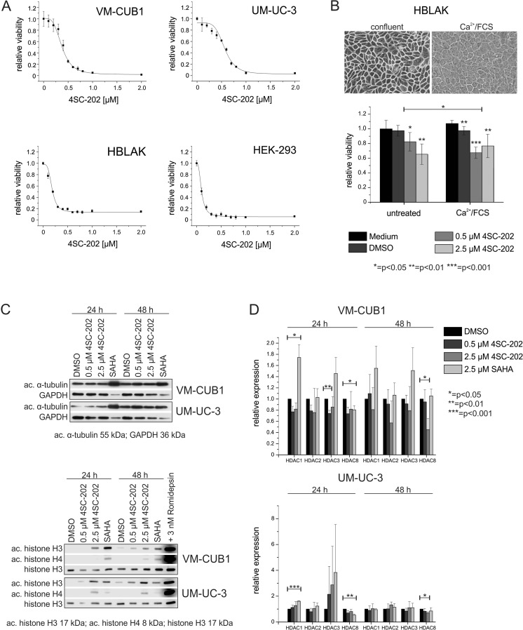
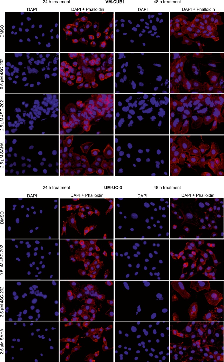
4SC-202 has an EC50 of 1.1 μM and causes hyperacetylation of histone H3 in HeLa cells. By interfering with the mitotic spindle's normal development and resulting in a collapsed spindle apparatus and multiple nucleation centers, 4SC-202 induces a G2/M cell cycle arrest. Furthermore, 4SC-202 exhibits a broad anti-proliferative activity with a mean IC50 of 0.7 μM against human cancer cell lines.[1]
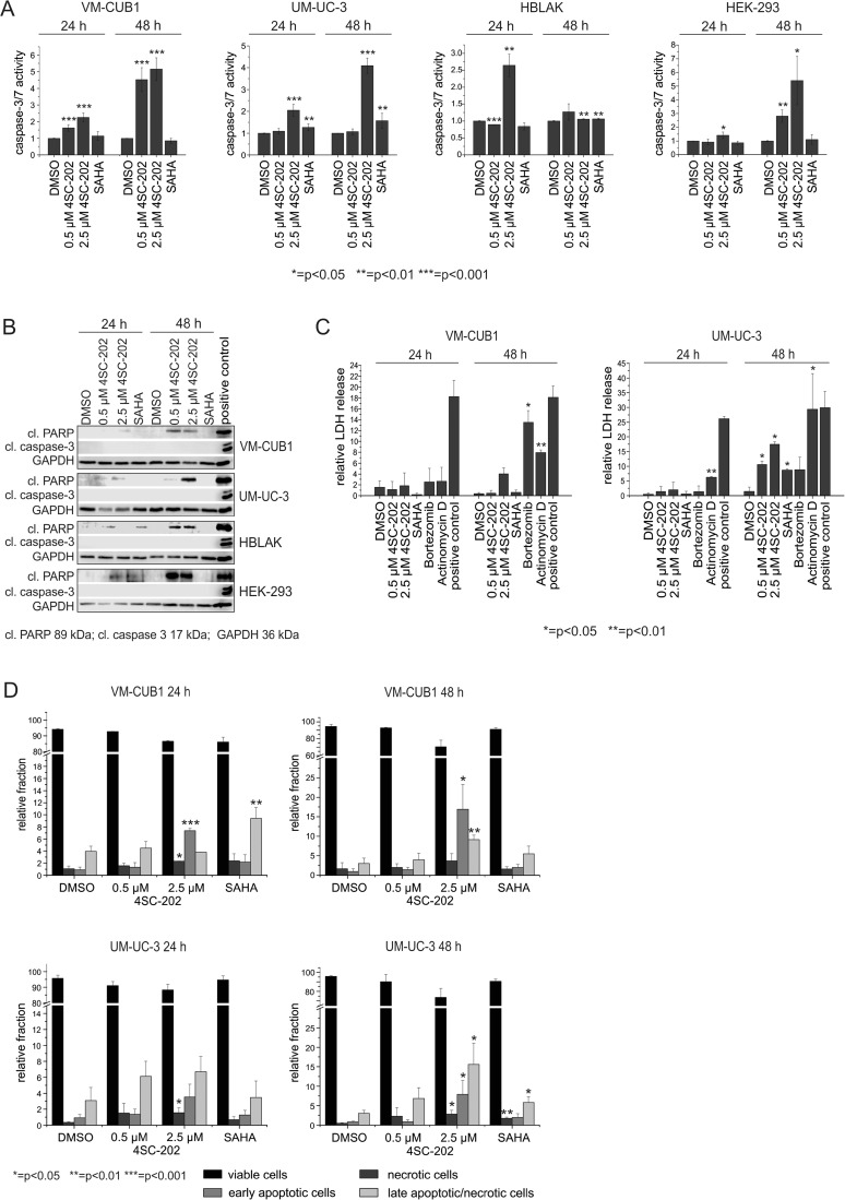
1. Antiproliferative activity: Domatinostat (4SC202) exhibited antiproliferative effects on multiple human cancer cell lines, with IC50 values of 0.3 μM (colon cancer HCT116), 0.5 μM (breast cancer MCF-7), and >5 μM (normal human dermal fibroblasts NHDF, indicating low toxicity to normal cells) [1] 2. Apoptosis induction: In HCT116 cells treated with Domatinostat (4SC202), caspase 3/7 activity (a marker of apoptosis) was significantly increased, confirming apoptosis induction [1] 3. Protein expression regulation: Western blot analysis showed that Domatinostat (4SC202) treatment increased the levels of acetylated histone H3 (Ac-H3) and acetylated α-tubulin in cancer cells, indicating effective HDAC inhibition [1] 1. Antiproliferative activity on melanoma cells: Domatinostat (4SC202) inhibited the proliferation of human melanoma cell lines, with IC50 values of 0.28 μM (A375), 0.45 μM (SK-MEL-28), and 0.32 μM (WM115) (detected by MTS assay) [2] 2. Apoptosis induction in melanoma cells: After treating A375 cells with 0.5 μM Domatinostat (4SC202) for 48 hours, Annexin V/PI staining showed that the apoptosis rate increased from 5% (control group) to 28% (flow cytometry analysis) [2] 3. Protein and gene expression regulation: Western blot revealed increased levels of Ac-H3 and Ac-H4 (acetylated histones), upregulated expression of p21 (a cell cycle inhibitor), and downregulated expression of Bcl-2 (an anti-apoptotic protein) in melanoma cells treated with Domatinostat (4SC202); real-time PCR confirmed that p21 mRNA levels were elevated and Bcl-2 mRNA levels were reduced [2] |
| ln Vivo |
4SC-202 exhibits strong metabolic stability, low plasma clearance, and high oral bioavailability in vivo. In both the RKO27 colon carcinoma model and the A549 NSCLC xenograft, 4SC-202 (120 mg/kg p.o.) exhibits strong and noticeable anti-tumor activity.[1]
1. Antitumor efficacy in colon cancer xenografts: In nude mice bearing HCT116 colon cancer xenografts, Domatinostat (4SC202) was administered orally at doses of 50 mg/kg and 100 mg/kg once daily for 21 days. Tumor growth inhibition (TGI) rates were approximately 50% (50 mg/kg) and 70% (100 mg/kg), with no significant body weight loss in mice [1] 2. Biomarker changes in tumors: Tumor tissues from treated mice showed increased levels of Ac-H3 (detected by immunohistochemistry), confirming in vivo HDAC inhibitory activity of Domatinostat (4SC202) [1] 1. Antitumor efficacy in melanoma xenografts: In nude mice bearing A375 melanoma xenografts, Domatinostat (4SC202) was administered orally at doses of 60 mg/kg and 100 mg/kg once daily for 28 days. TGI rates were 58% (60 mg/kg) and 75% (100 mg/kg) [2] 2. Biomarker changes in melanoma tumors: Tumor tissues from treated mice exhibited increased Ac-H3 and p21 expression, and decreased Ki-67 (a proliferation marker) expression (detected by immunohistochemistry and western blot) [2] 3. In vivo safety: No significant body weight loss was observed in treated mice; serum levels of alanine transaminase (ALT), aspartate transaminase (AST, liver function markers), and creatinine (renal function marker) were not significantly different from the control group [2] |
| Enzyme Assay |
1. Fluorescence-based HDAC activity assay: Recombinant HDAC enzymes (HDAC1, HDAC2, HDAC3, HDAC11) were incubated with a fluorogenic substrate and serial concentrations of Domatinostat (4SC202) in a reaction buffer at 37°C for 1 hour. After stopping the reaction with a trichloroacetic acid solution, the fluorescence intensity (excitation: 360 nm, emission: 460 nm) was measured. The inhibition rate was calculated by comparing with the solvent control group, and IC50 values were derived from dose-response curves [1]
1. HTRF-based HDAC activity assay: Recombinant HDAC enzymes (HDAC1, HDAC2, HDAC3, HDAC11) were mixed with biotin-labeled histone substrates and serial concentrations of Domatinostat (4SC202) in a reaction buffer, followed by incubation at 37°C for 2 hours. Eu³⁺-labeled anti-acetylated histone antibodies were added, and the mixture was incubated at room temperature for 1 hour. Homogeneous time-resolved fluorescence (HTRF) signals (donor emission: 620 nm, acceptor emission: 665 nm) were detected. The ratio of 665 nm/620 nm fluorescence was used to calculate the inhibition rate, and IC50 values were determined from four-parameter logistic regression [2] |
| Cell Assay |
Treatment with domatinostat (4SC-202 free base) tosylate has strong cytotoxic and proliferation-inhibitory effects on patient-derived primary HCC cells as well as established HCC cell lines (HepG2, HepB3, SMMC-7721). As a result of the activation of apoptosis signal-regulating kinase 1 (ASK1) induced by domatinostat (4SC-202 free base), it translocates to mitochondria and physically associates with Cyp-D.
1. Cell viability assay (MTT method): Human cancer cell lines (HCT116, MCF-7) and normal fibroblasts (NHDF) were seeded into 96-well plates at a density of 5×10³ cells/well and cultured overnight. Serial concentrations of Domatinostat (4SC202) (0.01–10 μM) were added, and the cells were incubated for 72 hours at 37°C with 5% CO₂. MTT reagent was added, and the plates were incubated for another 2 hours. The absorbance at 570 nm was measured, and the cell viability rate was calculated relative to the control group. IC50 values were obtained from dose-response curves [1] 2. Apoptosis assay (caspase 3/7 activity): HCT116 cells were seeded into 96-well plates and treated with Domatinostat (4SC202) for 48 hours. A caspase 3/7-specific fluorogenic substrate was added, and the plates were incubated for 1 hour at 37°C. Fluorescence intensity (excitation: 485 nm, emission: 535 nm) was measured, and the fold change in caspase activity was calculated compared to the control [1] 3. Western blot for acetylated proteins: HCT116 cells were treated with Domatinostat (4SC202) for 24 hours. Total proteins were extracted, separated by SDS-PAGE, and transferred to a PVDF membrane. The membrane was blocked with non-fat milk, incubated with primary antibodies against Ac-H3, Ac-α-tubulin, and β-actin (loading control) overnight at 4°C, then with a secondary antibody for 1 hour at room temperature. Enhanced chemiluminescence (ECL) reagent was used for signal detection, and band intensity was quantified by densitometry [1] 1. Cell viability assay (MTS method): Human melanoma cell lines (A375, SK-MEL-28, WM115) were seeded into 96-well plates at 4×10³ cells/well and cultured overnight. Domatinostat (4SC202) (0.01–10 μM) was added, and incubation continued for 72 hours. MTS reagent was added, and after 2 hours of incubation, absorbance at 490 nm was measured. Cell viability and IC50 values were calculated as described [2] 2. Apoptosis assay (Annexin V/PI staining): A375 cells were treated with Domatinostat (4SC202) for 48 hours, harvested, and stained with Annexin V-FITC and propidium iodide (PI) for 15 minutes at room temperature in the dark. Flow cytometry was used to analyze the percentage of apoptotic cells (Annexin V-positive/PI-negative and Annexin V-positive/PI-positive cells) [2] 3. Western blot for signaling proteins: Melanoma cells were treated with Domatinostat (4SC202) for 24 hours. Total proteins were extracted, and western blot was performed using primary antibodies against Ac-H3, Ac-H4, p21, Bcl-2, and β-actin. Signal detection and quantification were carried out with ECL reagent [2] 4. Real-time PCR: Total RNA was extracted from treated melanoma cells, reverse-transcribed into cDNA, and real-time PCR was performed using primers for p21, Bcl-2, and GAPDH (internal control). The relative mRNA levels were calculated using the 2⁻ΔΔCt method [2] |
| Animal Protocol |
A549 NSCLC xenograft model and RKO27 colon carcinoma model
120 mg/kg p.o. 1. Colon cancer xenograft model: Female nude mice (6–8 weeks old) were subcutaneously injected with 5×10⁶ HCT116 cells into the right flank. When tumors reached a volume of ~100 mm³, mice were randomly divided into three groups (n=6/group): control (solvent), 50 mg/kg Domatinostat (4SC202), and 100 mg/kg Domatinostat (4SC202). The drug was dissolved in physiological saline containing Cremophor EL (5% v/v) and administered orally once daily for 21 days. Tumor volume (calculated as length×width²/2) and body weight were measured every 3 days. At the end of the experiment, tumors were harvested for biomarker analysis [1] 1. Melanoma xenograft model: Female nude mice (6–8 weeks old) were subcutaneously implanted with 4×10⁶ A375 cells. When tumors reached ~120 mm³, mice were grouped (n=6/group): control (solvent), 60 mg/kg Domatinostat (4SC202), and 100 mg/kg Domatinostat (4SC202). The drug was dissolved in physiological saline containing 0.5% methylcellulose and 0.1% Tween 80, and administered orally once daily for 28 days. Tumor volume and body weight were measured every 4 days. Serum was collected for liver/kidney function tests, and tumors were excised for immunohistochemistry and protein analysis [2] |
| ADME/Pharmacokinetics |
1. Oral bioavailability in mice: After a single oral administration of 100 mg/kg domathinositol (4SC202) to mice, the peak plasma concentration (Cmax) was 2.8 μg/mL, the time to peak concentration (Tmax) was 1.5 hours, and the elimination half-life (t₁/₂) was 4.2 hours. The oral bioavailability was calculated to be 35% (compared to intravenous administration of the same dose) [2]
|
| Toxicity/Toxicokinetics |
1. In vitro toxicity to normal cells: Domatinostat (4SC202) showed low toxicity to normal human dermal fibroblasts (NHDF), with an IC50 >5 μM (far higher than the IC50 value of cancer cells)[1] 2. In vivo safety: Nude mice treated with 50–100 mg/kg domatinostat (4SC202) for 21 days did not show significant weight loss; histopathological examination did not reveal significant pathological changes in liver and kidney tissues[1] 1. In vitro toxicity to normal cells: The IC50 of Domatinostat (4SC202) in normal human keratinocytes (HaCaT) was 2.1 μM, which was 7–8 times higher than the IC50 in melanoma cells (A375: 0.28 μM)[2] 2. Plasma protein binding rate: The plasma protein binding rate was determined by balanced dialysis. The plasma protein binding rate of (4SC202) was 92% (mouse plasma, drug concentration: 1 μg/mL) [2]
3. Liver and kidney safety in vivo: Serum ALT level (control group: 25 U/L; 100 mg/kg group: 28 U/L), AST level (control group: 35 U/L; 100 mg/kg group: 28 U/L), hematocrit (mg/kg group: 39 U/L) and creatinine (control group: 0.8 mg/dL; 100 mg/kg group: 0.85 mg/dL) in the treatment group mice were not significantly different from those in the control group [2] |
| References | |
| Additional Infomation |
4SC-202 is being used in an investigational trial for the treatment of advanced hematologic malignancies. Domatinox is an orally bioavailable benzamide that inhibits human class I histone deacetylase (HDAC) isoenzymes 1, 2, and 3, exhibiting potential antitumor activity. Domatinox selectively binds to and inhibits class I HDAC, leading to the accumulation of highly acetylated histones. This may result in the induction of chromatin remodeling, selective transcription of tumor suppressor genes, and tumor suppressor protein-mediated inhibition of tumor cell division, ultimately inducing tumor cell apoptosis. This may inhibit the proliferation of susceptible tumor cells. HDACs are upregulated in various tumor types and are enzymes that deacetylate chromatin histones. 1. Domatinox (4SC202) is an orally potent selective HDAC inhibitor developed specifically for cancer treatment. At the time of the symposium in 2010, the drug was in the preclinical development stage and showed good anti-tumor activity in colon cancer and breast cancer models [1]. Domatenostat (4SC202) exerts its anti-melanoma effect by inhibiting HDACs, thereby leading to increased histone acetylation, upregulation of p21 (cell cycle arrest), downregulation of Bcl-2 (promoting apoptosis), and inhibition of tumor cell proliferation. When it was published in 2016, the drug was undergoing an early (Phase I/II) clinical trial for the treatment of advanced solid tumors, including melanoma [2].
|
| Molecular Formula |
C23H21N5O3S
|
|---|---|
| Molecular Weight |
447.51
|
| Exact Mass |
447.136
|
| Elemental Analysis |
C, 61.73; H, 4.73; N, 15.65; O, 10.73; S, 7.17
|
| CAS # |
910462-43-0
|
| Related CAS # |
1186222-89-8
|
| PubChem CID |
15985904
|
| Appearance |
White to khaki solid powder
|
| Density |
1.3±0.1 g/cm3
|
| Index of Refraction |
1.672
|
| LogP |
2.63
|
| Hydrogen Bond Donor Count |
2
|
| Hydrogen Bond Acceptor Count |
5
|
| Rotatable Bond Count |
6
|
| Heavy Atom Count |
32
|
| Complexity |
775
|
| Defined Atom Stereocenter Count |
0
|
| SMILES |
S(C1C=CC(C2C=NN(C)C=2)=CC=1)(N1C=CC(/C=C/C(=O)NC2C=CC=CC=2N)=C1)(=O)=O
|
| InChi Key |
PRXXYMVLYKJITB-IZZDOVSWSA-N
|
| InChi Code |
InChI=1S/C23H21N5O3S/c1-27-16-19(14-25-27)18-7-9-20(10-8-18)32(30,31)28-13-12-17(15-28)6-11-23(29)26-22-5-3-2-4-21(22)24/h2-16H,24H2,1H3,(H,26,29)/b11-6+
|
| Chemical Name |
(E)-N-(2-aminophenyl)-3-[1-[4-(1-methylpyrazol-4-yl)phenyl]sulfonylpyrrol-3-yl]prop-2-enamide
|
| Synonyms |
domatinostat; 4SC202; 4SC 202; 4SC-202
|
| HS Tariff Code |
2934.99.9001
|
| Storage |
Powder -20°C 3 years 4°C 2 years In solvent -80°C 6 months -20°C 1 month |
| Shipping Condition |
Room temperature (This product is stable at ambient temperature for a few days during ordinary shipping and time spent in Customs)
|
| Solubility (In Vitro) |
DMSO: ~89 mg/mL (~198.9 mM)
Water: 1 mg/mL (Insoluble) Ethanol: 1 mg/mL |
|---|---|
| Solubility (In Vivo) |
Solubility in Formulation 1: ≥ 2.5 mg/mL (5.59 mM) (saturation unknown) in 10% DMSO + 40% PEG300 + 5% Tween80 + 45% Saline (add these co-solvents sequentially from left to right, and one by one), clear solution.
For example, if 1 mL of working solution is to be prepared, you can add 100 μL of 25.0 mg/mL clear DMSO stock solution to 400 μL PEG300 and mix evenly; then add 50 μL Tween-80 to the above solution and mix evenly; then add 450 μL normal saline to adjust the volume to 1 mL. Preparation of saline: Dissolve 0.9 g of sodium chloride in 100 mL ddH₂ O to obtain a clear solution. Solubility in Formulation 2: ≥ 2.5 mg/mL (5.59 mM) (saturation unknown) in 10% DMSO + 90% (20% SBE-β-CD in Saline) (add these co-solvents sequentially from left to right, and one by one), clear solution. For example, if 1 mL of working solution is to be prepared, you can add 100 μL of 25.0 mg/mL clear DMSO stock solution to 900 μL of 20% SBE-β-CD physiological saline solution and mix evenly. Preparation of 20% SBE-β-CD in Saline (4°C,1 week): Dissolve 2 g SBE-β-CD in 10 mL saline to obtain a clear solution. View More
Solubility in Formulation 3: 2% DMSO+30% PEG 300+5% Tween 80+ddH2O: 10 mg/mL |
| Preparing Stock Solutions | 1 mg | 5 mg | 10 mg | |
| 1 mM | 2.2346 mL | 11.1729 mL | 22.3459 mL | |
| 5 mM | 0.4469 mL | 2.2346 mL | 4.4692 mL | |
| 10 mM | 0.2235 mL | 1.1173 mL | 2.2346 mL |
*Note: Please select an appropriate solvent for the preparation of stock solution based on your experiment needs. For most products, DMSO can be used for preparing stock solutions (e.g. 5 mM, 10 mM, or 20 mM concentration); some products with high aqueous solubility may be dissolved in water directly. Solubility information is available at the above Solubility Data section. Once the stock solution is prepared, aliquot it to routine usage volumes and store at -20°C or -80°C. Avoid repeated freeze and thaw cycles.
Calculation results
Working concentration: mg/mL;
Method for preparing DMSO stock solution: mg drug pre-dissolved in μL DMSO (stock solution concentration mg/mL). Please contact us first if the concentration exceeds the DMSO solubility of the batch of drug.
Method for preparing in vivo formulation::Take μL DMSO stock solution, next add μL PEG300, mix and clarify, next addμL Tween 80, mix and clarify, next add μL ddH2O,mix and clarify.
(1) Please be sure that the solution is clear before the addition of next solvent. Dissolution methods like vortex, ultrasound or warming and heat may be used to aid dissolving.
(2) Be sure to add the solvent(s) in order.
| NCT Number | Recruitment | interventions | Conditions | Sponsor/Collaborators | Start Date | Phases |
| NCT04393753 | Active Recruiting |
Drug: domatinostat in combination with avelumab |
Merkel Cell Carcinoma | 4SC AG | September 1, 2020 | Phase 2 |
| NCT04133948 | Active Recruiting |
Drug: Domatinostat Drug: Nivolumab |
Malignant Melanoma Stage III | The Netherlands Cancer Institute |
January 7, 2020 | Phase 1 Phase 2 |
| NCT04874831 | Withdrawn | Drug: domatinostat in combination with avelumab |
Merkel Cell Carcinoma | 4SC AG | November 1, 2021 | Phase 2 |
|
|---|
|
|
Products are for research use only; We do not sell to patients
Copyright 2020 InvivoChem LLC | All Rights Reserved


 COA
COA