| Size | Price | Stock | Qty |
|---|---|---|---|
| 2mg |
|
||
| 5mg |
|
||
| 10mg |
|
||
| 25mg |
|
||
| 50mg |
|
||
| 100mg |
|
||
| 250mg | |||
| Other Sizes |
Purity: ≥98%
| Targets |
KRAS-PDEδ Interaction: Deltarasin specifically inhibits the interaction between oncogenic KRAS (e.g., KRAS G12C, G12D) and PDEδ (a prenyl-binding protein). It binds to PDEδ with a dissociation constant (Ki) of 1.4 ± 0.1 nM (SPR assay) and inhibits KRAS-PDEδ complex formation with an IC50 of 2.3 ± 0.2 nM (HTRF assay). It shows no significant binding to other prenyl-binding proteins (e.g., PDE6δ, RabGDI) with Ki > 1000 nM [1]
|
|---|---|
| ln Vitro |
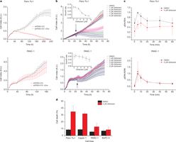
Deltarasin inhibits the interaction between RAS and PDEδ in liver cells, with a Kd of 41 nM. When deltarasin inhibits the PDEδ-KRAS interaction, human pancreatic ductal adenocarcinoma cells that rely on oncogenic KRAS proliferate less[1].
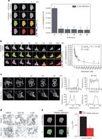
Inhibition of KRAS Membrane Localization & Oncogenic Signaling (Literature 1): 1. Human lung adenocarcinoma cells with KRAS mutations (H358: G12C; A549: G12S) were treated with Deltarasin (5–50 nM) for 24 hours. Immunofluorescence staining showed KRAS membrane localization reduced by 45% (10 nM), 65% (20 nM), 80% (50 nM) in H358 cells; Western blot detected decreased phosphorylation of downstream KRAS effectors: p-ERK (reduced by 50% at 20 nM) and p-AKT (reduced by 45% at 20 nM) in both cell lines. 2. Proliferation inhibition: MTT assay showed Deltarasin inhibited H358 cell proliferation with an IC50 of 15 ± 2 nM, A549 cells with IC50 = 22 ± 3 nM (72-hour treatment); clone formation assay revealed 20 nM Deltarasin reduced colony number by 70% (H358) and 60% (A549) vs. vehicle [1] - No Effect on KRAS Wild-Type Cells (Literature 1): Human bronchial epithelial cells (BEAS-2B, KRAS wild-type) treated with Deltarasin (5–50 nM) for 72 hours showed no significant changes in cell viability (>90% vs. vehicle) or ERK/AKT phosphorylation, confirming selectivity for KRAS-mutant cells [1] |
| ln Vivo |
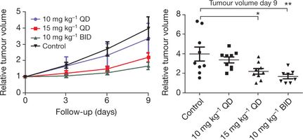
In nude mice with subcutaneous human Panc-Tu-I tumor cell xenografts, deltarasin (10 mg/kg, ip) reduces dose-dependent tumor growth[1].
Antitumor Efficacy in KRAS-Mutant Xenografts (Literature 1): Female nude mice (BALB/c nu/nu, 6–8 weeks old, n=6/group) were subcutaneously injected with 5×10⁶ H358 cells (KRAS G12C) to establish xenografts. When tumors reached ~100 mm³, mice were randomized to: 1. Vehicle: 5% DMSO + 95% saline (intraperitoneal injection, i.p.); 2. Deltarasin 10 mg/kg (i.p.); 3. Deltarasin 20 mg/kg (i.p.). Drugs were administered once daily for 21 days. Results: - Tumor volume: 10 mg/kg reduced by 40%, 20 mg/kg reduced by 65% vs. vehicle (day 21); - Tumor weight: 10 mg/kg reduced by 35%, 20 mg/kg reduced by 60% vs. vehicle; - Immunohistochemistry: Ki-67 (proliferation marker) positive cells reduced by 45% (10 mg/kg) and 70% (20 mg/kg); p-ERK positive cells reduced by 50% (20 mg/kg) [1] |
| Enzyme Assay |
SPR Assay for Deltarasin-PDEδ Binding (Literature 1):
The assay was performed using a BIAcore T200 system. Recombinant human PDEδ (residues 1–185) was covalently immobilized on a CM5 sensor chip via amine coupling (target density: 2000 response units). Deltarasin was serially diluted (0.1–20 nM) in running buffer (10 mM HEPES pH 7.4, 150 mM NaCl, 0.05% Tween 20, 1 mM DTT) and injected over the chip at a flow rate of 30 μL/min (association phase: 180 seconds; dissociation phase: 300 seconds). Sensorgrams were fitted to a 1:1 Langmuir binding model using BIAevaluation software to calculate the association rate constant (Ka = 3.8 × 10⁵ M⁻¹s⁻¹), dissociation rate constant (Kd = 5.3 × 10⁻¹¹ M), and Ki (1.4 ± 0.1 nM) [1] - HTRF Assay for KRAS-PDEδ Interaction Inhibition (Literature 1): The assay was conducted in 384-well plates with a 20 μL reaction volume. The mixture contained 50 mM Tris-HCl (pH 7.5), 10 mM MgCl₂, 2 mM DTT, 100 nM GST-KRAS G12C (N-terminally labeled with His-tag), 100 nM PDEδ (C-terminally labeled with biotin), serial dilutions of Deltarasin (0.1–10 nM), 2 nM Eu³⁺-labeled anti-His antibody (donor), and 10 nM streptavidin-conjugated XL665 (acceptor). After incubation at 25°C for 1 hour, time-resolved fluorescence was measured (excitation 337 nm, emission 620 nm for Eu³⁺, 665 nm for XL665). The 665/620 nm fluorescence ratio was used to quantify KRAS-PDEδ complex formation, and IC50 was calculated via nonlinear regression [1] |
| Cell Assay |
KRAS Membrane Localization Assay (Literature 1):
1. Cell Seeding: H358/A549 cells were seeded on coverslips in 6-well plates (2×10⁴ cells/well) and cultured in RPMI 1640 + 10% FBS overnight. 2. Treatment: Cells were treated with Deltarasin (5–50 nM) for 24 hours, then fixed with 4% paraformaldehyde (15 minutes, room temperature) and permeabilized with 0.1% Triton X-100 (10 minutes). 3. Staining & Imaging: Cells were blocked with 5% BSA (1 hour), incubated with anti-KRAS primary antibody (1:500, 4°C overnight) and Alexa Fluor 488-conjugated secondary antibody (1:1000, 1 hour, room temperature). Nuclei were stained with DAPI (5 minutes). KRAS membrane localization was analyzed via confocal microscopy (membrane/cytosol fluorescence intensity ratio quantified using ImageJ) [1] - Cell Proliferation & Signaling Assays (Literature 1): 1. MTT Proliferation Assay: Cells were seeded in 96-well plates (5×10³ cells/well), treated with Deltarasin (1–50 nM) for 72 hours. MTT (5 mg/mL) was added (20 μL/well) for 4 hours, DMSO dissolved formazan, and absorbance measured at 570 nm. 2. Western Blot: Cells were seeded in 6-well plates (2×10⁵ cells/well), treated with Deltarasin (5–50 nM) for 24 hours. RIPA buffer (with inhibitors) lysed cells; 30 μg protein separated by 10% SDS-PAGE, transferred to PVDF membranes, probed with anti-p-ERK (1:1000), anti-p-AKT (1:1000), anti-total ERK/AKT (1:1000), and anti-β-actin (1:5000) antibodies. ECL reagent visualized bands. 3. Clone Formation Assay: Cells (200 cells/well, 6-well plates) were treated with Deltarasin (5–20 nM) for 14 days. Colonies were stained with crystal violet, and colonies >50 cells were counted [1] |
| Animal Protocol |
Dissolved in 10% PLR and 5% DMSO; 10 mg/kg; Administered through i.p.
Mice bearing Panc-Tu-I xenografts H358 Xenograft Model (Literature 1): 1. Animal Preparation: Female BALB/c nu/nu mice (6–8 weeks old, 18–22g) were housed under SPF conditions (12h light/dark cycle, free access to food/water). 2. Tumor Induction: 5×10⁶ H358 cells (suspended in 0.2 mL PBS + 50% Matrigel) were subcutaneously injected into the right flank of each mouse. 3. Drug Preparation & Administration: Deltarasin was dissolved in 5% DMSO + 95% saline to concentrations of 1 mg/mL (10 mg/kg dose) and 2 mg/mL (20 mg/kg dose). When tumors reached ~100 mm³ (day 0), mice were randomized to groups (n=6/group) and received intraperitoneal injections once daily for 21 days (0.1 mL/10g body weight). Vehicle group received 5% DMSO + 95% saline. 4. Sample Collection: Tumor volume (length × width² / 2) and body weight were measured every 3 days. On day 21, mice were euthanized by cervical dislocation; tumors were excised (weighed, fixed in 10% formalin for immunohistochemistry or frozen for Western blot) [1] |
| Toxicity/Toxicokinetics |
In vitro cytotoxicity (Reference 1):
Normal human cells (BEAS-2B bronchial epithelial cells, HEK293 embryonic kidney cells) were treated with Deltarasin (1–50 nM) for 72 hours. MTT assay showed that cell viability was >90% at all concentrations, indicating no significant cytotoxicity to normal cells [1] - In vivo safety (Reference 1): - In the H358 xenograft model (Deltarasin 10–20 mg/kg, intraperitoneal injection, 21 days): - No death or abnormal behavior (e.g., somnolence, anorexia); - Weight change: 3.2 ± 0.5 g in the 10 mg/kg group and 2.8 ± 0.4 g in the 20 mg/kg group (3.5 ± 0.6 g in the control group), with a weight loss of <5%; - Serum biochemical indicators: ALT, AST, BUN and creatinine levels were all within the normal range (no significant difference compared with the control group); - Organ histopathology: No inflammation, necrosis or structural abnormalities were observed in the liver, kidney and spleen tissues (HE staining) [1] |
| References | |
| Additional Infomation |
Mechanism of action (Reference 1): Deltarasin specifically binds to the isoprene binding pocket of PDEδ, preventing PDEδ from chaperonering isoprene-modified KRAS to the plasma membrane. Reduced KRAS membrane localization inhibits downstream oncogenic signaling pathways (RAS-RAF-MEK-ERK, PI3K-AKT-mTOR), thereby inhibiting the proliferation and survival of KRAS-mutant cancer cells [1]
- Therapeutic potential (Reference 1): As the first KRAS-PDEδ interaction inhibitor, Deltarasin has shown efficacy in both in vitro and in vivo models of KRAS-mutant lung cancer, supporting its potential to treat KRAS-driven cancers (e.g., non-small cell lung cancer, pancreatic cancer), in which KRAS mutations are common and resistant to conventional therapies [1] - Selectivity advantage (Reference 1): Deltarasin does not bind significantly to other isoprenoid-binding proteins (e.g., PDE6δ, which is essential for visual function) or off-target kinases/enzymes, thereby minimizing the risk of off-target side effects (e.g., visual impairment) [1] |
| Molecular Formula |
C40H37N5O
|
|---|---|
| Molecular Weight |
603.75
|
| Exact Mass |
603.299
|
| CAS # |
1440898-61-2
|
| Related CAS # |
Deltarasin hydrochloride;1613404-76-4;Deltarasin trihydrochloride;1440898-82-7
|
| PubChem CID |
73292904
|
| Appearance |
Light yellow to yellow solid powder
|
| Density |
1.2±0.1 g/cm3
|
| Boiling Point |
824.3±75.0 °C at 760 mmHg
|
| Flash Point |
452.3±37.1 °C
|
| Vapour Pressure |
0.0±3.0 mmHg at 25°C
|
| Index of Refraction |
1.680
|
| LogP |
9.79
|
| Hydrogen Bond Donor Count |
1
|
| Hydrogen Bond Acceptor Count |
4
|
| Rotatable Bond Count |
9
|
| Heavy Atom Count |
46
|
| Complexity |
923
|
| Defined Atom Stereocenter Count |
1
|
| SMILES |
C1CNCCC1[C@@H](COC2=CC=C(C=C2)C3=NC4=CC=CC=C4N3CC5=CC=CC=C5)N6C7=CC=CC=C7N=C6C8=CC=CC=C8
|
| InChi Key |
NCIOVAYUMQEQEU-VKZSUDIWSA-N
|
| InChi Code |
InChI=1S/C40H37N5O.3ClH/c1-3-11-29(12-4-1)27-44-36-17-9-7-15-34(36)42-39(44)32-19-21-33(22-20-32)46-28-38(30-23-25-41-26-24-30)45-37-18-10-8-16-35(37)43-40(45)31-13-5-2-6-14-31;;;/h1-22,30,38,41H,23-28H2;3*1H/t38-;;;/m1.../s1
|
| Chemical Name |
2-[4-[(2S)-2-(2-phenyl-1H-benzimidazol-1-yl)-2-(4-piperidinyl)ethoxy]phenyl]-1-(phenylmethyl)-1H-benzimidazole, trihydrochloride
|
| Synonyms |
Deltarasin hydrochloride; Deltarasin HCl; Deltarasin;
|
| HS Tariff Code |
2934.99.9001
|
| Storage |
Powder -20°C 3 years 4°C 2 years In solvent -80°C 6 months -20°C 1 month |
| Shipping Condition |
Room temperature (This product is stable at ambient temperature for a few days during ordinary shipping and time spent in Customs)
|
| Solubility (In Vitro) |
DMSO: 100 mg/mL (165.6 mM)
Water: 1 mg/mL (Insoluble) Ethanol: 1 mg/L |
|---|---|
| Solubility (In Vivo) |
Solubility in Formulation 1: ≥ 1.25 mg/mL (2.07 mM) (saturation unknown) in 10% DMSO + 40% PEG300 + 5% Tween80 + 45% Saline (add these co-solvents sequentially from left to right, and one by one), clear solution.
For example, if 1 mL of working solution is to be prepared, you can add 100 μL of 12.5 mg/mL clear DMSO stock solution to 400 μL PEG300 and mix evenly; then add 50 μL Tween-80 to the above solution and mix evenly; then add 450 μL normal saline to adjust the volume to 1 mL. Preparation of saline: Dissolve 0.9 g of sodium chloride in 100 mL ddH₂ O to obtain a clear solution. Solubility in Formulation 2: 1.25 mg/mL (2.07 mM) in 10% DMSO + 90% (20% SBE-β-CD in Saline) (add these co-solvents sequentially from left to right, and one by one), suspension solution; with ultrasonication. For example, if 1 mL of working solution is to be prepared, you can add 100 μL of 12.5 mg/mL clear DMSO stock solution to 900 μL of 20% SBE-β-CD physiological saline solution and mix evenly. Preparation of 20% SBE-β-CD in Saline (4°C,1 week): Dissolve 2 g SBE-β-CD in 10 mL saline to obtain a clear solution. View More
Solubility in Formulation 3: ≥ 1.25 mg/mL (2.07 mM) (saturation unknown) in 10% DMSO + 90% Corn Oil (add these co-solvents sequentially from left to right, and one by one), clear solution. |
| Preparing Stock Solutions | 1 mg | 5 mg | 10 mg | |
| 1 mM | 1.6563 mL | 8.2816 mL | 16.5631 mL | |
| 5 mM | 0.3313 mL | 1.6563 mL | 3.3126 mL | |
| 10 mM | 0.1656 mL | 0.8282 mL | 1.6563 mL |
*Note: Please select an appropriate solvent for the preparation of stock solution based on your experiment needs. For most products, DMSO can be used for preparing stock solutions (e.g. 5 mM, 10 mM, or 20 mM concentration); some products with high aqueous solubility may be dissolved in water directly. Solubility information is available at the above Solubility Data section. Once the stock solution is prepared, aliquot it to routine usage volumes and store at -20°C or -80°C. Avoid repeated freeze and thaw cycles.
Calculation results
Working concentration: mg/mL;
Method for preparing DMSO stock solution: mg drug pre-dissolved in μL DMSO (stock solution concentration mg/mL). Please contact us first if the concentration exceeds the DMSO solubility of the batch of drug.
Method for preparing in vivo formulation::Take μL DMSO stock solution, next add μL PEG300, mix and clarify, next addμL Tween 80, mix and clarify, next add μL ddH2O,mix and clarify.
(1) Please be sure that the solution is clear before the addition of next solvent. Dissolution methods like vortex, ultrasound or warming and heat may be used to aid dissolving.
(2) Be sure to add the solvent(s) in order.
|
|---|
|
|
Products are for research use only; We do not sell to patients
Copyright 2020 InvivoChem LLC | All Rights Reserved


 COA
COA