| Size | Price | Stock | Qty |
|---|---|---|---|
| 5mg |
|
||
| 25mg |
|
||
| 50mg |
|
||
| 100mg |
|
||
| 250mg |
|
||
| 500mg | |||
| Other Sizes |


Purity: ≥98%
| Targets |
Aurora A (IC50 = 13 nM); Aurora B (IC50 = 79 nM); Aurora C (IC50 = 61 nM)
From [2] (Aurora kinase & Bcr-Abl kinase inhibition assays): - Danusertib (PHA739358) is a multi-kinase inhibitor with potent activity against Aurora kinases (A/B/C) and Bcr-Abl kinase (including imatinib-resistant mutants); - IC50 values for recombinant human Aurora kinases: - Aurora A = 1.2 nM, Aurora B = 3.0 nM, Aurora C = 2.5 nM (broad-spectrum Aurora inhibition); - IC50 values for Bcr-Abl kinase (recombinant human): - Wild-type (WT) Bcr-Abl = 4.5 nM; - Imatinib-resistant T315I mutant Bcr-Abl = 30 nM; - Other mutants (G250E, M351T) = 5.2–8.0 nM; - Weak inhibition of non-target kinases (e.g., CDK1: IC50 > 800 nM; PLK1: IC50 > 1000 nM) [2] - From [1], [3]: No new target data; focus on in vitro/in vivo efficacy via Aurora/Bcr-Abl inhibition [1,3] |
|---|---|
| ln Vitro |
The viability of C13 and A2780cp cells is severely reduced by denusertib (0.01 to 50 μM). After 24 and 48 hours of treatment, the IC50s for C13 cells are 10.40 and 1.83 μM, and for A2780cp cells, they are 19.89 and 3.88 μM. In C13 and A2780cp cells, dunusertib promotes a cell arrest cycle in the G2/M phase. After being treated with denusertib, the proportion of cells arrested in the G2/M phase rises noticeably, and polyploidy accumulates in C13 and A2780cp cells. Danusertib increases the expression of p21 Waf1/Cip1, p27 Kip1, and p53 while depressing CDK1/CDC2 and cyclin B1 expression. Danusertib activates the PI3K/Akt/mTOR signaling pathway in C13 and A2780cp cells to cause autophagy[1]. All examined leukemic cell lines are significantly inhibited from proliferating by PHA-739358, with IC50 values ranging from 0.05 μM to 3.06 μM. IM-resistant M351T, E255K, and T315I mutants are among the BaF3-p210 cells in which PHA-739358 exerts antiproliferative effects. BaF3 -p210 wt cells and IM-resistant mutants exhibit decreased phosphorylation of CrkL in response to PHA-739358 (5 μM)[2]. In vitro, dacartib totally prevents GEP-NET cell proliferation and induces cell-cycle arrest[3].
Activity in human ovarian cancer cells (from [1]): - In SKOV3 and OVCAR3 ovarian cancer cell lines: 1. Danusertib (0.1–50 nM) dose-dependently inhibited proliferation: - SKOV3: IC50 = 2.8 nM (72 h MTT assay); - OVCAR3: IC50 = 3.5 nM (72 h MTT assay); 2. 10 nM induced G2/M cell-cycle arrest: G2/M phase cells increased from 15% (vehicle) to 70% (SKOV3, PI staining, flow cytometry); 3. 20 nM induced apoptosis: Annexin V-positive cells = 52% (SKOV3) vs. 6% (vehicle); western blot: cleaved caspase-3 upregulated 4.0-fold, cleaved PARP upregulated 3.5-fold; 4. 15 nM induced autophagy: LC3-II/LC3-I ratio increased 3.2-fold (western blot); autophagosome formation increased 5-fold (transmission electron microscopy); 5. 25 nM inhibited epithelial-mesenchymal transition (EMT): E-cadherin (epithelial marker) upregulated 2.5-fold, vimentin (mesenchymal marker) downregulated 60%; PI3K/Akt/mTOR pathway: p-Akt (Ser473) reduced 80%, p-mTOR (Ser2448) reduced 75% (western blot) [1] - Activity in Bcr-Abl-positive leukemia cells (from [2]): - In K562 (WT Bcr-Abl) and K562-T315I (imatinib-resistant) cells: 1. Danusertib (0.5–100 nM) inhibited proliferation: - K562: IC50 = 5.0 nM (72 h CCK-8 assay); - K562-T315I: IC50 = 35 nM (72 h CCK-8 assay); 2. 20 nM reduced p-Bcr-Abl (Tyr412) by 90% (K562) and 85% (K562-T315I) (western blot); 3. 30 nM induced apoptosis: K562-T315I cells showed 45% Annexin V positivity vs. 7% (vehicle) [2] - Activity in gastroenteropancreatic neuroendocrine tumor (GEP-NET) cells (from [3]): - In BON-1 (pancreatic NET) and QGP-1 (gastric NET) cells: 1. Danusertib (1–50 nM) inhibited proliferation: - BON-1: IC50 = 4.2 nM (72 h MTT assay); - QGP-1: IC50 = 5.8 nM (72 h MTT assay); 2. 10 nM reduced colony formation by 75% (BON-1, 14-day methylcellulose assay); western blot: p-Aurora A (Thr288) reduced 90% [3] |
| ln Vivo |
PHA-739358 (15 mg/kg twice day, ip) and IM are well tolerated; over the course of the 10-day treatment period, they virtually suppressed tumor growth and significantly inhibited K562 cell proliferation[2]. When compared to controls or mice given streptozotocine/5-fluorouracil, danusertibsertib (2×15 mg/kg/d, ip) dramatically slows down the growth of tumors in vivo in a subcutaneous murine xenograft model[3].
Danusertib treatment inhibits growth of subcutaneous GEP-NET xenografts and lowers serum chromogranin levels [3] Treatment of tumor-bearing mice with danusertib at a dose of 2 × 15 mg/kg/d decreased growth of BON1 and QGP xenografts (Fig. 3A and B). BON1 tumor growth was significantly inhibited from day 4 (P < 0.001) until the end of the experiment (P < 0.001). The mean absolute tumor volume was reduced by 88.2%, and the mean percental tumor growth was also reduced by 88.2%, compared with vehicle-treated controls (Fig. 3A, Table 2). In mice with QGP xenografts, treatment with danusertib led to a virtual shrinkage of QGP tumors from day 4 (P < 0.001), until the end of the experiment (P < 0.001). The final tumor volume was only 6.3 ± 8.2% of the original tumor volume (Fig. 3B, Table 2). Mean absolute and percental tumor growth in danusertib-treated mice was reduced by 98.4% and 98.6%, respectively, compared with vehicle-treated controls. When compared with treatment with streptozotocine/5-fluoruracil (STZ/5-FU), which is a frequently used cytostatic therapy for GEP-NETs, the antiproliferative effect of danusertib was significantly higher from day 12 (P < 0.001), in both BON1 and QGP tumors (Fig. 3A and B, Table 2). Treatment of GEP-NET liver metastases with danusertib [3] In transplanted mice, treatment with danusertib or vehicle was commenced when liver metastases became clearly detectable and were eligible for volumetric analysis, which was after 2 to 3 weeks following cell transplantation. Average size of BON1 metastases (single nodules) before treatment was 2.9 ± 1.3 mm3 (n = 20). Although liver metastases of BON1 tumors in vehicle-treated controls continued to grow until large areas of the liver were replaced by tumor tissue, treatment with danusertib significantly inhibited growth of liver metastases (day 12 after start of treatment 55.9 ± 39.7 mm3, n = 10 vs. 5.2 ± 3.8 mm3; n = 10; P < 0.01; Fig. 6A and C, Table 3). This corresponds to a relative tumor volume compared with day 0 (100%) of 1,495.3 ± 1,277.9% in controls versus 164.5 ± 50.2% in danusertib-treated mice (P < 0.01). Mean absolute and percental tumor growth in danusertib-treated mice was reduced by 90.7% and 89.0%, respectively, compared with vehicle-treated controls. Average size of QGP metastases in the liver before treatment was 6.8 ± 3.7 mm3 (n = 9). Danusertib entirely inhibited growth of QGP metastases, although no shrinkage was observed, as in subcutaneous tumors (control: day 12, 51.7 ± 35.0 mm3; n = 4 vs. danusertib 6.1 ± 4.3 mm3; n = 5, P < 0.05; Fig. 6B and C, Table 3). This corresponds to a relative tumor volume of 810.7 ± 355.4% versus 108.7 ± 58.9%, P < 0.01. The mean absolute tumor volume was reduced by 88.2%, and the mean percental tumor growth was reduced by 86.6%, compared with vehicle-treated controls.[3] Efficacy in orthotopic GEP-NET liver metastasis model (from [3]): - Animal model: Female nude mice (6–8 weeks old, n=8/group) with orthotopic BON-1 xenografts (intrapancreatic injection of 1×10⁶ cells, day 0) to induce liver metastases; - Treatment groups: 1. Vehicle: 5% DMSO + 95% normal saline (intraperitoneal injection, IP, 3 times/week for 4 weeks); 2. Danusertib 25 mg/kg: IP, 3 times/week for 4 weeks; 3. Danusertib 50 mg/kg: IP, 3 times/week for 4 weeks; - Efficacy (day 28): 1. 50 mg/kg reduced pancreatic primary tumor weight by 80% (0.2 g vs. 1.0 g vehicle); 2. Liver metastasis nodules reduced from 15 ± 3 (vehicle) to 3 ± 1 (50 mg/kg); 3. Tumor lysates: p-Aurora A reduced 85%, p-mTOR reduced 70% (western blot); serum chromogranin A (GEP-NET marker) reduced 75% (ELISA) [3] |
| Enzyme Assay |
The emergence of resistance to imatinib (IM) mediated by mutations in the BCR-ABL domain has become a major challenge in the treatment of chronic myeloid leukemia (CML). Here, we report on studies performed with a novel small molecule inhibitor, PHA-739358, which selectively targets Bcr-Abl and Aurora kinases A to C. PHA-739358 exhibits strong antiproliferative and proapoptotic activity against a broad panel of human BCR-ABL–positive and –negative cell lines and against murine BaF3 cells ectopically expressing wild-type (wt) or IM-resistant BCR-ABL mutants, including T315I. Pharmacologic synergism of IM and PHA-739358 was observed in leukemia cell lines with subtotal resistance to IM. Treatment with PHA-739358 significantly decreased phosphorylation of histone H3, a marker of Aurora B activity and of CrkL, a downstream target of Bcr-Abl, suggesting that PHA-739358 acts via combined inhibition of Bcr-Abl and Aurora kinases. Moreover, strong antiproliferative effects of PHA-739358 were observed in CD34+ cells derived from untreated CML patients and from IM-resistant individuals in chronic phase or blast crisis, including those harboring the T315I mutation. Thus, PHA-739358 represents a promising new strategy for treatment of IM-resistant BCR-ABL-positive leukemias, including those harboring the T315I mutation. Clinical trials investigating this compound in IM-resistant CML have recently been initiated[2].
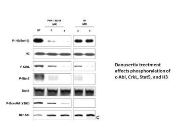
PHA-739358 shares activity of both Aurora and Abl kinase inhibitors [2] The influence of PHA-739358 on Aurora kinase activity was assessed by the phosphorylation status of histone H3 at Ser10 (Figures 4,5C). K562 cells exposed to PHA-739358 showed strong reduction of phosphorylation with only 0.1% phospho-H3 (Figure 4C) compared with 3.5% in untreated (Figure 4A) and 3.7% in IM-treated cells (Figure 4B). Inhibitory activity of PHA-739358 on Bcr-Abl kinase was evaluated by determining phosphorylation of CrkL and Stat5, 2 well-known downstream targets of Bcr-Abl (Figure 5). PHA-739358 treatment produced distinct inhibition of c-Abl autophosphorylation on Tyr393 (Figure 5C). In addition, pronounced inhibition of phosphorylation of CrkL (28% residual phosphorylation) and Stat5 (37% residual phosphorylation) was observed to a similar degree as for IM (19% and 22% residual phosphorylation, respectively; Figure 5A,B). Aurora kinase activity assay (radioactive-based, from [2]): 1. Purified recombinant human Aurora A/B/C (0.2 μg/mL each) was incubated with biotinylated histone H3 peptide (Ser10 motif, 1 μg/mL) and [γ-³²P]ATP (5 μCi, 10 μM) in kinase buffer (50 mM Tris-HCl pH 7.5, 10 mM MgCl₂, 1 mM DTT) at 30°C for 15 min. 2. Serial concentrations of Danusertib (0.1–100 nM) were added, and incubation continued for 30 min. 3. Reaction mixtures were spotted onto P81 phosphocellulose paper, washed 3 times with 1% phosphoric acid to remove unincorporated ATP. 4. Radioactivity was measured via liquid scintillation counter; IC50 was calculated using four-parameter logistic regression [2] - Bcr-Abl kinase activity assay (HTRF-based, from [2]): 1. Purified recombinant human WT/Bcr-Abl-T315I (0.2 μg/mL) was incubated with biotinylated STAT5 peptide (Tyr694 motif, 1 μg/mL) and ATP (10 μM) in assay buffer (50 mM HEPES pH 7.4, 5 mM MgCl₂, 0.1 mM Na₃VO₄) at 37°C for 20 min. 2. Serial concentrations of Danusertib (0.5–100 nM) were added, and incubation continued for 30 min. 3. Reaction was terminated with 20 mM EDTA; anti-phospho-STAT5 cryptate antibody and streptavidin-europium conjugate were added. 4. Time-resolved fluorescence (excitation 340 nm, emission 665 nm/620 nm ratio) was measured; IC50 was calculated [2] |
| Cell Assay |
Ovarian carcinoma (OC) is one of the most common gynecological malignancies, with a poor prognosis for patients at advanced stage. Danusertib (Danu) is a pan-inhibitor of the Aurora kinases with unclear anticancer effect and underlying mechanisms in OC treatment. This study aimed to examine the cancer cell killing effect and explore the possible mechanisms with a focus on proliferation, cell cycle progression, apoptosis, autophagy, and epithelial to mesenchymal transition (EMT) in human OC cell lines C13 and A2780cp. The results showed that Danu remarkably inhibited cell proliferation, induced apoptosis and autophagy, and suppressed EMT in both cell lines. Danu arrested cells in G₂/M phase and led to an accumulation of polyploidy through the regulation of the expression key cell cycle modulators. Danu induced mitochondria-dependent apoptosis and autophagy in dose and time-dependent manners. Danu suppressed PI3K/Akt/mTOR signaling pathway, evident from the marked reduction in the phosphorylation of PI3K/Akt/mTOR, contributing to the autophagy inducing effect of Danu in both cell lines. In addition, Danu inhibited EMT. In aggregate, Danu exerts potent inducing effect on cell cycle arrest, apoptosis, and autophagy, but exhibits a marked inhibitory effect on EMT. PI3K/Akt/mTOR signaling pathway contributes, partially, to the cancer cell killing effect of Danu in C13 and A2780cp cells[1].\
\Cell analysis, immunostaining, and flow cytometry [3] \Cells were seeded in DMEM, fixed and immunostained for aurora-A, aurora-B, and histone H3 phosphorylation, as described. For analysis of cell proliferation, increasing concentrations of danusertib (dissolvent control; 5 nmol/L–5 μmol/L) were added after 24 hours of cell culture. Forty-eight to 120 hours later, the number of viable cells was determined. Analysis of DNA content and H3 phosphorylation were carried out by flow cytometry as described.\ \Short-term expansion of CD34+ cells [2] \For short-term expansion assays, 103 CD34+ cells were plated in triplicates in 96-well plates containing 100 μL of serum-free medium per well supplemented with human stem-cell factor (100 ng/mL), human Flt-3 Ligand (100 ng/mL), human thrombopoietin (50 ng/mL), human interleukin-3 and -6 (IL-3 and IL-6, respectively, both 20 ng/mL), and granulocyte colony-stimulating factor (20 ng/mL) plus Danusertib (PHA739358)or IM at the indicated concentrations. After 5 days, an additional 100 μL of cytokine and PHA-739358 or IM containing medium were added. Cell numbers within each individual well were evaluated at days 3, 6, and 9 or at days 3, 6, and 12 for healthy donor samples.\ \MTT assay [2] \Cells were seeded in triplicates in 96-well flat-bottomed microtiter plates at a density of 1.5 × 10⁴ cells/well in 150 μL of their respective media. After 24 hours increasing concentrations of Danusertib (PHA739358) (0-5.0 μM) or/and IM (0-20 μM) were added. After 48 hours of treatment, the viable cells were assayed for their ability to transform 3-(4,5-dimethylthiazol-2-yl)-2,5-diphenyltetrazolium bromide (MTT) into purple formazan, as described previously.23 The compound concentration that inhibits response by 50% (IC50) is defined as the concentration resulting in 50% growth inhibition that corresponds to the fraction affected (Fa value) of 0.5. All tested cells were exposed to 10 different concentrations of each compound. Fraction affected (Fa) and dose-effect relationship at the point of IC50 were analyzed by CalcuSyn Software.\ \Analysis of DNA content and apoptosis by flow cytometry [2] \Cells were plated in triplicates in 6-well plates and cultured in 2 mL of their respective media. After 24 hours, cells were exposed to increasing concentrations of Danusertib (PHA739358) for 48 hours, then washed with phosphate-buffered saline (PBS), and fixed in cold 70% ethanol overnight at −20°C. For flow cytometric analysis, cells were washed twice with PBS, resuspended in PBS containing RNAse A (100 μg/mL) and propidium iodide (10 μg/mL), and incubated for 30 minutes on ice; 10 000 cells were analyzed from each sample.\ \Assessment of phosphorylation status by intracellular flow cytometry [2] \Cells exposed to 5 μM Danusertib (PHA739358) or 5 μM IM for 2 hours or 24 hours were collected, fixed in 2% formaldehyde for 10 minutes at 37°C, chilled on ice for 1 minute, and then permeabilized with ice-cold 90% methanol for 30 minutes on ice. From each sample, 5 × 10⁵ cells were washed with 2 mL incubation buffer (PBS/0.5% bovine serum albumin), centrifuged at 50g for 5 minutes, and resuspended in 100 μL of incubation buffer with 2.0 μL of Phospho-CrkL, Phospho-Stat5, or Phospho-Histone H3-(Ser10) specific antibody. After 45 minutes of incubation at RT, cells were washed twice, resuspended in 100 μL incubation buffer with 0.5 μL of the secondary antibody, and incubated at RT for 30 minutes in the dark. Again, cells were washed twice with washing buffer. Samples stained with specific Phospho-Histone H3-(Ser10) antibody were also stained with propidium iodide.\\All samples were analyzed by flow cytometry, and isotype controls were included for cytometer setup. The amount of phosphorylated proteins (P-CrkL and P-Stat5) was determined as the geometric mean fluorescence intensity, and the changes of the phosphorylation status were expressed as a percentage of the no-drug control.\CML CD34+ cells were incubated with either 5 μM Danusertib (PHA739358) for 72 hours and 96 hours or with 5 μM IM for 96 hours. After collection, cells were prepared as described above.\ \Western blotting [2] \K562 cells cultured under standard conditions were exposed for 1 hour to 2 μM and 5 μM of Danusertib (PHA739358) or IM. Collected cells were lysed in sodium dodecyl sulfate (SDS) buffer (125 mM Tris-HCl, pH 6.8, 2% SDS) and after sonication and boiling, 20 μg total extract of the indicated samples was loaded on SDS-polyacrylamide gel electrophoresis precast gels and immunoblotted. The following antibodies were used: c-Abl, Phospho-H3-(Ser10; Upstate), Histone-H3, Stat5. Anti-PathScan Bcr-Abl activity assay was used for the detection of Phospho-Bcr-Abl, Phospho-Stat5, and Phospho-CrkL. Ovarian cancer cell assay (from [1]): 1. SKOV3/OVCAR3 cells (5×10³ cells/well) were seeded in 96-well plates, incubated overnight at 37°C (5% CO₂). Serial concentrations of Danusertib (0.1–50 nM) were added, cultured for 72 h; MTT reagent (10 μL/well) was added, absorbance at 570 nm measured for IC50. 2. Apoptosis: SKOV3 cells (1×10⁵ cells/mL) treated with 20 nM Danusertib for 48 h, stained with Annexin V-FITC/PI, analyzed via flow cytometry. 3. Autophagy: Cells treated with 15 nM Danusertib for 24 h, stained with LC3 antibody (immunofluorescence) or lysed for western blot (LC3-II/LC3-I ratio). 4. EMT detection: Cells treated with 25 nM Danusertib for 72 h, lysed for western blot (E-cadherin, vimentin) or analyzed for migration (Transwell assay) [1] - Leukemia cell assay (from [2]): 1. K562/K562-T315I cells (5×10³ cells/well) were seeded in 96-well plates, treated with Danusertib (0.5–100 nM) for 72 h; CCK-8 reagent (10 μL/well) was added, absorbance at 450 nm measured for IC50. 2. Western blot: Cells treated with 20 nM Danusertib for 24 h, lysed; 30 μg protein probed with anti-p-Bcr-Abl (Tyr412) and anti-total Bcr-Abl antibodies [2] - GEP-NET cell assay (from [3]): 1. BON-1/QGP-1 cells (5×10³ cells/well) were seeded in 96-well plates, treated with Danusertib (1–50 nM) for 72 h; MTT assay measured proliferation. 2. Colony formation: BON-1 cells (1×10³ cells/well) seeded in 6-well plates, treated with 10 nM Danusertib for 14 days; colonies fixed with 4% paraformaldehyde, stained with 0.1% crystal violet, counted [3] |
| Animal Protocol |
DMSO; 15 mg/kg; i.p. Female SCID mice
In a subcutaneous murine xenograft model, danusertib significantly reduced tumor growth in vivo compared with controls or mice treated with streptozotocine/5-fluorouracil. As a consequence, decreased levels of tumor marker chromogranin A were found in mouse serum samples. In a newly developed orthotopic model for GEP-NET liver metastases by intrasplenic tumor cell transplantation, dynamic MRI proved significant growth inhibition of BON1- and QGP-derived liver metastases.[3]
Experimental protocol [3] Mice bearing BON1 or QGP tumors were divided into a treatment group receiving Danusertib (PHA739358) intraperitoneally at a dose of 2 × 15 mg/kg/d or a control group receiving the same volume of vehicle solution (5% dextrose). Streptozotocine and 5-fluorouracil were applied intraperitoneally at a dose of 1 × 15 mg/kg/d. Evaluation of Danusertib (PHA739358) and IM in vivo [2] To evaluate the efficacy and toxicity of Danusertib (PHA739358) in vivo, a subcutaneous animal model for CML was used; 5 × 10⁷ K562 cells were injected into the flanks of female SCID mice and tumor growth was monitored daily by palpation. On day 7, when tumors reached an estimated weight of 100 to 150 mg, animals were assigned to 3 experimental groups by random selection and received the following treatment for a period of 10 days: group 1, control, vehicle solution (7 mice); group 2, PHA-739358 twice a day intraperitoneally at a dose of 15 mg/kg (7 mice); and group 3, IM twice a day per os at 100 mg/kg. Tumor growth was assessed by caliper, and tumor weight was calculated according to the following formula: Tumor weight = [length (mm) × width2 (mm)]/2. Toxicity was monitored by changes in body weight and vitality of the animals. Orthotopic GEP-NET liver metastasis protocol (from [3]): 1. Animals: Female nude mice (6–8 weeks old, 18–20 g, n=8/group). 2. Xenograft establishment: Day 0: Intrapancreatic injection of 1×10⁶ BON-1 cells (100 μL 1:1 PBS-matrigel) to induce liver metastases. 3. Treatment initiation: Day 7 (confirmed pancreatic tumor via ultrasound). 4. Treatment groups: - Vehicle: 5% DMSO + 95% normal saline, intraperitoneal injection (IP), 3 times/week (Monday/Wednesday/Friday) for 4 weeks. - Danusertib 25 mg/kg: Dissolved in vehicle, IP, same frequency. - Danusertib 50 mg/kg: Dissolved in vehicle, IP, same frequency. 5. Monitoring & sampling: - Weekly body weight and ultrasound (tumor size); day 28: Euthanize mice, harvest pancreatic tumors and liver; weigh tumors, count liver metastasis nodules; tumor lysates for western blot; serum for chromogranin A ELISA [3] |
| Toxicity/Toxicokinetics |
In vivo safety (cited from [3]): - Intraperitoneal injection of danusertib in mice at a maximum dose of 50 mg/kg for 4 weeks: 1. Weight change ≤5% (compared to solvent control group); no obvious toxicity (drowsiness, diarrhea); 2. Serum ALT/AST/creatinine levels within the normal range (day 28); 3. No abnormalities found in liver, kidney or pancreas histopathological examination (HE staining) [3]
- In vitro safety in normal cells (cited from [1]): - Human normal ovarian epithelial cells (IOSE-80) treated with danusertib (≤50 nM) for 72 hours: 1. Cell viability >85% (MTT method), compared to 98% in the solvent control group; 2. Apoptosis rate <10% (Annexin V staining), compared to 5% in the solvent control group [1] |
| References |
|
| Additional Infomation |
N-[5-[(2R)-2-methoxy-1-oxo-2-phenylethyl]-4,6-dihydro-1H-pyrrolo[3,4-c]pyrazol-3-yl]-4-(4-methyl-1-piperazinyl)benzamide belongs to the piperazine class of compounds. Danusertinib has been used in clinical trials for the treatment of leukemia. Danusertinib is a small molecule 3-aminopyrazole derivative with potential antitumor activity. Danusertinib binds to and inhibits the activity of Aurora kinase, leading to growth arrest and apoptosis in tumor cells overexpressing Aurora kinase. The drug may preferentially bind to and inhibit the activity of Aurora B kinase. Aurora kinase is a serine/threonine kinase and an important regulator of cell proliferation and division. The emergence of imatinib (IM) resistance mediated by BCR-ABL domain mutations has become a major challenge in the treatment of chronic myeloid leukemia (CML). This article reports on a novel small molecule inhibitor, PHA-739358, which selectively targets Bcr-Abl and Aurora kinases A through C. PHA-739358 exhibited potent anti-proliferative and pro-apoptotic activity against various human BCR-ABL-positive and negative cell lines, as well as mouse BaF3 cells ectopically expressing wild-type (wt) or IM-resistant mutants (including T315I). A synergistic pharmacological effect between IM and PHA-739358 was observed in leukemia cell lines with subtotal imatinib (IM) resistance. PHA-739358 treatment significantly reduced the phosphorylation levels of histone H3 (a marker of Aurora B activity) and CrkL (a downstream target of Bcr-Abl), suggesting that PHA-739358 exerts its effects through the combined inhibition of Bcr-Abl and Aurora kinases. Furthermore, potent antiproliferative activity of PHA-739358 was observed in CD34+ cells of untreated chronic myeloid leukemia (CML) patients and IM-resistant patients in chronic or blast crisis (including those with the T315I mutation). Therefore, PHA-739358 represents a promising new strategy for treating IM-resistant BCR-ABL-positive leukemia (including leukemia with the T315I mutation). A clinical trial of this compound in imatinib-resistant chronic myeloid leukemia (CML) patients has recently been initiated. [2]
Objective: Aurora kinase plays a key role in cell cycle regulation. Aberrant expression of Aurora kinase leads to aneuploidy and tumor growth. Given the poor efficacy of conservative treatment for advanced gastrointestinal neuroendocrine tumors (GEP-NET), Aurora kinase may be a potential target for novel therapeutic strategies. Experimental design: To detect the expression of Aurora kinase in human GEP-NETs. The efficacy of the novel Aurora kinase inhibitor danusertib in two human GEP-NET cell lines (BON1 and QGP) was evaluated in vitro and in vivo. Results: Despite generally high cell differentiation, most of the 10 insulinoma patients and 33 nonfunctional pancreatic or midgut GEP-NET patients expressed Aurora A. Both human gastrointestinal pancreatic neuroendocrine tumor (GEP-NET) cell lines expressed Aurora kinases A and B, with high phosphorylation levels at histone H3 Ser10 indicating enhanced Aurora kinase B activity. Notably, danusertib induced cell cycle arrest and completely inhibited GEP-NET cell proliferation in vitro. Decreased histone H3 phosphorylation levels indicated effective inhibition of Aurora kinase B. In a subcutaneous mouse xenograft model, danusertib significantly inhibited tumor growth in vivo compared to control or streptozotocin/5-fluorouracil-treated mice. Consequently, decreased levels of the tumor marker chromogranin A were detected in mouse serum samples. In a novel orthotopic GEP-NET liver metastasis model established through intrasplenic tumor cell transplantation, dynamic MRI confirmed a significant inhibition of the growth of BON1- and QGP-derived liver metastases. Conclusion: These results suggest that danusertib may provide a novel therapeutic strategy for metastatic GEP-NETs expressing Aurora kinase. [3] Mechanism of action (cited from [1,2,3]): 1. Inhibition of Aurora kinase: Danusertib inhibits Aurora A/B/C, blocking spindle assembly and chromosome segregation, leading to G2/M phase arrest, apoptosis and reduced proliferation [2,3]; 2. Inhibition of Bcr-Abl: Inhibition of wild-type and imatinib-resistant Bcr-Abl (e.g., T315I), reducing p-Bcr-Abl and downstream STAT5 activation, inhibiting leukemia cell growth [2]; 3. In ovarian cancer: In addition, it can inhibit the PI3K/Akt/mTOR pathway and EMT, thereby inhibiting tumor cell migration/invasion; inducing autophagy as a secondary antitumor effect [1] - Therapeutic potential (cited from [1,2,3]): 1. Targeting imatinib-resistant Bcr-Abl-positive leukemia (e.g., chronic myeloid leukemia with T315I mutation) [2]; 2. Effectively combats ovarian cancer through multi-pathway inhibition (Aurora + PI3K/Akt/mTOR + EMT) [1]; 3. Inhibits the growth and liver metastasis of gastrointestinal pancreatic neuroendocrine tumors (GEP-NET), a refractory neuroendocrine tumor subtype [3] - Drug category (from [2]): Danusertinib belongs to the pyrazolo[1,5-a]pyrimidine multi-kinase inhibitor class [2] |
| Molecular Formula |
C26H30N6O3
|
|
|---|---|---|
| Molecular Weight |
474.55
|
|
| Exact Mass |
474.237
|
|
| Elemental Analysis |
C, 65.80; H, 6.37; N, 17.71; O, 10.11
|
|
| CAS # |
827318-97-8
|
|
| Related CAS # |
|
|
| PubChem CID |
11442891
|
|
| Appearance |
Typically exists as Off-white to yellow solids at room temperature
|
|
| Density |
1.3±0.1 g/cm3
|
|
| Boiling Point |
664.1±55.0 °C at 760 mmHg
|
|
| Flash Point |
355.5±31.5 °C
|
|
| Vapour Pressure |
0.0±2.0 mmHg at 25°C
|
|
| Index of Refraction |
1.663
|
|
| LogP |
2.03
|
|
| Hydrogen Bond Donor Count |
2
|
|
| Hydrogen Bond Acceptor Count |
6
|
|
| Rotatable Bond Count |
6
|
|
| Heavy Atom Count |
35
|
|
| Complexity |
731
|
|
| Defined Atom Stereocenter Count |
1
|
|
| SMILES |
N(C1=NNC2CN(CC1=2)C(=O)[C@@H](C1C=CC=CC=1)OC)C(C1C=CC(N2CCN(C)CC2)=CC=1)=O
|
|
| InChi Key |
XKFTZKGMDDZMJI-HSZRJFAPSA-N
|
|
| InChi Code |
InChI=1S/C26H30N6O3/c1-30-12-14-31(15-13-30)20-10-8-19(9-11-20)25(33)27-24-21-16-32(17-22(21)28-29-24)26(34)23(35-2)18-6-4-3-5-7-18/h3-11,23H,12-17H2,1-2H3,(H2,27,28,29,33)/t23-/m1/s1
|
|
| Chemical Name |
N-[5-[(2R)-2-methoxy-2-phenylacetyl]-4,6-dihydro-1H-pyrrolo[3,4-c]pyrazol-3-yl]-4-(4-methylpiperazin-1-yl)benzamide
|
|
| Synonyms |
Danusertib; 827318-97-8; PHA-739358; Danusertib (PHA-739358); (R)-N-(5-(2-methoxy-2-phenylacetyl)-1,4,5,6-tetrahydropyrrolo[3,4-c]pyrazol-3-yl)-4-(4-methylpiperazin-1-yl)benzamide; PHA 739358; Danusertib [INN]; CHEMBL402548;
|
|
| HS Tariff Code |
2934.99.9001
|
|
| Storage |
Powder -20°C 3 years 4°C 2 years In solvent -80°C 6 months -20°C 1 month |
|
| Shipping Condition |
Room temperature (This product is stable at ambient temperature for a few days during ordinary shipping and time spent in Customs)
|
| Solubility (In Vitro) |
DMSO: 95 mg/mL (200.2 mM)
Water: 1 mg/mL (Insoluble) Ethanol: 1 mg/mL (slightly soluble or insoluble) |
|---|---|
| Solubility (In Vivo) |
Solubility in Formulation 1: ≥ 2.08 mg/mL (4.38 mM) (saturation unknown) in 10% DMSO + 40% PEG300 + 5% Tween80 + 45% Saline (add these co-solvents sequentially from left to right, and one by one), clear solution.
For example, if 1 mL of working solution is to be prepared, you can add 100 μL of 20.8 mg/mL clear DMSO stock solution to 400 μL PEG300 and mix evenly; then add 50 μL Tween-80 to the above solution and mix evenly; then add 450 μL normal saline to adjust the volume to 1 mL. Preparation of saline: Dissolve 0.9 g of sodium chloride in 100 mL ddH₂ O to obtain a clear solution. Solubility in Formulation 2: ≥ 2.08 mg/mL (4.38 mM) (saturation unknown) in 10% DMSO + 90% (20% SBE-β-CD in Saline) (add these co-solvents sequentially from left to right, and one by one), clear solution. For example, if 1 mL of working solution is to be prepared, you can add 100 μL of 20.8 mg/mL clear DMSO stock solution to 900 μL of 20% SBE-β-CD physiological saline solution and mix evenly. Preparation of 20% SBE-β-CD in Saline (4°C,1 week): Dissolve 2 g SBE-β-CD in 10 mL saline to obtain a clear solution. View More
Solubility in Formulation 3: ≥ 2.08 mg/mL (4.38 mM) (saturation unknown) in 10% DMSO + 90% Corn Oil (add these co-solvents sequentially from left to right, and one by one), clear solution. Solubility in Formulation 4: 1% DMSO +30% polyethylene glycol+1% Tween 80 : 30mg/mL |
| Preparing Stock Solutions | 1 mg | 5 mg | 10 mg | |
| 1 mM | 2.1073 mL | 10.5363 mL | 21.0726 mL | |
| 5 mM | 0.4215 mL | 2.1073 mL | 4.2145 mL | |
| 10 mM | 0.2107 mL | 1.0536 mL | 2.1073 mL |
*Note: Please select an appropriate solvent for the preparation of stock solution based on your experiment needs. For most products, DMSO can be used for preparing stock solutions (e.g. 5 mM, 10 mM, or 20 mM concentration); some products with high aqueous solubility may be dissolved in water directly. Solubility information is available at the above Solubility Data section. Once the stock solution is prepared, aliquot it to routine usage volumes and store at -20°C or -80°C. Avoid repeated freeze and thaw cycles.
Calculation results
Working concentration: mg/mL;
Method for preparing DMSO stock solution: mg drug pre-dissolved in μL DMSO (stock solution concentration mg/mL). Please contact us first if the concentration exceeds the DMSO solubility of the batch of drug.
Method for preparing in vivo formulation::Take μL DMSO stock solution, next add μL PEG300, mix and clarify, next addμL Tween 80, mix and clarify, next add μL ddH2O,mix and clarify.
(1) Please be sure that the solution is clear before the addition of next solvent. Dissolution methods like vortex, ultrasound or warming and heat may be used to aid dissolving.
(2) Be sure to add the solvent(s) in order.
| NCT Number | Recruitment | interventions | Conditions | Sponsor/Collaborators | Start Date | Phases |
| NCT00872300 | Terminated | Drug: PHA-739358 | Multiple Myeloma | Nerviano Medical Sciences | October 2008 | Phase 2 |
| NCT00766324 | Completed | Drug: PHA-739358 | Metastatic Hormone Refractory Prostate Cancer |
Nerviano Medical Sciences | September 2007 | Phase 2 |
|
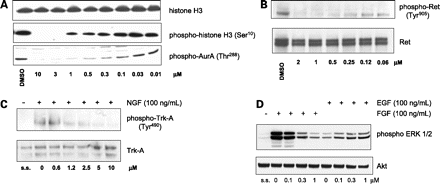
Inhibition of cellular pathways by PHA-739358. Mol Cancer Ther. 2007 Dec;6(12 Pt 1):3158-68. |
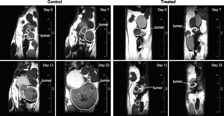
PHA-739358 treatment results in the inhibition of tumor growth in TRAMP mice as assessed by MRI evaluation. Mol Cancer Ther. 2007 Dec;6(12 Pt 1):3158-68. |
Products are for research use only; We do not sell to patients
Copyright 2020 InvivoChem LLC | All Rights Reserved


 COA
COA