| Size | Price | Stock | Qty |
|---|---|---|---|
| 5mg |
|
||
| Other Sizes |
Purity: ≥98%
| Targets |
rRNA synthesis, MIA PaCa-2 cells ( IC50 = 54 nM ); (rRNA synthesis, A375 cells ( IC50 = 113 nM ); rRNA synthesis, HCT-116 cells ( IC50 = 142 nM )
CX-5416 hydrochloride targets RNA polymerase I (Pol I), the enzyme responsible for transcribing ribosomal RNA (rRNA) genes. Pol I is a 14-subunit enzyme complex that localizes to the nucleolus and drives the synthesis of precursor rRNA, which is essential for ribosome biogenesis. In cancer cells, increased Pol I activity supports the elevated protein synthesis required for rapid cell proliferation. CX-5416 selectively inhibits Pol I-mediated rRNA synthesis without significantly affecting Pol II (mRNA synthesis) or Pol III (tRNA synthesis), making it a valuable tool for studying the role of ribosome biogenesis in cancer cell growth and survival. |
|---|---|
| ln Vitro |
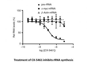
CX-5461 inhibits Pol I-mediated rRNA synthesis potently and orally (IC50s of 142 nM in HCT-116, 113 nM in A375, and 54 nM in MIA PaCa-2 cells). It has negligible or no effect on Pol II (IC50, ≥25 μM). CX-5461 has a slight inhibitory effect on the translation of proteins and DNA. At a mean EC50 of 147 nM, CX-5461 demonstrates extensive antiproliferative activity against a panel of human cancer cell lines. However, its minimal impact on the viability of nontransformed human cells is evident, as its EC50 values are approximately 5000 nM. The CX-5461 EC50 values for the HCT-116, A375, and MIA PaCa-2 cell lines are 167, 58, and 74 nM, in that order. Solid tumor cancer cells are subjected to a p53-independent process by CX-5461 that causes autophagy and senescence instead of apoptosis[1]. With an IC50 of 27.3 nM ± 8.1 nM for Pol I transcription after 1 hour and IC50 of 5.4 nM ± 2.1 nM for cell death after 16 hours, Eμ-Myc lymphoma cells from tumor-bearing mice exhibit exceptional sensitivity to CX-5461. Through the nucleolar stress response in Eμ-Myc Lymphoma Cells, CX-5461 activates p53[2].
CX-5416 hydrochloride demonstrates potent in vitro activity as a Pol I inhibitor. The compound inhibits rRNA synthesis in cancer cell lines with IC50 values of 142 nM in HCT-116 colorectal cancer cells, 113 nM in A375 melanoma cells, and 54 nM in MIA PaCa-2 pancreatic cancer cells. CX-5416 shows little or no effect on Pol II-mediated transcription (IC50 ≥25 μM), confirming its selectivity for Pol I. The compound's ability to selectively inhibit rRNA synthesis makes it a valuable tool for studying the role of ribosome biogenesis in cancer cell proliferation. Detailed cellular activity data are available in the primary literature. |
| ln Vivo |
CX-5461 exhibits antitumor activity against solid tumors of humans in murine xenograft models. Significant MIA PaCa-2 growth inhibition is demonstrated by CX-5461 (50 mg/kg, p.o. ), with TGI equal to 69% on day 31 and 79% on day 32 on A375[1]. At one hour after treatment, CX-5461 (50 mg/kg, p.o.) inhibits Eμ-Myc tumor cells with 84% repression in Pol I transcription in C57BL/6 mice. Additionally, CX-5461 causes the tumor burden in the lymph nodes to rapidly decrease, and it also causes the spleen's size to decrease to within normal limits[2].
In vivo antitumor activity of CX-5461 [1] The antitumor activity of CX-5461 was evaluated in two murine xenograft models of human cancers, pancreatic carcinoma (MIA PaCa-2) and melanoma (A375). In these xenograft models, CX-5461 was administered orally (50 mg/kg) either once daily or every 3 days. Untreated animals received vehicle orally on the equivalent schedule, whereas the positive control gemcitabine was administered intraperitoneally once every 3 days at 120 mg/kg. CX-5461 demonstrated significant MIA PaCa-2 TGI with TGI equal to 69% on day 31 (Fig. 7A), comparable to that of gemcitabine (63% TGI). Likewise, CX-5461 demonstrated significant A375 TGI (Fig. 7B) with TGI equal to 79% on day 32. Unpaired t test revealed statistically significant differences between the vehicle-treated and CX-5461–treated groups throughout both studies. CX-5461 was well tolerated at all tested schedules as judged by the absence of significant changes in animal body weights. These data demonstrate that a selective inhibitor of Pol I transcription can produce in vivo antitumor responses against solid tumors, with a favorable therapeutic window. Inhibition of Pol I Transcription by CX-5461 Selectively Induces p53-Mediated Cell Death of Lymphoma Cells In Vivo while Sparing Normal B Cells [2] We next examined whether inhibition of Pol I transcription and thus activation of the Rp-MDM2-p53 nucleolar stress pathway could be used to selectively kill malignant B cells in vivo. C57BL/6 mice with established disease from transplanted Eμ-Myc lymphoma that is wild-type for p53 (Clone 4242) were treated with a single oral dose of CX-5461 (50 mg/kg) or vehicle. The Eμ-Myc tumor cells infiltrating the lymph nodes showed marked sensitivity to CX-5461, with cells exhibiting an 84% repression in Pol I transcription at 1 hr posttreatment (Figure 5A), also confirmed by RNA chromogenic in situ hybridization (CISH) for 47S pre-rRNA levels in the spleen (Figures S5A and S5B). Moreover, CX-5461 induced a rapid reduction in tumor burden in the lymph nodes (3.1% GFP-tagged malignant cells ± 0.20 for CX-5461-treated versus 34% GFP-tagged malignant cells ± 5.5 for vehicle-treated mice at 24 hr post therapy, p < 0.01) (Figures 5B and S5C) and a concomitant reduction of spleen size to within the normal range (0.14 g ± 0.01 for CX-5461-treated versus 0.47 g ± 0.01 for vehicle-treated mice at 24 hr posttherapy, p < 0.001) (Figure 5C). Consistent with the in vitro studies, reduction in malignant B cell numbers in vivo in response to CX-5461 therapy was preceded by the rapid activation of p53 and p21 and markers of apoptosis within 6 hr of drug administration (Figures 5D–5F and S5D). Although CX-5461 activated p53 and induced apoptosis among malignant cells, it did not trigger these responses in the normal spleen cells of wild-type nontumor bearing mice (Figures 6A and 6B) and did not affect either spleen size or B cell numbers (Figures 6C and 6D). The lack of a cytotoxic effect in normal spleens cells was not due to lack of inhibition of Pol I transcription as CISH demonstrated robust reductions in 47S pre-rRNA levels in the spleens of wild-type nontumor bearing mice as observed in tumor bearing mice (Figures 6E and S6A) Further-more, the nucleolar integrity of normal bone marrow cells from mice treated with CX-5461 was maintained, as determined by FBL and B23 immunofluorescence (Figure 6F). By comparison, exposure of nontumor bearing mice to 5Gy of γ-irradiation, aclinically appropriate dose for hematologic malignancies, resulted in marked elevation of p53 levels, apoptosis, reduced spleen weight, and B cell numbers (Figures 6A–6D). We also examined the response of B220+ B cells from the spleens and bone marrow of 4- to 6-week-old premalignant Eμ-Myc mice. These cells also demonstrated profound sensitivity to CX-5461 as characterized by p53 activation and markers of apoptotic cell death (increased CASP3 cleavage and sub-G1 cells) whereas the normal B cells from age-matched littermate mice failed to show p53 activation or increased cell death (Figures S6B–S6E). Together these data demonstrate the therapeutic response to inhibition of Pol I transcription by CX-5461 in vivo is highly selective for premalignant and malignant cells, which differentiates CX-5461 from genotoxic therapies such as γ-irradiation. To further demonstrate the therapeutic effect of CX-5461 in this model, mice with established transplanted p53 wild-type Eμ-Myc lymphoma (clone 107) were treated with three doses of CX-5461 at 50 mg/kg once every 3 days. This regimen significantly prolonged the survival of the tumor-bearing mice (Figure 7A, p < 0.0001) and restored the white blood cell count to within the normal range (Figure 7B). Indeed, the day following the last dose of CX-5461, there were few identifiable GFP-tagged tumor cells in the peripheral blood (Figures 7C and 7D). Notably, there was preservation and restoration of the recipient-derived nonmalignant normal B cell population, again indicating selective eradication of malignant disease (Figure 7C, bottom panel). Moreover, more prolonged dosing with CX-5461 also showed minimal effects on normal B cells (data not shown). CX-5461 had no discernible adverse effect on the health of the mice and the measured body weights showed minimal deviation below that at the commencement of therapy and in comparison to vehicle-treated mice (Figure 7E). To assess the ability of prolonged dosing of CX-5461 to extend survival, mice transplanted with p53 wild-type Eμ-Myc tumors (clone 4242) were maintained on continuous repeat dosing of CX-5461 at 40 mg/kg once every 3 days (Figure S7A). This treatment regimen reproducibly increased the average survival compared to the three repeat dose regimen with the mice going through a stage of apparent disease remission (no detectable tumor cells in peripheral blood at day 7/8) before eventually relapsing (Figures S7B– S7E). CX-5461 was also evaluated in a murine genetic model of acute myeloid leukemia (AML1/ETO9a+Nras) that reproduces many of the clinical aspects of leukemias in human patients that carry AML1/ETO fusion genes (Zuber et al., 2009). As with therapy in the Eμ-Myc model, transplanted AML1/ETO9a+Nras leukemic cells showed marked sensitivity to CX-5461 in vivo, undergoing p53-dependent apoptotic cell death within 6 hr of treatment of a single 40 mg/kg dose of CX-5461 (Figures S7F– S7I). This apoptotic response was comparable to 100 mg/kg cytarabine, a cytotoxic drug frequently used in treatment of patients with AML. In vivo efficacy data for CX-5416 hydrochloride are not extensively documented in publicly available sources. Based on its potent in vitro activity and oral bioavailability, the compound is expected to have potential utility in cancer models driven by elevated Pol I activity. CX-5416 hydrochloride is described as orally bioavailable, making it suitable for oral administration in animal studies. The compound has been characterized as a Pol I inhibitor with potential anticancer activity. Further in vivo studies are needed to fully characterize the compound's therapeutic potential, including its efficacy in tumor xenograft models, pharmacokinetic properties, and safety profile. The in vitro enzyme inhibition assay for CX-5416 hydrochloride measures the inhibition of Pol I-mediated rRNA synthesis. The assay typically involves incubating cancer cells with varying concentrations of CX-5416 hydrochloride (ranging from nanomolar to micromolar) and measuring the incorporation of radiolabeled uridine or other nucleotide precursors into rRNA. Alternatively, rRNA synthesis can be quantified by qRT-PCR of nascent rRNA transcripts or by measuring the expression of rRNA processing intermediates. IC50 values are determined by fitting dose-response curves to the inhibition data. The compound is dissolved in DMSO and diluted in assay medium to achieve the desired final concentrations, with DMSO concentration kept constant across all wells. Appropriate positive controls and negative controls are included in each assay run. |
| Enzyme Assay |
CX-5461-related effects on transcription are measured by qRT-PCR, which quantifies two short-lived RNA transcripts (half-lives ~20-30 minutes), one produced by Pol I and another by Pol II. As the Pol I transcript, the 45S pre-rRNA functioned, while the comparator Pol II transcript was the protooncogene c-myc mRNA. Cell stress in general is known to impact both Pol I and Pol II transcription. Cells are only exposed to test agents for a brief amount of time (2 hours) in order to reduce any possible effects of such stress. There is enough time for CX-5461 to impact the synthesis of these transcripts, resulting in a reduction of more than 90%.
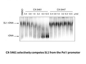
Cell-free Pol I transcription assay [1] A reaction mixture consisting of 30 ng/μL DNA template corresponding to (−160/+379) region on rDNA and 3 mg/mL nuclear extract isolated from HeLa S3 cells in a buffer containing 10 mmol/L Tris-HCl, pH 8.0, 80 mmol/L KCl, 0.8% polyvinyl alcohol, 10 mg/mL α-amanitin was combined with different amounts of test compounds and incubated at ambient for 20 minutes. Transcription was initiated by addition of rNTP mix to a final concentration of 1 mmol/L and was incubated for 1 hour at 30°C. Afterward, DNase I was added and the reaction was further incubated for 2 hours at 37°C. DNase digestion was terminated by the addition of EDTA to final concentration of 10 mmol/L, followed immediately by 10-minute incubation at 75°C, and then samples were transferred to 4°C. The levels of resultant transcript were analyzed by qRT-PCR on 7900HT Real Time PCR System. Chromatin immunoprecipitation [1] Cells were treated with 2 μmol/L of CX-5461 for 1 hour and chromatin immunoprecipitation (ChIP) assay was performed as previously described. Electrophoretic mobility shift assay [1] 32P-labeled DNA probe corresponding to the rDNA promoter was produced by PCR using prHu3 plasmid as a template. The SL1 complex was isolated from HeLa S3 cells nuclear extract as described previously. For the competition studies, a mixture of 5 nmol/L of DNA probe and 0.6 μg SL1 complex was incubated in binding buffer for 15 minutes at ambient temperature with 0.4 to 12.5 μmol/L CX-5461 or CX-5447. The resulting complexes were resolved using Novex TBE DNA retardation PAGE. The gels were dried and exposed to X-ray film [1]. The in vitro cellular assay for CX-5416 hydrochloride is performed using cancer cell lines such as HCT-116, A375, and MIA PaCa-2. Cells are cultured in appropriate medium and treated with varying concentrations of CX-5416 hydrochloride or vehicle control (DMSO) for specified time points (typically 48-72 hours). Cell viability and proliferation are assessed using assays such as MTT, CellTiter-Glo, or by direct cell counting. rRNA synthesis inhibition is confirmed by measuring the incorporation of radiolabeled uridine or by qRT-PCR of rRNA transcripts. The compound's effects on Pol II-mediated transcription are assessed as a control for selectivity. IC50 values are determined for each cell line, and dose-response relationships are established by analyzing cell viability and rRNA synthesis inhibition across different compound concentrations. |
| Cell Assay |
The following day, cells are treated with CX-5461 dose response for 96 hours after being plated on 96-well plates. Alamar Blue and CyQUANT assays are used to measure cell viability[1].
Detection and quantification of acidic vesicular organelles with acridine orange staining [1] Cells were plated on 10-cm dishes and treated the next day with indicated doses of CX-5461 for 24 or 48 hours. At the end of treatment, cells were stained for 15 minutes with 1 μg/mL of acridine orange, trypsinized, washed twice with ice-cold PBS, and analyzed on a BD LSR II flow cytometer. The analysis was performed as previously described. The following day, cells are treated with CX-5461 dose response for 96 hours after being plated on 96-well plates. Alamar Blue and CyQUANT assays are used to measure cell viability. In vivo animal experiments with CX-5416 hydrochloride are not extensively described in publicly available sources. Based on its in vitro activity and oral bioavailability, potential in vivo studies would likely use immunocompromised mice bearing human cancer xenografts (e.g., HCT-116, A375, or MIA PaCa-2). Tumor cells would be implanted subcutaneously into the flank of nude or SCID mice. When tumors reach a predetermined size, animals would be randomized into treatment groups receiving CX-5416 hydrochloride or vehicle control. The compound would be administered orally due to its oral bioavailability. Tumor volume would be measured twice weekly using calipers, and body weight monitored to assess tolerability. At study endpoint, tumors would be harvested for analysis of rRNA synthesis inhibition, Pol I target engagement, and markers of proliferation and apoptosis. |
| Animal Protocol |
Mice: Female Balb/c athymic mice (NCr nu/nu fisol) aged 5 to 6 weeks are used in animal experiments. Hypnic (NCr nu/nu fisol) mice are injected subcutaneously in the right flank of the mice with 100 μL of cell suspension. Calculating the tumor volume involves applying the formula (l×w2)/2, where w represents the tumor's width and l its length in millimeters. Tumor measurements are carried out through caliper analysis. randomized to treatment groups with either vehicle (50 mM NaH2PO4, pH 4.5), NSC 613327, or CX-5461 for established tumors (approx 110-120 mm3). On the last day of the study, tumor growth inhibition (TGI) is calculated using the following formula: TGI (%)=[100 − (VfD− ViD)/ (VfV − ViV) × 100], where ViV represents the initial mean tumor volume in the vehicle-treated group, VfV represents the final mean tumor volume in the vehicle-treated group, ViD represents the initial mean tumor volume in the drug-treated group, and VfD represents the final mean tumor volume in the drug-treated group on the study day.
In vivo efficacy in murine xenograft model [1] Mice were inoculated with 5 × 10⁶ in 100 μL of cell suspension subcutaneously in the right flank. Tumor measurements were performed by caliper analysis, and tumor volume was calculated using the formula (l × w2)/2, where w = width and l = length in mm of the tumor. Established tumors (∼110–120 mm3) were randomized into vehicle (50 mmol/L NaH2PO4, pH 4.5), gemcitabine, or CX-5461 treatment groups. Tumor growth inhibition (TGI) was determined on the last day of study according to the formula: TGI (%) = [100 − (VfD − ViD)/ (VfV − ViV) × 100], where ViV is the initial mean tumor volume in vehicle-treated group, VfV is the final mean tumor volume in vehicle-treated group, ViD is the initial mean tumor volume in drug-treated group, and VfD is the final mean tumor volume in drug-treated group. MV 4;11Xenograft Model [1] Female immunocompromised mice CrTac:Ncr-Foxn1nu (6–8 weeks old) were maintained under clean room conditions in sterile filter top cages. Xenografts were initiated by subcutaneous injection of 5 × 10⁶ MV4;11 cells into the right hind flank region of each mouse. When tumors reached a designated volume of ∼150 mm3, animals were randomized and divided into Vehicle (50 mM NaH2PO4, pH 4.5) or CX-5461 treatment groups of 10 mice per group. CX-5461 was administered intraperitoneally once a week at 125 mg/kg for the length of 25 days. Tumor volumes and body weights were measured twice weekly. The length and width of the tumor were measured with calipers and the volume calculated using the following formula:tumor volume = (length × width2)/2. Tumor growth inhibition (TGI) was determined on the last day of study according to the formula: TGI (%) = [100 – (VfD – ViD)/(VfV – ViV) × 100], where ViV is the initial mean tumor volume in vehicle-treated group, VfV is the final mean tumor volume in vehicle-treated group, ViD is the initial mean tumor volume in drug-treated group, and VfD is the final mean tumor volume in drug-treated group. Detailed pharmacokinetic (PK) parameters for CX-5416 hydrochloride are not extensively documented in publicly available sources. The compound is described as orally bioavailable, indicating that it has adequate oral absorption for in vivo studies. CX-5416 hydrochloride has a molecular weight of 550.07 and a chemical formula of C27H28ClN7O2S. The compound is soluble in DMSO for formulation purposes. For in vivo oral administration, the compound would need to be formulated using appropriate vehicles to ensure adequate solubility and stability. The compound should be stored at -20°C under dry, sealed conditions, away from moisture. Detailed PK parameters including half-life, clearance, volume of distribution, and maximum concentration (Cmax) are not available from the current search results and would require consultation of the primary literature. |
| ADME/Pharmacokinetics |
Comprehensive toxicological data for CX-5416 hydrochloride are not extensively documented in publicly available sources. As a research-grade compound, CX-5416 hydrochloride is intended for laboratory research purposes only and is not approved for human therapeutic use. Standard laboratory safety practices should be followed when handling this compound, including the use of appropriate personal protective equipment and working in a well-ventilated area. The compound should be stored according to the manufacturer's recommendations to maintain stability and prevent degradation. Comprehensive toxicological profiling (e.g., LD50, maximum tolerated dose, organ-specific toxicity) is not available from the current search results and would require consultation of the primary literature or safety data sheets.
|
| Toxicity/Toxicokinetics |
CX-5416 hydrochloride is a research compound developed for studying the role of Pol I-mediated rRNA synthesis in cancer cell proliferation and ribosome biogenesis. The compound's potent inhibition of Pol I (IC50 values of 54-142 nM across cancer cell lines) and selectivity over Pol II (IC50 ≥25 μM) make it a valuable tool for dissecting the contributions of rRNA synthesis to cancer cell growth and survival. CX-5416 hydrochloride is orally bioavailable, supporting its utility for in vivo studies. The compound is not currently in clinical trials nor approved for therapeutic use; it remains an investigational tool compound for preclinical cancer research. CX-5416 hydrochloride is available from various chemical suppliers for research purposes.
|
| References |
|
| Additional Infomation |
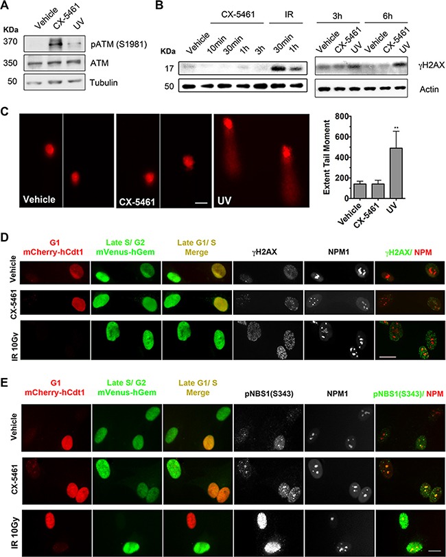
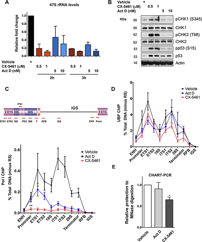
CX-5461 is an organic heterotetracyclic compound with the structure 5-oxo-5H-[1,3]benzothiazo[3,2-a][1,8]naphthidine-6-carboxylic acid, where the 2-position is substituted with a 4-methyl-1,4-diazacycloheptane-1-yl group and the 6-carboxyl group is substituted with a [(5-methylpyrazin-2-yl)methyl]nitrile group. CX-5461 is a ribosomal RNA (rRNA) synthesis inhibitor that specifically inhibits RNA polymerase I-driven rRNA transcription in various cancer cell lines. Currently, CX-5461 is undergoing Phase I/II clinical trials for advanced hematologic malignancies and triple-negative breast cancer with BRCA1/2 mutations. CX-5461 exhibits antitumor activity, induces apoptosis, and inhibits EC 2.7.7.6 (RNA polymerase). It is a diazaphene compound, belonging to the organic heterotetracyclic class of compounds, and is a pyrazine, secondary amide, and naphthidine derivative. Pirnalutrex is an orally bioavailable RNA polymerase I (Pol I) inhibitor with potential antitumor activity. After oral administration, pinenalutrex selectively binds to and inhibits Pol I, blocking Pol I-mediated ribosomal RNA (rRNA) synthesis, inducing apoptosis, and inhibiting tumor cell growth. Pol I is a multi-protein complex that synthesizes rRNA, is upregulated in cancer cells, and plays a crucial role in cell proliferation and survival. Excessive activation of rRNA transcription is associated with uncontrolled proliferation of cancer cells. Dysregulation of ribosomal RNA synthesis is associated with uncontrolled proliferation of cancer cells. RNA polymerase (Pol) I is a multi-protein complex that synthesizes rRNA and is widely activated in cancer. Therefore, selective Pol I inhibitors may provide a universal therapeutic strategy for blocking cancer cell proliferation. By combining medicinal chemistry research with tandem screening based on cells and molecules, CX-5461, a potent small-molecule rRNA synthesis inhibitor, was designed. CX-5461 selectively inhibits Pol I-driven transcription, while having no significant effect on Pol II-driven transcription, DNA replication, and protein translation. Molecular studies have shown that CX-5461 inhibits the initiation of rRNA synthesis and induces senescence and autophagy in solid tumor cell lines through a p53-independent pathway, but does not induce apoptosis. CX-5461 has good oral bioavailability and has shown in vivo antitumor activity against human solid tumors in a mouse xenograft model. Our results suggest that CX-5461 is a promising, potent, selective and orally available cancer treatment that could be used in clinical trials. [1] RNA polymerase I-mediated enhanced transcription of ribosomal RNA genes (rDNA) is a common feature of human cancers, but it is unclear whether it is necessary for malignant phenotypes. We found that the small molecule CX-5461 can act as a targeted therapy for rDNA transcription, selectively killing B lymphoma cells in vivo while maintaining the survival of wild-type B cell populations. This therapeutic effect is due to nucleolar destruction and activation of p53-dependent apoptosis signaling. Human leukemia and lymphoma cell lines also show high sensitivity to rDNA transcription inhibitors, and this sensitivity depends on the mutation status of p53. These results suggest that selective inhibition of rDNA transcription can be used as a therapeutic strategy to specifically activate p53 and treat hematologic malignancies. [2] RNA polymerase I (Pol I)-mediated transcription of ribosomal RNA genes (rDNA) is confined to the nucleolus and is the rate-limiting step in cell growth and proliferation. CX-5461 inhibition of Pol I selectively induces p53-mediated apoptosis in tumor cells in vivo. Currently, CX-5461 is undergoing clinical trials at Peter McKenna Hospital in Melbourne for patients with advanced hematologic malignancies. This paper demonstrates that CX-5461 can induce p53-independent cell cycle checkpoints mediated by the ATM/ATR signaling pathway without DNA damage. Furthermore, our data show that the combination of drugs targeting the ATM/ATR signaling pathway with CX-5461 can significantly improve the therapeutic effect of p53-deficient tumors in vivo, which are usually unresponsive to single drugs. Mechanistically, we found that CX-5461 induces an aberrant chromatin structure in which transcriptionally active relaxed rDNA repeat sequences lack transcription of Pol I, thereby activating the ATM signaling pathway within the nucleolar. Therefore, we propose that the acute inhibition of Pol transcription initiation by CX-5461 induces a novel nucleolar stress response, which could serve as a target to enhance therapeutic efficacy. (Oncotarget. 2016 Aug 2;7(31):49800-49818.)
|
| Molecular Formula |
C27H28CLN7O2S
|
|
|---|---|---|
| Molecular Weight |
550.074922561646
|
|
| Exact Mass |
549.171372
|
|
| Elemental Analysis |
C, 58.95; H, 5.13; Cl, 6.44; N, 17.82; O, 5.82; S, 5.83
|
|
| CAS # |
2101314-20-7
|
|
| Related CAS # |
1138549-36-6
|
|
| PubChem CID |
131699687
|
|
| Appearance |
Light yellow to yellow solid powder
|
|
| Hydrogen Bond Donor Count |
2
|
|
| Hydrogen Bond Acceptor Count |
9
|
|
| Rotatable Bond Count |
4
|
|
| Heavy Atom Count |
38
|
|
| Complexity |
915
|
|
| Defined Atom Stereocenter Count |
0
|
|
| SMILES |
C(C1C(C2=CC=C(N3CCCN(C)CC3)N=C2N2C3=CC=CC=C3SC=12)=O)(=O)NCC1=NC=C(C)N=C1.Cl
|
|
| InChi Key |
LXNKBUVQVKWAHI-UHFFFAOYSA-N
|
|
| InChi Code |
InChI=1S/C27H27N7O2S.2ClH/c1-17-14-29-18(15-28-17)16-30-26(36)23-24(35)19-8-9-22(33-11-5-10-32(2)12-13-33)31-25(19)34-20-6-3-4-7-21(20)37-27(23)34;;/h3-4,6-9,14-15H,5,10-13,16H2,1-2H3,(H,30,36);2*1H
|
|
| Chemical Name |
2-(4-methyl-1,4-diazepan-1-yl)-N-[(5-methylpyrazin-2-yl)methyl]-5-oxo-[1,3]benzothiazolo[3,2-a][1,8]naphthyridine-6-carboxamide;dihydrochloride
|
|
| Synonyms |
|
|
| HS Tariff Code |
2934.99.9001
|
|
| Storage |
Powder -20°C 3 years 4°C 2 years In solvent -80°C 6 months -20°C 1 month Note: Please store this product in a sealed and protected environment, avoid exposure to moisture. |
|
| Shipping Condition |
Room temperature (This product is stable at ambient temperature for a few days during ordinary shipping and time spent in Customs)
|
| Solubility (In Vitro) |
|
|||
|---|---|---|---|---|
| Solubility (In Vivo) |
Solubility in Formulation 1: 6.25 mg/mL (10.66 mM) in PBS (add these co-solvents sequentially from left to right, and one by one), clear solution; with sonication (<60°C).
(Please use freshly prepared in vivo formulations for optimal results.) |
| Preparing Stock Solutions | 1 mg | 5 mg | 10 mg | |
| 1 mM | 1.8179 mL | 9.0896 mL | 18.1792 mL | |
| 5 mM | 0.3636 mL | 1.8179 mL | 3.6358 mL | |
| 10 mM | 0.1818 mL | 0.9090 mL | 1.8179 mL |
*Note: Please select an appropriate solvent for the preparation of stock solution based on your experiment needs. For most products, DMSO can be used for preparing stock solutions (e.g. 5 mM, 10 mM, or 20 mM concentration); some products with high aqueous solubility may be dissolved in water directly. Solubility information is available at the above Solubility Data section. Once the stock solution is prepared, aliquot it to routine usage volumes and store at -20°C or -80°C. Avoid repeated freeze and thaw cycles.
Calculation results
Working concentration: mg/mL;
Method for preparing DMSO stock solution: mg drug pre-dissolved in μL DMSO (stock solution concentration mg/mL). Please contact us first if the concentration exceeds the DMSO solubility of the batch of drug.
Method for preparing in vivo formulation::Take μL DMSO stock solution, next add μL PEG300, mix and clarify, next addμL Tween 80, mix and clarify, next add μL ddH2O,mix and clarify.
(1) Please be sure that the solution is clear before the addition of next solvent. Dissolution methods like vortex, ultrasound or warming and heat may be used to aid dissolving.
(2) Be sure to add the solvent(s) in order.
| NCT Number | Recruitment | interventions | Conditions | Sponsor/Collaborators | Start Date | Phases |
| NCT04890613 | Recruiting | Drug: CX-5461 | COVID-19 | Senhwa Biosciences, Inc. | September 8, 2021 | Phase 1 |
| NCT02719977 | Completed | Drug: CX5461 | Cancer | Canadian Cancer Trials Group | June 13, 2016 | Phase 1 |
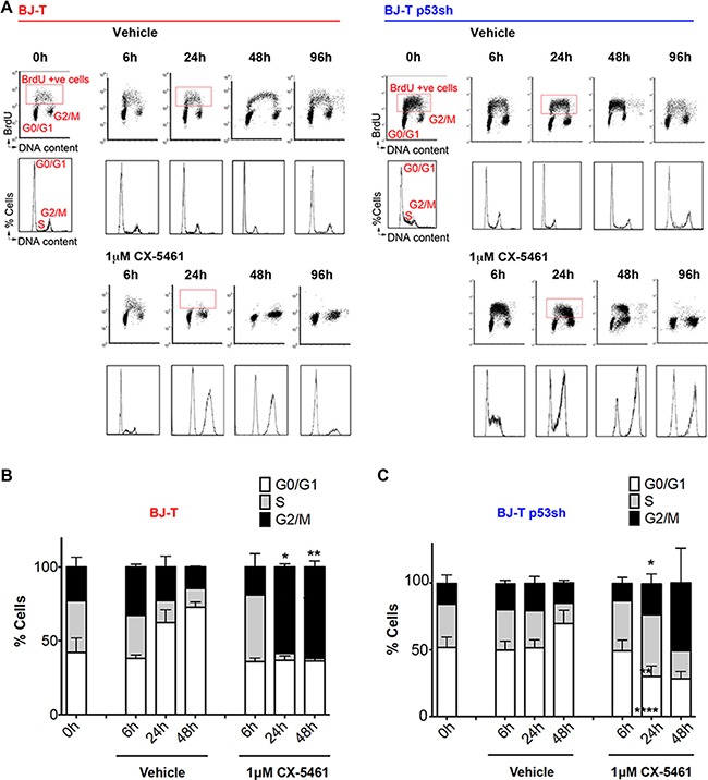
BJ-T and BJ-T p53sh cells exhibit G1 arrest, S-phase delay and G2 cell cycle arrest in response to CX-5461.Oncotarget.2016 Aug 2;7(31):49800-49818. |
|---|
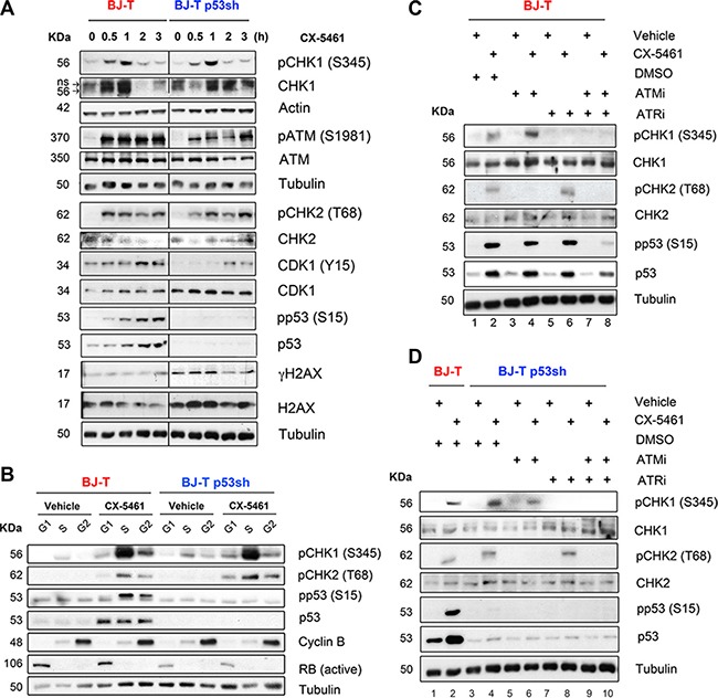
Inhibition of Pol I transcription initiation by CX-5461 activates the ATM/ATR signaling pathway.Oncotarget.2016 Aug 2;7(31):49800-49818. |
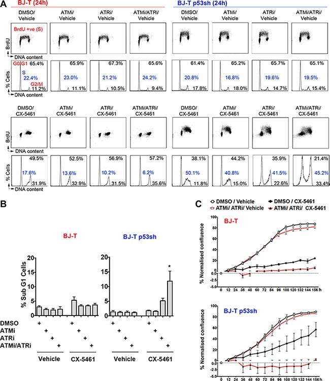
Combination treatment of ATM and ATR inhibitors with CX-5461 induces cell death in the absence of p53.Oncotarget.2016 Aug 2;7(31):49800-49818. |
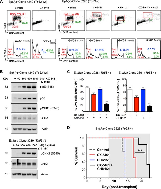
CX-5461 combination with a dual CHK1/CHK2 inhibitor induces cancer cell death ofTp53-null (Tp53−/−)Eμ-Myclymphoma cellsin vitroandin vivo.Oncotarget.2016 Aug 2;7(31):49800-49818. |
|---|
CX-5461 activates ATM signaling within the nucleoli in the absence of DNA damage.Oncotarget.2016 Aug 2;7(31):49800-49818. |
Inhibition of Pol I transcription initiation by CX-5461 results in rDNA repeats that are devoid of Pol I, but maintain an exposed chromatin state that associates with ATM/ATR pathway activation.Oncotarget.2016 Aug 2;7(31):49800-49818. |
|
|---|
|
|
Products are for research use only; We do not sell to patients
Copyright 2020 InvivoChem LLC | All Rights Reserved


 COA
COA